Investigating the reassortment potential and pathogenicity of the S segment in Akabane virus using a reverse genetics system - PubMed (original) (raw)
Investigating the reassortment potential and pathogenicity of the S segment in Akabane virus using a reverse genetics system
Eun-Jee Na et al. BMC Vet Res. 2025.
Abstract
Background: Akabane virus (AKAV) is an arthropod-borne virus that causes congenital malformations and neuropathology in cattle and sheep. In South Korea, AKAVs are classified into two main genogroups: K0505 and AKAV-7 strains. The K0505 strain infects pregnant cattle, leading to fetal abnormalities, while the AKAV-7 strain induces encephalomyelitis in post-natal cattle. The pathogenicities of K0505 and AKAV-7 strains differ significantly; however, the specific gene in the AKAV-7 strain that drives its pathogenicity remains unidentified. In this study, changes in viral replication and pathogenicity were investigated, particularly when the S segment of AKAV-7 was mutated using a T7 RNA polymerase-based reverse genetics (RG) system.
Results: The rAKAV-7ΔNSs virus, with a deletion in the NSs protein of the wild-type AKAV-7 virus (wtAKAV-7), and the rAKAV-7(S-K0505) virus, where the S segment of wtAKAV-7 was reassorted with that from the wild type K0505 strain (wtK0505), were successfully rescued. The rAKAV-7ΔNSs virus demonstrated impaired replication in Vero cells and exhibited reduced mortality and RNA viral load in the organs of suckling mice compared to the wtAKAV-7. The rAKAV-7(S-K0505) virus displayed similar growth kinetics in Vero cells and showed no significant reduction in mortality rate in suckling mice compared to wtAKAV-7.
Conclusions: These observations suggest that the S segment, especially the NS protein, is associated with the pathogenicity of AKAV-7. Also, the results imply that the L and M segments might explain the differences in pathogenicity between the AKAV-7 and K0505 strains. Moreover, our findings indicate the potential for reassortment between distinct genogroups of AKAVs.
Keywords: Akabane virus; Pathogenicity; Reassortment; Reverse genetics system; S segment; Suckling mouse.
© 2025. The Author(s).
Conflict of interest statement
Declarations. Ethics approval and consent to participate: All animal experiments were conducted in an animal biosafety level 2 laboratory. The procedures were performed in accordance with the Guide for the Care and Use of the National Institutes of Health (Edition, 2011). The experimental protocols were approved by the Ethics Committees of Jeonbuk National University (approval number: NON2024-020–001). Consent for publication: Not applicable. Competing interests: The authors declare no competing interests.
Figures
Fig. 1
Scheme for the reverse genetics (RG) system of AKAVs. For construction of the transcription plasmid, full-length cDNA fragments of the L, M, and S segments were cloned into the TVT7R vector, containing T7 promoter and HDV sequence. To construct the helper plasmids, ORFs of the RdRp and N were cloned into the pTM1 vector. Three transcription plasmids and two helper plasmids were co-transfected into BHK/T7-9 cells. The supernatants of BHK/T7-9 cells were passaged in Vero cells, and the rAKAV-7, rAKAV-7(S-K0505), and rAKAV-7ΔNSs viruses were rescued
Fig. 2
IFA identification of wild type and rescued AKAVs in Vero cells. Vero cells were infected using a MOI of 0.01. At 18 h post infection, fluorescence of FITC (green) was observed, and nuclei were counterstained with NucBlue (blue). The DMEM (A), wtAKAV-7 (B), wtK0505 (C), rAKAV-7 (D), rAKAV-7(S-K0505) (E), and rAKAV-7ΔNSs (F) are displayed. Images were captured using an EVOS M5000 Imaging System. Scale bars represent 100 μm
Fig. 3
Multi-step growth curves of wild type and rescued AKAVs in Vero cells. Vero cells were infected using a MOI of 0.01. Supernatants from Vero cells were collected at 12, 24, 36, 48, 60, and 72 hpi. Dagger (†) and double dagger (‡) symbols indicate significant differences between AKAV-7ΔNSs and the other viruses (p < 0.05). Data points represent means, and error bars indicate standard deviation (SD). Statistical significance was determined by two-way ANOVA (Tukey's test)
Fig. 4
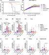
Body weight, body temperature, survival rate, and AKAV copy numbers of BALB/c suckling mice infected with wild type and rescued AKAVs were monitored over 14 days post-infection. Survival rate (A) and body weight (%) (B) were analyzed. Additionally, brain (C), spinal cord (D), lung (E), liver (F), spleen (G), kidney (H), and small intestine (I) were harvested for AKAV copy number analysis, which was conducted using qPCR. Data are presented as means ± SD. Viral RNA levels were analyzed using one-way ANOVA (Tukey’s test). The viral RNA load for euthanized or succumbed mice is highlighted with a red border. *, p < 0.05; **, p < 0.01; ***, p < 0.001; ****, p < 0.0001
Fig. 5
Histopathological findings in the brain (cerebrum) and lumbar spinal cord sections of mice infected with wild type and rescued AKAVs: DMEM (A, F); wtK0505 (B, G), wtAKAV-7 (C, H); rAKAV-7(S-K0505) (D, I) and rAKAV-7ΔNSs (E, J) (H&E). An arrow indicates neuronal necrosis, an arrowhead points to gliosis, asterisks denote inflammatory cell infiltration into perivascular and neuronal areas, and a crosshatch signifies spongiosis. Scale bars measure 100 μm. Immunohistochemistry (IHC) detected anti-AKAV N protein in brain (cerebrum) and lumbar spinal cord sections from infections with wild type and ruscued AKAVs; sections include: DMEM, brain (K); wtK0505, brain (L); wtAKAV-7, brain (M); rAKAV-7(S-K0505), spinal cord (N) and rAKAV-7ΔNSs, spinal cord (O). Positive staining appears brown in the cytoplasm. Scale bars measure 50 μm
Similar articles
- Erratum: Eyestalk Ablation to Increase Ovarian Maturation in Mud Crabs.
[No authors listed] [No authors listed] J Vis Exp. 2023 May 26;(195). doi: 10.3791/6561. J Vis Exp. 2023. PMID: 37235796 - Viromics-based precision diagnosis of reproductive abnormalities in cows reveals a reassortant Akabane disease virus.
Sun Y, Zhang R, Wang H, Sun Z, Yi L, Tu C, Yang Y, He B. Sun Y, et al. BMC Vet Res. 2024 Nov 29;20(1):539. doi: 10.1186/s12917-024-04400-5. BMC Vet Res. 2024. PMID: 39614255 Free PMC article. - Bioluminescent and fluorescent reporter-expressing recombinant Akabane virus (AKAV): an excellent tool to dissect viral replication.
Liu J, Wang F, Zhao J, Qi Y, Chang J, Sun C, Jiang Z, Ge J, Yin X. Liu J, et al. Front Microbiol. 2024 Nov 14;15:1458771. doi: 10.3389/fmicb.2024.1458771. eCollection 2024. Front Microbiol. 2024. PMID: 39611086 Free PMC article. - Depressing time: Waiting, melancholia, and the psychoanalytic practice of care.
Salisbury L, Baraitser L. Salisbury L, et al. In: Kirtsoglou E, Simpson B, editors. The Time of Anthropology: Studies of Contemporary Chronopolitics. Abingdon: Routledge; 2020. Chapter 5. In: Kirtsoglou E, Simpson B, editors. The Time of Anthropology: Studies of Contemporary Chronopolitics. Abingdon: Routledge; 2020. Chapter 5. PMID: 36137063 Free Books & Documents. Review. - Impact of residual disease as a prognostic factor for survival in women with advanced epithelial ovarian cancer after primary surgery.
Bryant A, Hiu S, Kunonga PT, Gajjar K, Craig D, Vale L, Winter-Roach BA, Elattar A, Naik R. Bryant A, et al. Cochrane Database Syst Rev. 2022 Sep 26;9(9):CD015048. doi: 10.1002/14651858.CD015048.pub2. Cochrane Database Syst Rev. 2022. PMID: 36161421 Free PMC article. Review.
References
- De Regge N. Akabane, Aino and Schmallenberg virus—where do we stand and what do we know about the role of domestic ruminant hosts and Culicoides vectors in virus transmission and overwintering? Curr Opin Virol. 2017;27:15–30. - PubMed
- Murata Y, Uchida K, Shioda C, Uema A, Bangphoomi N, Chambers J, Akashi H, Nakayama HJJoCP: Histopathological studies on the neuropathogenicity of the Iriki and OBE-1 strains of Akabane virus in BALB/cAJcl mice. J Comp Pathol. 2015;153(2–3):140–149.
- Abudurexiti A, Adkins S, Alioto D, Alkhovsky SV, Avšič-Županc T, Ballinger MJ, Bente DA, Beer M, Bergeron É, Blair CD. Taxonomy of the order Bunyavirales: update 2019. Adv Virol. 2019;164:1949–65.
- Oem J-K, Yoon H-J, Kim H-R, Roh I-S, Lee K-H, Lee O-S, Bae Y-C. Genetic and pathogenic characterization of Akabane viruses isolated from cattle with encephalomyelitis in Korea. Vet Microbiol. 2012;158(3–4):259–66. - PubMed
MeSH terms
LinkOut - more resources
Full Text Sources
Medical
Miscellaneous


