Macular microvascular and structural changes on optical coherence tomography angiography in atypical optic neuritis (original) (raw)
Observational Study
Copyright ©The Author(s) 2025. Published by Baishideng Publishing Group Inc. All rights reserved.
World J Methodol. Mar 20, 2025; 15(1): 98482
Published online Mar 20, 2025. doi: 10.5662/wjm.v15.i1.98482
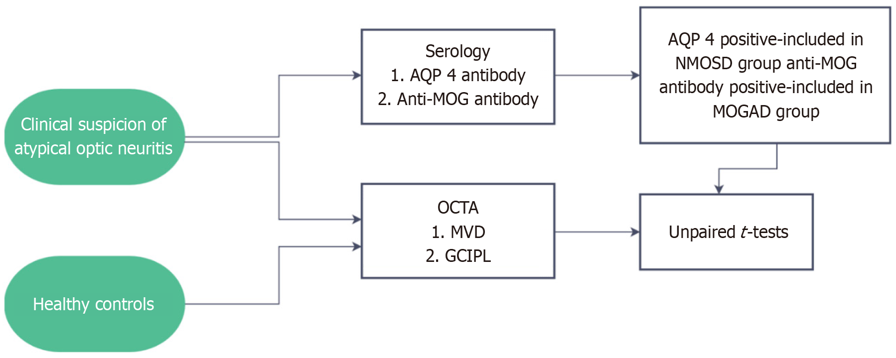
Figure 1 Schematic representation of the methodology for the study. NMOSD: Neuromyelitis optica spectrum disorders; MOGAD: Myelin oligodendrocyte glycoprotein antibody disorder; MVD: Macular vascular density; AQP 4: Aquaporin-4; OCTA: Optical coherence tomography angiography; GCIPL: Ganglion cell + inner plexiform layer.
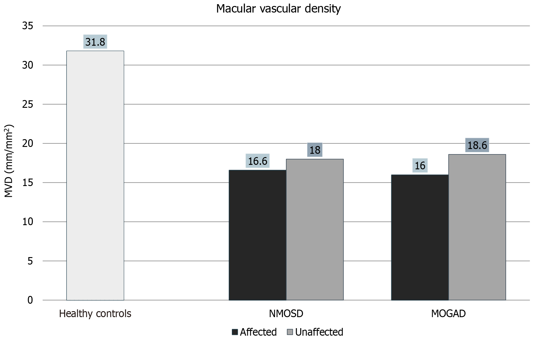
Figure 2 Reduction in macular vascular density in neuromyelitis optica spectrum disorders and myelin oligodendrocyte glycoprotein antibody disorder affected as well as unaffected eyes compared to controls. NMOSD: Neuromyelitis optica spectrum disorders; MOGAD: Myelin oligodendrocyte glycoprotein antibody disorder; MVD: Macular vascular density.
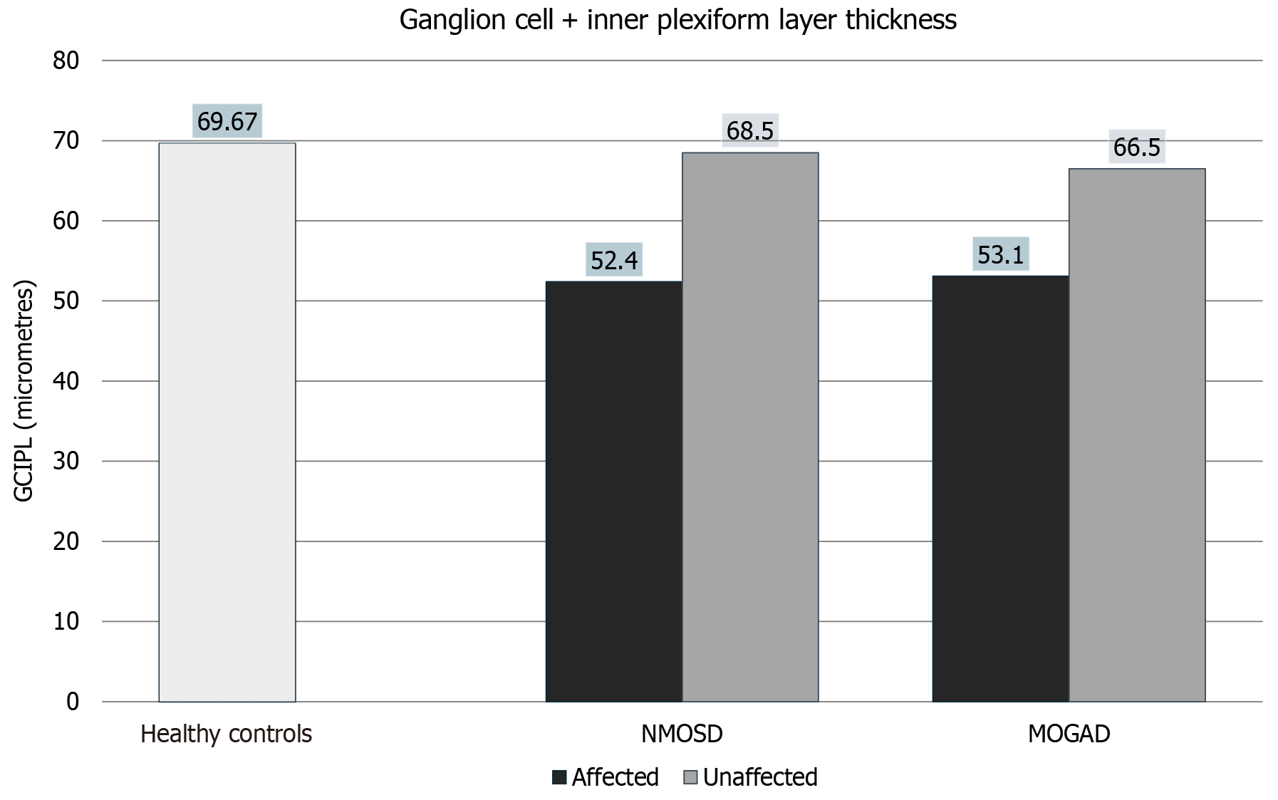
Figure 3 Ganglion cell + inner plexiform layer thickness in neuromyelitis optica spectrum disorders and myelin oligodendrocyte glycoprotein antibody disorder eyes compared with controls. NMOSD: Neuromyelitis optica spectrum disorders; MOGAD: Myelin oligodendrocyte glycoprotein antibody disorder; GCIPL: Ganglion cell + inner plexiform layer.
- Citation: Mahatme C, Kaushik M, Saravanan VR, Kumar K, Shah VM. Macular microvascular and structural changes on optical coherence tomography angiography in atypical optic neuritis. World J Methodol 2025; 15(1): 98482
- URL: https://www.wjgnet.com/2222-0682/full/v15/i1/98482.htm
- DOI: https://dx.doi.org/10.5662/wjm.v15.i1.98482


