Options available for profiling small samples: a review of sample amplification technology when combined with microarray profiling (original) (raw)
Abstract
The possibility of performing microarray analysis on limited material has been demonstrated in a number of publications. In this review we approach the technical aspects of mRNA amplification and several important implicit consequences, for both linear and exponential procedures. Amplification efficiencies clearly allow profiling of extremely small samples. The conservation of transcript abundance is the most important issue regarding the use of sample amplification in combination with microarray analysis, and this aspect has generally been found to be acceptable, although demonstrated to decrease in highly diluted samples. The fact that variability and discrepancies in microarray profiles increase with minute sample sizes has been clearly documented, but for many studies this does appear to have affected the biological conclusions. We suggest that this is due to the data analysis approach applied, and the consequence is the chance of presenting misleading results. We discuss the issue of amplification sensitivity limits in the light of reports on fidelity, published data from reviewed articles and data analysis approaches. These are important considerations to be reflected in the design of future studies and when evaluating biological conclusions from published microarray studies based on extremely low input RNA quantities.
MICROARRAY TECHNOLOGY
In 2005 we reached the decade mark for the microarray technique, one of the most powerful high throughout gene expression technologies. Microarray technology was introduced in a seminal paper by Schena et al. (1) and was an initiating step towards a microrevolution in the field of molecular biology. Genomic advances, particularly in sequencing projects, have provided the basis for microarray construction applicable to a wide range of species. The technique provides a snapshot of the repertoire of genes expressed by a cell or tissue at the time of harvest and RNA purification. A series of samples can be compared horizontally, gene by gene for all the genes, to obtain an expression profile. In biomedicine, detection of differences and alterations in expression patterns holds the potential to yield valuable insight into a broad range of biological processes accompanying normal or diseased tissue, disease prediction, diagnostics and treatment, cellular differentiation, development and drug discovery. The technology has become widespread for investigational purposes, particularly within some fields of research, and availability is now less of an issue. Production facilities are commonly found at many academic institutions, and a wide range of products from necessary laboratory equipment to experimental reagents are commercially available.
The development of the technology is still progressing. However, the main focus has shifted from the technology itself to the pre- and post-processes related to the experiments, more specifically, to study-design and material preparation, in addition to data management and mining. The shift in focus is partially driven by the urge to expand the use of microarrays and also expand extractable knowledge and to avoid misinterpretation. It is important to emphasize that although microarrays are powerful biological tools, there are potential pitfalls that can attenuate their power. Critical evaluation of what we put into the experiment followed by relevant processing of data out is essential. Despite of the pitfalls, microarray technology is certainly making an impact, not least within cancer research. The numerous exploratory trials to scan for robust patterns of data (signatures) within patient groups that classify and/or correlate with clinical data, support the vision of the use of arrays as a routine clinical tool. However, realization of this vision has been burdened with validation issues regarding the lists of genes included in a molecular signature (2). To date, only one array-based analysis tool, the DNA-based AmpliChip CYP450 genotyping test, has been cleared for clinical use in the US and the EU. Through standardization and validation, customized RNA-based arrays are likely to eventually follow.
MATERIAL REQUIREMENT IN THE EARLY YEARS OF THE MICROARRAY ERA
One of the main reasons why the microarray technology initially failed to enter a number of research fields was the large amount of material that each experiment required. In the introductory microarray technology paper, the amount of mRNA used for target preparation was 5 µg (1). In terms of total RNA, this figure converts to 165–500 µg assuming 1–3% of total RNA consists of mRNA. In a collection of microarray review papers from 1999, the total RNA quantity requirement was noted to be 50–200 µg (3). As one cell contains 10–30 pg total RNA, the number of cells required to obtain 50–200 µg spans from 1.6 × 10⁶ to 2 × 10⁷. Needless to say, not all investigators could provide those quantities of their cells of interest and were thus prevented from using micorarray technology. One solution to investigate homogenous cells from a specific cell type or diseased tissue, was to culture cells from freshly taken clinical samples. The drawback of using short-term cultures is that cells are separated from the natural microenvironment, and changes in gene expression due to handling could potentially confound biological findings. The advantage with cancer research has been that material from tumor tissue or cell lines could often be supplied in large quantities. However, for investigators who can provide bulk tissue, the question is whether it is possible to decipher the complex expression patterns and extract the gene expression profile from cells of interest, considering the heterogeneity of cell types present in the tissue. Studies have shown that gene expression profiling of bulk tissue yields the profile of the dominating cell type while the minor cell subsets are washed out (4). In addition, high-abundance messages from cells of interest, as well as contaminating cells, can easily obscure measurements from low abundance transcripts. Hence, profiling bulk tissue has its limitations. However, advances in technology designed for selective collection of specialized cells, such as laser capture microdissection (LCM), could not be fully exploited in combination with microarray analysis. Although LCM allowed the fine precision of laser dissection of single cells, the number of laser firings had to be increased to answer to the dilemma of sufficient material for gene expression profiling. When regarding the hopes and intentions to use of microarrays as routine diagnostic tools, it is in fact ironic that the best choice of feasible patient sampling, biopsies, were excluded by standard microarray protocols due to limited material. Thus to exploit the potential of microarray technology and to profile purified cell populations of interest, this large material requirement had to be overcome in a quantitatively valid manner. Strategies to reduce the required sample sizes have been technically relatively successful. However, the lower sensitivity limits (lower input quantities) with respect to accurate, reliable expression monitoring are rarely investigated, and pose a potential limitation to the use microarray technology that cannot be overlooked.
TWO APPROACHES TO OVERCOME THE SUBSTANTIAL MATERIAL REQUIREMENT
Efforts to reduce the amount of RNA needed for target preparation have focused on two main strategies, signal amplification and sample amplification. The first strategy entails improving and optimizing the labeling reaction in order to increase the number of signal molecules per transcript. Augmenting the number of signals per transcript can be achieved by technologies such as dendrimer (5) or tyramide signal amplification (TSA) (6). The dendrimer technology uses a two-step hybridization procedure. First, cDNA is synthesized using an oligo dT primer containing a capture sequence and subsequently hybridized to the array. Second, dendrimers, containing a multitude of fluorescent molecules per dendrimer, are hybridized to the capture sequence. The claims of this technology is less material needed, no dye bias and improved signal to background ratio compared to conventional labeling. Improvements increasing signal output can also be achieved by applying enhanced reagents in the standard protocol, alternative labeling strategies, or alternative signal molecules. Considerable optimization of the target preparation has in fact occurred in the commercial sector. A probable driving force is the advantage gain in terms of sensitivity claims of the various reagents or labeling kits. For indirect labeling, the latest version of a commonly used kit (Fairplay II, Stratagene) claims that as little as 2 µg of total RNA (a minimum of ∼7 × 10⁴ cells) is required. In addition, another brand allows the use of 3 µg total RNA (Atlas Powerscript Kit, Clontech). A dendrimer technology based product (Genisphere) claims that the use of 0.25–1 µg total RNA (2.5 × 10⁴–1 × 10⁵) is sufficient for a target preparation leading to good quality arrays.
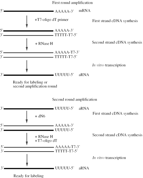
The minimum number of cells required by the commercial products mentioned above was 2.5 × 10⁴. This number still exceeds by far the quantity of cells found in certain samples [biopsies and fine needle aspirates (FNA)] or small, purified cell populations (cell sorted, immunomagnetically selected or laser captured). The second strategy to reduce material requirements has therefore focused on global amplification of the sample. Before the microarray era, Van Gelder et al. (7), devised a strategy to linearly amplify mRNA from limited quantities of heterogeneous cDNA in their studies of gene expression in the brain. Their method, commonly referred to as the Eberwine method, has provided the basis of the procedures used today. The general steps involve RT of mRNA with an oligo dT primer, bearing a T7 RNA polymerase promoter site (Figure 1). After conversion of the mRNA–cDNA hybrid to double stranded (ds) cDNA, antisense RNA (aRNA) was transcribed in vitro by T7 polymerase. This method was used even down to single cell analysis of gene expression of neurons in several studies (8–13). Radioactively labeled aRNA was hybridized to either known genes on dot blots or screened libraries of unknown mRNAs from selected tissues. The complexity (the number of unique RNA sequences) of the aRNA products was in several cases used as a measure of the success of amplification of a heterogeneous RNA pool (8,11,13). Using mRNA amplification for quantitative analysis of gene expression was possible under the assumption that the multistep procedure maintained the relative abundance of the gene transcripts. An examination of whether this assumption holds and that the aRNA actually represents the initial mRNA sample, was only first investigated by Poirier et al. (14). In this case, mRNA was not amplified from single cells, but from 5 µg of total RNA from HeLa cells. The study compared aRNA versus poly(A) RNA differential screening and they found equivalent patterns, indicating that the gene expression pattern was maintained throughout the amplification step.
Figure 1.
Flowchart of a global, linear mRNA amplification procedure generating antisense RNA (aRNA). This figure is based on the classical Eberwine method presented by Van Gelder et al. (7). An oligo dT primer containing a T7 polymerase binding site is used to prime the first strand cDNA synthesis. Digestion of the mRNA strand in the mRNA-cDNA hybrid by RNase H leaves small fragments of RNA, which are used to prime second strand cDNA synthesis. Antisense RNA is then transcribed by T7 RNA polymerase. Second and subsequent rounds of amplification are initiated by random priming.
Within an interval of input material, an investigator has the option of using either signal amplification, such as the dendrimer technology based kit or sample amplification. The dendrimer assay is more streamlined. However, there are fewer quality checkpoints and the entire sample is applied in one hybridization experiment. Using sample amplification, although laborious, the procedure can be monitored at several steps and the amount of material generated is generally sufficient for several hybridizations. Further, the use of proprietary technology exemplified by the dendrimer based kit limits flexibility while the reagents needed for mRNA amplification are common molecular research products that easily can be assembled by the investigator if not purchasing one of the many commercially available sample amplification kits. Flexibility is also an issue regarding the input amount and since sample amplification has shown potential down to one cell, this approach has an increased overall applicability compared to current signal amplification technology.
mRNA AMPLIFICATION AND MICROARRAY ANALYSES
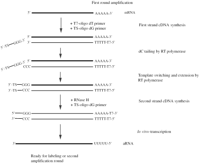
Despite the lack of systematic assessments of the potential distortion to the relative transcript abundance or other limitations of global mRNA amplification, the method rapidly became a tool of choice for the profiling of small samples. The Affymetrix platform (high density oligonucleotide arrays) integrated at an early stage the use of mRNA amplification and hybridization with biotinylated complementary RNA (cRNA) products (15–17). The procedure was rapidly considered a standard step for all samples, even large. Due to the widespread use of the Affymetrix oligo array platform, numerous papers have been published containing data obtained with at least one round of in vitro transcription. Wang et al. (18) and Baugh et al. (19) presented modified versions of the Eberwine protocol and were the first to quantitatively inspect the differences in gene expression profiles before and after amplification using cDNA and Affymetrix chips, respectively. The amplification protocol found in Baugh et al. (19) follows closely the classical Eberwine method, but includes modifications directed towards optimization. The classical antisense mRNA amplification is 3′ end biased, due to the use of oligo dT primers to initiate first strand cDNA synthesis at the poly(A) 3′ end of the mRNA transcript and complete coverage of the 5′ end is not ensured. For that particular reason, Wang et al. (18) exploited a template-switching effect at the 5′ end of the mRNA transcript to ensure the synthesis of full length ds cDNA (Figure 2). This template-switching effect is based on the terminal transferase activity of the reverse transcriptase that adds additional, non-template residues, primarily cytosines, to the 3′ end of the cDNA. The reverse transcript buffer mixture also contains a primer containing an oligo(G) sequence at its 3′ end which will base pair with the newly synthesized dCTP stretch. Reverse transcriptase then switches templates and continues replicating the defined sequence of the annealed primer. The result is full-length cDNA. The impact of 5′ end loss of the mRNA transcript, however, is dependent on the probe design as discussed in a later paragraph. Both studies concluded that the level of concordance between amplified and non-amplified material was high, although there were some discrepancies that increased as the input of mRNA into the amplification reaction decreased (18,19).
Figure 2.
Overview of a linear mRNA amplification based on the procedure described by Wang et al. (18). Following oligo dT priming, the method exploits the template switching effect of the reverse transcriptase enzyme. The RT enzyme incorporates non-template dCTPs at the 3′ end of the transcript, then switches templates and continues replication to the end of the primer. The result is full length cDNA. For the second strand, a primer with bases complementary to the dCTP stretch is applied. Antisense RNA is transcribed by the T7 RNA polymerase.
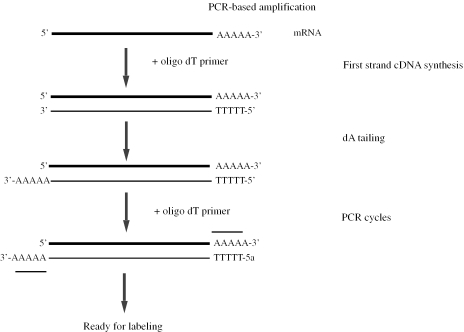
Following these two seminal amplification papers, a number of protocol variations for sample amplification have appeared, all promising improvements either in terms of technical issues, fidelity or sensitivity of the procedure (20–22). As an alternative to the conventional T7-based linear approach, PCR-based exponential strategies were introduced. The PCR-based methods share the feature of introducing PCR-priming sites at both ends of each reverse transcribed cDNA molecule, followed by global amplification of cDNA by PCR cycles (Figure 3) (23,24). Li et al. (25) proposed the inclusion of a cDNA amplification step using the SMART™ PCR technology, which is based on the template switching principle described above. Another alternative strategy uses the template switching feature to generate ds cDNA, from which only one strand is copied throughout consecutive cycles (26). A separate reaction synthesizes the second strand cDNA and the resulting double stranded products serve as templates for in vitro transcription.
Figure 3.
Schematic illustration based on a reported exponential amplification method (24). The mRNA transcript is reverse transcribed with an oligo dT primer. The cDNA is tailed by terminal transferase to create an oligo dA tail. Addition of a poly dA stretch allows the use of one oligo dT- (or oligo dT adaptor-) primer to be used in subsequent PCR cycles.
Amplification of sample material using PCR techniques does have some advantages. PCR-based amplification yields amplification rates exceeding by far the efficiency of linear amplification (24). This implies that the amount of input material can be further reduced compared to most linear amplification procedures. The method is less labor intensive than the multistep protocols for linear mRNA amplification, which directly implies better cost-effectiveness. The ds cDNA products are suitable for hybridization to array probes of either strand orientation, unlike aRNA products as discussed below. Further, the double stranded products are more stable than RNA products. However, there are great concerns regarding relevant properties inherent in DNA polymerase such as misincorporation of bases, bias toward shorter transcripts and differential amplification efficiencies of different templates based on GC composition. Effects of these properties, especially the two latter which give rise to non-linear amplification, may lead to the misrepresentation of the quantitative transcript values in a sample after multiple PCR cycles.
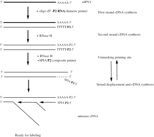
DNA polymerase can also be applied in linear amplification as in a single primer amplification (SPA) method described by Smith et al. (27). Extension of this technology has resulted in an isothermal amplification of cDNA using a single primer (Figure 4) (28). Only one purification step is required, hence the method is streamlined and rapid. The single stranded (ss) cDNA generated is complementary to conventional oligo array probes.
Figure 4.
In the first step of this isothermal linear amplification procedure, a DNA–RNA chimeric primer hybridizes to the poly (A) tail of the mRNA where both segments are extended by the reverse transcriptase. The first strand cDNA is template for the second strand synthesis, which leaves an RNA–DNA heteroduplex at one end. RNase H unmasks the priming site by digestion of the RNA P2 segment of the chimeric primer. Isothermal amplification of cDNA is performed using a single DNA–RNA primer that binds to the revealed single strand segment and is extended by a strand-displacing DNA polymerase. Once the extension is initiated, the RNase again digests the RNA P2 segment allowing a new primer molecule to bind again and extend leading to a continuous isothermal generation of ss cDNA copies. The illustration was adapted from Dafforn et al. (28).
For a more detailed review of amplification procedures and protocols that have been applied to cDNA arrays including practical suggestions, we refer to a recent publication by Wang (29).
STRAND SPECIFIC AMPLIFICATION—ANTISENSE AND SENSE STRANDS
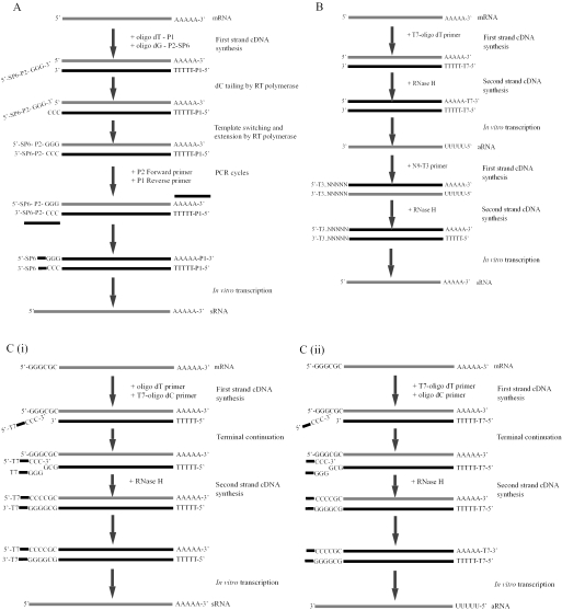
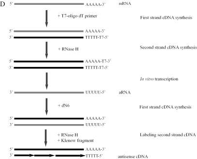
Unlike amplification strategies that generate ds cDNA as final products, such as PCR-based procedures, the in vitro based procedures generate RNA with either sense or antisense strand orientation. The general linear mRNA amplification protocols result in antisense RNA production. Strand specification has no implications when using cDNA array. However, hybridization to oligonucleotide arrays is strand specific and requires the antisense strand of the nucleic acids (RNA or DNA). The strategy chosen by Affymetrix involves the synthesis of biotin-labeled cRNA (or amino allyl incorporation for indirect fluorescent labeling) which is directly suitable for hybridizations. A potential disadvantage of labeled RNA probes is the reduced specificity of RNA–DNA interactions compared to DNA–DNA interactions due to increased stability of binding energy and thus less sensitive to mismatches. Besides the commonly used method of cRNA synthesis, there are a number of strategies reported to obtain the correct strand for labeling. The template switch effect can be applied to incorporate a second primer with an RNA polymerase binding sequence at the 3′ end of the first strand cDNA product (Figure 5A) (30). In that way, the ds cDNA products serve as templates for sense strand directed in vitro transcription, which can be reverse transcribed into labeled cDNA suitable for oligonucleotide arrays. Alternatively, the first round of amplification can follow a standard procedure, while in the second round it is possible to prime the aRNA with random 9mers bearing a T3 polymerase promoter site (Figure 5B) (31). The use of a T3 RNA polymerase will then lead to synthesis of sense-stranded RNA. In an additional strategy, either transcript orientation during RNA amplification can be chosen (Figure 5Ci and ii) (32). Schlingemann et al. (33) showed how the classical Eberwine procedure could be modified for strand specific hybridization on oligoarrays (Figure 5D). In this protocol, the final RNA in vitro reaction is followed by dye incorporation using a Klenow fragment, generating labeled antisense cDNA.
Figure 5.
(A) First strand cDNA is initiated by priming with an oligo dT primer containing and anchoring primer site. The template switch effect is applied to incorporate a primer containing both an anchoring primer site and a RNA polymerase binding site. The anchored priming sites are used in a limited PCR cycling step. Sense RNA (sRNA) is transcribed by SP6-RNA polymerase during an in vitro reaction. Adapted figure from Rajeevan et al. (30). (B) The first round of this procedure is equivalent to the first round of the classical Eberwine procedure and RNA in the antisense direction is synthesized in the in vitro reaction. At the start of the second round of amplification, aRNA is primed with random nonamer primers modified by the addition of an upstream T3 polymerase promoter site. Second strand cDNA is synthesized as in the Eberwine protocol. The RNA transcripts produced in this second amplification round are oriented in the sense direction. Modified figure from Kaposi-Novak et al. (31). (Ci and Cii). An oligo dT primer and a terminal continuation (TC) primer containing a T7 promoter sequence in the sense oriented transcription are added to the mRNA sample for first strand cDNA synthesis. TC is based on the observation of the reverse transcriptase enzyme adds a few Cs and also Gs nonspecifically at the end of mRNA templates. The TC primer anneals with this stretch and provides a binding site for second strand cDNA synthesis. RNA in vitro transcription can be driven using a promoter sequence attached to either the 3′ or the 5′ oligo primers and in thus generates either sense or antisense RNA transcripts. For further methodological details of the terminal continuation strategy see Che and Ginsberg (32). (D) The first and subsequent rounds of amplification follow the same procedure as the classical Eberwine method. The final aRNA is reverse transcribed into sense cDNA and used as a template for Klenow labeling, yielding fluorescently labeled antisense cDNA, which are in the correct orientation for hybridization to oligo arrays. Adapted figure from Schlingemann et al. (33).
COMMONLY INVESTIGATED FEATURES OF SAMPLE AMPLIFICATION
The most common aspects arising from the use of sample amplification, irrespective of whether the method confers linear or exponential amplification, include; amplification efficiency, 3′ bias and length of aRNA/cDNA products, reproducibility, fidelity of maintaining relative transcript abundance, benefits of using amplified material versus non-amplified and disadvantages with amplification procedures.
Amplification efficiency
Amplification efficiency is often noted as the amplification fold or factor. This is calculated by dividing the final aRNA yield by the estimated initial mRNA input. Generally, it is the total RNA that is measured and the content of mRNA is calculated based on a certain fraction composition of total RNA. The general assumption is a 1–3% mRNA content in total RNA, dependent on the cell type. Accurate quantification of total RNA may be difficult, especially when dealing with small samples and low RNA concentrations. Hence, besides technical aspects inherent in the amplification protocol, the amplification fold factor is affected by RNA measurements and theoretically estimated mRNA content of the cell type in the sample. In other words, the amplification efficiency reported is potentially different from the actual value due to error prone measurements and incorrect assumptions of true mRNA content. A facilitated comparison of amplification efficiencies between protocols and laboratories would require standardization of e.g. an assay using a commercially available RNA source with either a documented and stable mRNA content or a mix of known quantities of transcripts. For linear amplification, the efficiency range for two rounds of linear amplification that has generally been reported lies between 103 and 105. It is advisable to critically evaluate the basis of this calculation for comparison between studies or protocols. Typically, cell line material and serial cell dilutions to obtain sequentially smaller samples yield better amplification efficiencies than clinical material.
With respect to the technical issues, increase in the amplification efficiency can possibly be achieved by optimization of the individual steps, for example by choice of enzymes or temperature settings. It is worth noting that the yield of aRNA increases as the in vitro reaction extends in time. However, prolonged synthesis is not recommended, as degradation of near full length aRNA products are observed past 4 h (34).
The amplification factor using a PCR based method has been reported to be 3 × 10¹¹ (24). This figure was obtained by dividing the cDNA yield by the initial estimated mRNA content in the sample. Strategies combining both linear and PCR amplification procedures have been described in the literature (35–37). The reported amplification factor of 106–107 lies between that of two rounds of linear amplification and exponential amplification (36).
3′ bias and aRNA product lengths
The conventional Eberwine based amplification protocols involve directional priming from the 3′ ends of mRNA transcripts in the first round of amplification and random priming in the second and subsequent rounds, leading to shortened products. The transcript lengths are dependent on the processive features of the specific reverse transcriptases and polymerases applied as well as reaction conditions. Product lengths after two rounds of amplification can easily be evaluated on an Agilent Bioanalyzer, and are typically in the range of 200–1000 bp, peaking at 400–500 bp. Baugh et al. (19) showed that signal obtained from oligo probes that are 5′ biased are markedly reduced, due to 3′ bias and the lack of full length products. Hence, the consequence of these two features is dependent on the probe design on the arrays. However, the effect is minimal as the probe sets for cDNA- and oligoarrays are generally 3′ end biased and so are also conventional labeling protocols of non-amplified targets.
Reproducibility
In general, the reproducibility of amplification experiments and the subsequent hybridizations is reported to be high. Parallel amplifications yield highly correlated expression profiles (18,19). A common observation is that the reproducibility of replicate hybridizations is higher than for experiments using total RNA (38,39). These observations demonstrate consistency and indicate that amplification is reproducible even for genes whose relative transcript levels are not maintained. Baugh et al. (19) also showed that reproducibility was high, even if diluting the input RNA down to tens of nanograms (r ≈ 0.94). In a comparable dilution experiment the correlation value was slightly reduced (r ≈ 0.87) showing that variable reproducibility increased as the quantity of RNA amplified was reduced (40). In a comparison between linear and PCR amplification, the results showed that reproducibility was very high for linear amplification, and slightly lower for a SMART PCR-based amplification (41). In contrast, Klur et al. (42) showed that their PCR-based protocol was slightly more reproducible than the linear approach.
Fidelity of maintaining relative transcript abundance
Faithful preservation of the abundance levels of gene transcripts is the most important issue regarding the use of any amplification procedure in combination with quantitative microarray studies. If the up-scaling procedure introduces variability by inaccurate maintenance of relative transcript copy numbers, then quantitative measures of gene expression levels are rendered invalid. Hence, in order to combine amplification protocols with microarray technology, it is necessary to document that the procedure conserves the quantitative features of the input RNA source. In early reports introducing global mRNA amplification for microarray studies, the documentation of the degree of fidelity was generally conducted by comparing profiles between amplified and non-amplified material, comparing calls present or absent (Affymetrix platform), use of internal RNA standards, or northern blot or real time RT–PCR verification. These evaluations covered linear based procedures (18–20,43–45), exponential based procedures (46,47), or compared both amplification strategies (41,48). When comparing profiles, calculation of Pearson correlation coefficients was the most common statistical approach applied. Evaluation of the consistency of outliers between amplified and non-amplified material was a widely used parameter in the early papers, due to the popular differential expression analysis, where any ratio greater than 2-fold was conventionally accepted as relevant for further analysis. The study by Wang et al. (18) only focused on the maintenance of differentially expressed genes (outliers) between two RNA sources. When aRNA was generated from 0.25–3.0 µg of total RNA, 85–92% of the outliers from the control experiments using total RNA, were also identified as differentially expressed genes after aRNA hybridization analysis. Scheidl et al. (43) presented all genes in common between aRNA and total RNA, and not only the outliers. Although the intensity levels were not preserved, the relative abundances of transcripts were maintained, giving rise to a relatively high correlation factor (0.84). However, as signal intensities approached background levels, the correlation coefficients dropped. Independent studies achieved comparable correlation coefficients (0.82 and 0.8) (20,49). The general conclusion drawn in these studies was that microarray data from amplified material is comparable to non-amplified material but there is a slight decrease in correlation coefficients, reflecting changes in transcript ratios. Superficial examination would characterize many of these reports showing the reliability of sample amplification. The studies are informative, but limited. Notably, one study presented components of a variance model in their study (20). The variance of true expression and measurement errors were estimated for both amplified arrays based on aRNA obtained with different amplification protocols and non-amplified material. The authors found a decreased variance in gene expression after amplification, and concluded that amplification had a dampening effect on the true expression of some genes. Nygaard et al. (38) presented a quantitative study to examine the effects of amplification on ratio preservation. They assessed the number of genes that showed differential expression when comparing amplified versus non-amplified to be 10%. Here, a two-sample _t_-test, using the Benjamini-Hochberg multiple testing procedure, to find genes consistently or inconsistently expressed between amplified and non-amplified material from two RNA sources was applied. As microarray experiments are subjected to a substantial amount of variability, the contribution to noise due to the amplification procedure was estimated by ANOVA analysis. Using the estimated ANOVA parameters, they calculated the signal to noise ratios both with inclusion of variations due to amplification, and without. A decrease in the signal to noise ratio due to amplification was reported.
In recent literature regarding mRNA amplification, Pearson correlation coefficient calculations to estimate inconsistency of data generated from amplified material compared to non-amplified material or from different amplification protocols, have consistently been a main choice of statistically based analysis (40,50–54). The reported results from these analyses have been restricted to outliers or genes with a fold change of >2. However, it is necessary to comment on the use of the 2-fold change criterion as a measure of significance. Many published microarray studies have utilized the 2-fold criterion. The method is straightforward, but it is apparent that this is not informative in all settings. Utilization of this criterion is diminishing and is being replaced by the application of _t_-tests. In addition, there is a larger focus on genes co-regulated in pathways or signatures rather than single, differentially expressed genes. These features strongly indicate that the 2-fold criterion to detect outliers between two RNA samples is not a suitable end-point for evaluations of RNA amplification fidelity.
An alterative approach to strictly using microarray datasets for evaluation of fidelity was presented by Goff et al. (55). They chose to compare a subgroup of amplified data against real time RT–PCR data and calculate the correlation value. Their reason for using real time RT–PCR data as the true standard was the common use of this latter technique to validate microarray data.
Besides the continuous use of Pearson correlation coefficients, more sophisticated, statistical analytical methods have also recently appeared (44,56–58). To analyze the degree of fidelity of amplification on differential gene expression between two different samples, a comparison of _t_-scores or posterior distribution of fold change for individual genes have been applied (44,57,58). Again, the documentation from these analyses has been restricted to a small subset of genes, more specifically the outliers, e.g. the top 10 ranked genes.
As the amplification protocols are tested to cover even lower ranges of input RNA quantities it is likewise important to investigate the maintenance of relative transcript abundance levels at these settings. This has been done by less informative approaches such as correlation studies and not surprisingly, correlation drops. Baugh et al. (19) calculated gene-specific _t_-scores for the observed difference between two different RNA sources that were amplified from serially diluted amounts of material (10 µg, 200 ng and 10 ng). They then derived the correlation coefficient of the _t_-scores from the different datasets and found a good correlation, although the top ranked genes (highest _t_-scores) did not extensively overlap when comparing 10 µg with 10 ng. A poor overlap between outliers was also found in another study, where only 44% of differentially expressed genes in the 50 ng amplified RNA specimens matched the 5 µg non-amplified RNA material (50). Without generating a specific subset, Schlingemann et al. (33) used a linear model to assign _P_-values to differences in ratios found across a data series from a range of RNA dilutions. There was an increase in number of genes that displayed differential ratios, compared to the standard protocol, as material was gradually reduced. Further investigations of the use of scarce material and the effects of amplification on the preservation of transcript levels, was recently studied (59). The hybridization design was based on the use of only one source of RNA and co-hybridizing a reference cell quantity with smaller test samples. The authors globally estimated the portion of genes differentially expressed in the reference and test samples, based on _P_-values calculated from a moderated _t_-test. The number of differentially expressed genes increased when amplifying from minute samples. By using a novel model to determine sensitivity limits of amplified material with respect to reliable microarray data, it was shown that the accuracy of maintaining relative transcript abundance was transcript copy number dependent. The authors found that only moderate/high expression genes were quantitatively reliable in experiments ≤1000 cells. Low expression genes were subjected to stochastic fluctuations, thus limiting the precision of gene expression measurements. Hence, to summarize this section, these published studies show that the use of RNA amplification on small-size samples confer the risk of generating unreliable data, and the validity of biological conclusions drawn from such data may therefore be poor. This risk increases as sample size decreases.
For profiling of scarce material, exponentially based global mRNA amplification methods have been developed. However, more concern is raised regarding ratio preservation in exponential amplification procedures due to non-linearity caused by DNA polymerase enzymatic function as this may contribute to reduced validity when used in quantitative expression studies. Over-amplification is also an issue for PCR-based strategies. Endege et al. (60) suggested that the reaction should be terminated one cycle prior to saturation of highly expressed genes to ensure that the majority of gene transcripts are in the exponential phase. Iscove et al. (24) was the first group to focus their study on the fidelity of exponential amplification by comparing ratios obtained from amplified and non-amplified targets. Outliers were defined within a specific interval and the results from exponentially and linearly amplified targets were compared to the true ratios (non-amplified). From the data, they concluded that their method was superior to one round of linear amplification. In contrast, Nagy et al. (61) found that 21 cycles in their PCR amplification lead to overamplification [in comparison Iscove _et al_. (24) applied 65 cycles] which resulted in major distortion of ratios. However, they claimed that their method of real-time PCR, halted after 13–15 cycles, preserved the ratios. The non-amplified control samples used in this study were labeled using dendrimer-based signal amplification (Genisphere). In a SMART–PCR setting, the maintenance of transcript abundance was estimated using real time PCR on initial number of cDNA copies before amplification, and on cDNA copies after amplification. The mean amplification factor was calculated for four genes with different copy number levels (62). The factor ranged between 28–40, which was considered sufficient accuracy, according to the authors. A study comparing the classical T7-based method with SMART-PCR, found that on cDNA arrays, gene expression measurements of linearly amplified material showed better correlation with non-amplified samples than material from SMART-PCR (41).
A critical mind is necessary in evaluation of the informative value of the contribution to the topic of RNA amplification fidelity provided by published studies. First of all, is the starting point in terms of material quantities relevant and is the experimental design reasonable? There are many reports in the literature where the authors have applied serial dilutions of RNA to investigate different input amounts into the amplification protocol of choice. The results are certainly informative, but the question is how close they are to reality. Starting from diluted RNA and starting from what is roughly the equivalent in cell material, are two entirely different settings. The additional handling of material in order to isolate RNA imposes technical variability to the composition of the transcript pools in the samples.
Most of the comparisons are performed against non-amplified total RNA experiments, following a standard protocol. Whether total RNA reflects the ‘true’ standard is debatable. Increased variability of detection of low expressing genes is a feature common to both amplified RNA- and total RNA-labeled targets and altered expression profiles are commonly found in this expression range. It is important to have in mind that targets made from total RNA are based on the 1–3% mRNA content of the initial material, and thus there is normally a great difference between the amount of target applied for the amplified and non-amplified arrays. The difference in amount can be up to ten times in a comparison experiment and result in a doubling of absolute intensity measured on the amplified arrays (38). A likely outcome in this case is that some genes with low expression are scored as differentially expressed in the amplified target compared to the reference, non-amplified target, but in reality the amplified products are closer to the true expression as these transcripts are well within the detection range when amplified from an optimal amount of RNA. Hence, it is difficult to extract which differentially expressed genes between the amplified material and the total RNA are the result of poor performance of the amplification protocol or from imprecise measurements of the true expression obtained from the total RNA arrays. Replicate arrays may reveal consistent or variable results, where consistency indicates a true measurement of data. An alternative approach to verify the source of the variability would be to compare against results from other high-throughput methods. However, access to other methods is limited, and thus many resort to low-throughput real-time RT–PCR for verification of gene expression or ratio levels. Quantitative real time RT–PCR is considered more sensitive than standard microarray procedures. However, it is important to remember that real-time RT–PCR also suffers the same inconsistencies at low copy numbers as global RNA amplification and may similarly not represent true measurements.
One feature of these studies of transcript and ratio preservation is that authors confer a subjective opinion of what defines sufficient amplification accuracy and in several cases the analysis is limited to a subgroup of genes. Correlation coefficients are generally found to be good and acceptable. An unanswered question is: at what correlation value should one reject a given method? At what level does the output data contain too much uncertainty that further biological interpretation is questionable? It is certainly advisable to carefully read the details of the comparisons or analysis to critically evaluate the basis of the author's conclusions. For example, it is likely that other investigators would not perceive an amplification factor varying between 28–40 among genes in the global reaction to yield sufficiently accurate quantitative data.
Benefits from using amplified material versus non-amplified
The main benefit of sample amplification is obviously that less material is needed to perform the microarray hybridization, but there are also other advantages. When following standard labeling procedures, the quality of RNA is a crucial factor influencing array quality. When applying mRNA amplification, however, the aRNA products are normally of very high quality after purification, and the signal to noise ratios on the arrays are significantly increased (38). In addition, the number of genes detected by fluorescent signaling using amplified material is significantly higher compared to non-amplified samples (38,39,41,44). One study confirmed by other molecular techniques that these genes were in fact expressed in the cells and not the result of unspecific binding or artifacts (44). This indicated that amplified RNA was more sensitive to low abundance transcripts than the standard method using total RNA. Due to improved signal to background levels using aRNA, Feldman et al. (49) not only scored more genes on arrays with amplified material, but also observed a doubling in the number of outliers. Increased sensitivity appears to be greatest for low copy number genes. A probable reason is that the amount aRNA used for labeling is roughly 3–10 times higher than the corresponding mRNA content in the total RNA targets.
Partially degraded RNA can be used without disturbing the conservation of relative expression levels (63). In partially degraded samples, only the transcripts missing the original 3′ end are lost.
The amplification efficiency generally results a surplus of aRNA material, so that multiple hybridization experiments can be performed. However, this may not apply for extremely small samples.
Disadvantages with amplification procedures
A disadvantage with amplification procedures is that they are generally multistep and laborious, often taking 3–5 days to complete two rounds of linear amplification. This is an issue taken seriously by kit providers, knowing that users appreciate rapid, streamlined protocols.
Products of certain linear amplification protocols are not compatible with the conventional strand orientation of the probes on oligoarrays.
Reduced hybridization specificity has been observed using commonly applied cRNA targets compared to use of ss cDNA targets (64).
Furthermore, a slight distortion of relative transcript abundance maintenance has been identified when comparing against data from non-amplified material (38,41).
Finally, according to Nygaard et al. (59) reliable quantitative data are limited for small samples. Hence, there still remain restrictions to the microarray experiments, and due to the nature of these restrictions (stochastic processes) it is unlikely that they will be surpassed with the current microarray technology and are further discussed in a latter section.
COMMERCIAL AMPLIFICATION KITS
Instead of using discrete reagents, it is now possible to choose between a range of amplification kits from vendors such as Affymetrix, Arcturus, Ambion, Clontech, Telechem Int., Roche Applied Biosciences and NuGen (Table 1). Table 1 displays information regarding the minimum input material amount specified in the respective manuals found on the manufacturers' web site. In general, two rounds of amplification are required to generate sufficient amplified RNA when starting with minimum total RNA quantities. A few manufacturers provide a minimum value of input material that is different from their recommended input, indicating that they have a sensitive assay, but the rate of success is variable for the lowest input range.
Table 1.
An overview of commercially available target amplification kits
| Kit | Manufacturer | Principle | Range of input total RNA | Minimum input total RNA | Recommended minimum input total RNA |
|---|---|---|---|---|---|
| GeneChip two cycle target labeling | Manufactured by Invitrogen for Affymetrix | Linear amp | 10–100 ng | 10 ng | 10 ng |
| RiboAmp | Arcturus | Linear amp | 1–40 ng | 1–10 ng (250–500 cells) | 10–40 ng (500–2000 cells) |
| RiboAmp HS | Arcturus | Linear amp | 100 pg–1 ng | 100–500 pg (10–50 cells) | 500 pg–1 ng |
| MessageAmp II | Ambion | Linear amp | 0.1–100 ng | 0.1 ng | 100 ng |
| Low RNA Input Fluorescent Linear Amplification | Agilent Technologies | Linear amp | 50 ng–5 µg | 50 ng | 50 ng |
| BD SMART mRNA Amplification | Clontech | Linear amp (Template switch mechanism) | 0.1–5 µg | 100 ng | 100 ng |
| BD Atlas SMART Fluorescent Probe Amplification | Clontech | PCR-based (Template switch mechanism applied) | 10 ng–1 µg | 10 ng | 10 ng |
| ArrayIt MiniAmp | TeleChem Inc | PCR-based (5–10 cycles) and one round linear amp | 50 ng–1 µg | 50 ng | 50 ng |
| RAS Microarray Target Amplification | Roche | PCR-based | 50 ng–1 µg | 50 ng (1000 cells) | 50 ng (1000 cells) |
| Ovation (Ribo-SPIA) Aminoallyl RNA Amplification | NuGen | Linear isothermal amp | 5–100 ng | 5 ng | 5 ng |
Recent articles have validated amplification procedures and kits provided by commercial manufacturers (64–66).
LITERATURE SURVEY: WHAT STRATEGY HAS BEEN USED FOR SMALL SAMPLES?
So which strategy have investigators chosen to convert their small, limited samples to material that is sufficient for microarray analysis? To answer this question, the articles surveyed below were divided into groups according to how the samples were collected, starting with fine needle biopsies, followed by LCM, cultivation of specific cells and finally microaspiration.
Fine needle biopsies represent a feasible, minimally invasive method to collect patient tissue specimens while the latter techniques are commonly used to obtain relatively homogenous cell populations from various types of tissues sources. FNA or fine needle core biopsies are in many settings borderline cases regarding the need for amplification or not. Total RNA yield is typically less than 5 µg and commonly reported to be ±1 µg. The use of signal amplification is one option if all the samples are within the range of total RNA quantity specified by the signal amplification procedure. Another strategy to bypass the use of an amplification step is the use of radioactive labeled targets and nylon membranes, reported to yield increased sensitivity (67,68). In a breast cancer study, 1–2 µg total RNA obtained from FNA was labeled with 33P and hybridized to high-density cDNA microarray nylon membranes (67). The analysis was restricted to genes expressed at higher levels than the typical mean expression value for the individual arrays and showed that profiling of FNA material could assess estrogen receptor and HER2 receptor status. However, the array industry has generally switched away from nylon membrane array to glass arrays. The use of radiolabeled targets on small membrane arrays or glass arrays in attempts to increase sensitivity have been problematic due to the general lack of appropriate detection systems that can handle the fine resolution of isotopes. For the use of FNA, glass arrays and conventional procedures, Assersohn et al. (69) showed that the success rate for adequate quality hybridization of non-amplified material was 15%. This low percentage indicated the necessity of amplification. Wang et al. (70) conducted a prospective study when amplifying total RNA extracted from FNA from melanoma metastases and followed the history of the lesions in order to correlate transcript patterns with clinical outcomes. Biopsies were taken before and after treatment, and RNA was amplified according to their modified Eberwine protocol. Genes that discriminated treatment response from non-responders were generated by statistical analysis. In a similar study, total RNA isolated from FNAs from breast cancer patients undergoing neoadjuvant chemotherapy were sampled (71). The amplification procedure followed the Eberwine protocol with minor modifications. Specific profile features were found to distinguish responders from non-responders of treatment. Rouzier et al. (72) investigated whether the different molecular subtypes of breast cancer displayed different responses to preoperative chemotherapy. A minimum of 1 µg total RNA was amplified and profiled on Affymetrix chips.
Another use of the combination FNA and RNA amplification has been to elucidate the progression of cancers by profiling samples at different stages of the disease. This can be exemplified in a study by Mazzanti et al. (73). The amplification approach applied was the modified Eberwine method and the purpose was to find diagnostic genes for the different states of the disease.
The beneficial factors of using FNAs, are first of all that the sampling technique is feasible for large studies, and that multiple aspirations from same patient/individual are possible. Further, as long as the quality of the RNA isolated is satisfactory, the amount is usually in excess with respect to most amplification protocols and thus reducing the technical challenge of up-scaling the material. As mentioned, the FNA may represent borderline cases with respect to the need to amplify, but rather than performing hybridizations in the lower end of the sensitivity range with respect to amount non-amplified target labeled, the advantage resides in the ability to increase the number of genes detected through amplification of transcripts. The studies presented above exemplify that non-invasive sampling procedures, amplification of RNA and expression profiling together form a powerful combination for cancer care management. Material can be collected preoperatively, and expression profiles may be analyzed for disease stage, sub-classification of cancer type, treatment response and clinical outcome. However, currently a large part of gene expression profiling is performed on surgically removed tumor tissue where neoadjuvant chemotherapy treatment has already been selected and exerted.
In contrast to fine needle biopsies, LCM requires an RNA amplification step prior to gene expression profiling, unless the investigator spends a substantial amount of time vastly increasing the sample size by multiple captures. There are a number of published reports combining laser microdissection with microarray technology. The first reported study collected 1000 neurons, performed three rounds of linear amplification, and hybridized in a single channel design to cDNA arrays containing 477 clones (16). To get a view of more recently used experimental protocols (specifically amplification protocols and array types) and the extent of findings reported, selected papers from a literature search were divided into two groups according to initial amount of input material, moderate range (3000–50 000 cells) [Table 2 (A)] and minute range (1–2500 cells) [Table 2 (B)]. The papers were selected by searching in PubMed using the following terms and combinations thereof: gene expression, microarray, profiling, RNA amplification, laser capture, microdissection, LCM and small samples. Papers where the main focus was demonstrating the feasibility of combining small samples and microarray technology were discarded, as the aim was to look specifically at the application of these technologies in a biological study. The selection was biased towards journals available online through our institution subscriptions, and towards relatively recently published papers (2002–2005). The experimental protocols have been retrieved from the respective materials and methods sections, while the reported findings based on amplified material were primarily extracted from the abstracts. Frequent output from the datasets were, a handful of genes that were subjectively chosen for validation by real time RT–PCR, and/or validated by supporting literature connecting some biological significance between the chosen genes and the samples. More specific analysis of differentially expressed genes or other strategies applied are noted.
Table 2.
The collected information extracted from the published reports employed are displayed
| Author | Year | Journal | Subject | Samples | Material amount | Amp method | Array type and target label | Data reported |
|---|---|---|---|---|---|---|---|---|
| (A) Moderate amount of RNA | ||||||||
| Ernst et al. (85) | 2002 | Am. J. Path. | Prostate carcinoma | Epithelial and stromal cells from both cancerous and normal tissue | ∼20 000 cells | Modified Eberwine based method [Baugh _et al_. (19)] | Affymetrix MG-U74A Biotinylated cRNA targets | SOP 15-fold change |
| Miura et al. (86) | 2002 | Cancer Res. | Lung adeno-carcinoma | Lung carcinoma stages I and II | Not presented in article but from the text deduce a moderate range | Eberwine based method [Luo _et al_. (16)] | In house cDNA slides, 4 slide sets, in total 18 432 cDNAs Fluorescently labeled targets | SOP test statistic of the differentially expressed genes, those related to loci frequently reported to be altered, were emphasized |
| Mori et al.(87) | 2002 | Surgery | Gastric carcinoma | Tumor, lymph nodes and normal tissue | 10–20 000 cells | Eberwine based method | Takara human cancer cDNA chip 2.0 Fluorescently labeled targets | SOP 2-fold change |
| Nakazono et al. (88) | 2003 | Plant Cell | Epidermis and vascular tissue of plants | Epidermal cells and vascular cells | >10 000 cells or ∼40 ng RNA | Eberwine based method [Luo _et al_. (16)] | In house cDNA arrays 9984 cDNA clones Fluorescently labeled targets | SOP 2-fold change Differentially expressed genes sorted into functional categories |
| Datson et al. (89) | 2004 | Eur. J. Neurosci. | Hippocampus in brain | Hippocampal subregions | 42–83 ng | MessageAmp (Ambion) for 1st round amp and BioArray (Enzo) for 2nd | Affymetrix Rat U34A 8000 sequences Biotinylated cRNA targets | SOP SAM Sorted the differentially expressed genes into gene ontology classes |
| Hoang et al. (90) | 2004 | J. Thorac. Cardiovasc. Surg. | Lung carcinoma | NSCLC Tumor tissue | 3000–5000 cells | RiboAmp | MicroMax Human 2400-gene cDNA chip | Clustering analysis 15 specimens clustered into 3 groups. A gene subset of 75 genes may identify genotypes prone to metastasize. |
| Matsuzaki et al.(91) | 2004 | Mol. Hum. Reprod. | Endometriosis | Epithelial and stromal cells in both endometriosis and matched eutopic endomentrium | 50 ng | Eberwine based method [Baugh _et al_. (19)] | Clontech Atlas human 1.2 cDNA nylon array. 1186 genes 32P–labeled cDNA targets | SOP 2-fold change |
| Buchstaller et al. (92) | 2004 | J. Neurosci. | Developing Schwann cells | Neural crest stem cells, Schwann cells | 7000–50 000 cells | Eberwine based method | Affymetrix mouse genome U74Av2 Biotinylated cRNA targets | Clustering analysis In addition to SOP and gene ontology classification of differentially expressed genes |
| Mimori et al. (93) | 2005 | Clin. Exp. Metastasis | Breast cancer | Primary carcinoma cells, metastatic cells and normal cells | — | Eberwine based method | Takara Human Carcinoma chip 2.0. 624 genes Fluorescently labeled targets | SOP Investigated methylation status of 3 genes of interest |
| (B) Minute amount of RNA | ||||||||
| Huang et al. (94) | 2003 | Biochem. Biophys. Res. Commun. | Prostate cancer | Invasive and in situ cancer cells | 50 cells | RNA–PCR | Affymetrix U95A2 12 558 genes Biotinylated cRNA targets | SOP 4-fold change No lists. Focus on one gene only, a truncated Bcl-2 |
| Kamme et al. (79) | 2003 | J. Neurosci. | Neurons | Single cells from hippocampus CA1 subregion, | Single cells | Eberwine based method | In house cDNA array 4529 clones Fluorescently labeled targets | Cluster analysis Identified two different cell types. Single cells within one group displayed variability in gene expression |
| Seshi et al. (78) | 2003 | Blood Cells Mol. Dis. | Bone marrow stromal cells | Mesenchymal progenitor cells | Single cells | RiboAmp | Affymterix U95Av2 12 625 probes Biotinylated cRNA targets | Found a ‘master list’ of genes expressed in the different stromal cell populations |
| Ma et al. (95) | 2003 | Proc. Natl Acad. Sci. | Breast cancer | Different stages of breast cancer | 2000–2500 cells | RiboAmp | In house cDNA arrays 12 000 genes Fluorescently labeled cDNA targets | Clustering and test statistic Tumor grade but not tumor stage was found to correlated with distinct gene expression signature |
| Mohr et al. (82) | 2004 | Biochimie | Pleural mesothelioma cells | Whole tumor, pleural mesothelioma and normal pleural mesothelial cells | 1000 cells | RiboAmp | MWG Pan Human 10 k oligo array 9850 genes Fluorescently labeled targets | SOP 2-fold change |
| Cristobal et al. (96) | 2005 | Brain Res. Mol. Brain Res. | Inner ear sensory cells | Cell types from inner ear sensory epithelia | ∼0.5–1 ng | RiboAmp HS | GE Healthcare, CodeLink Rat Whole Genome Bioarrays 33 849 probes Biotinylated cRNA targets | SOP 5-fold difference |
| Ivanov et al. (97) | 2005 | FEBS Lett. | Mammalian eye lens | Cells from different stages of fiber cell maturation | A minimum of 200 zone specific cells | MessageAmp | Agilent, 22K mouse Oligo arrays Fluorescently labeled targets | SOP SAM analysis Selected genes were classified according to gene ontology |
| Moore et al. (98) | 2005 | Int. J. Cancer | Prostate carcinoma | Normal and neoplastic epithelium | ∼2000 cells | MessageAmp | In house cDNA arrays 6200 clones Fluorescently labeled targets | SOP 20-fold change Searched profiles for genes involved in lipid metabolism pathway— found one particular gene of interest |
One aspect that distinguishes the studies in Table 2 (A) compared to (B), is the frequent application of mRNA amplification kits in 2 (B). One possible reason is the easier choice to apply a kit that claims successful amplification from minute materials, rather than testing a procedure using self-assembled products. With respect to the results, there are a few points to critically consider. The use of a high fold change criterion such as 15-, 5- or 4-fold change may indicate at least two possibilities. Firstly, that the samples are from very different cell type sources and hence many genes are differentially expressed. Secondly, the variability in gene expression between closely related samples is high so that too many genes are above the generally used 2-fold change. The latter may indicate randomness inherent in the amplification procedure when starting from minute samples. Also notable in a few minute sample-studies is that the focus of a high-throughput analysis is placed on just a few subjectively chosen genes. It is ambiguous to the reader whether there was no other findings in the rest of the data or not. Lack of further biologically significant data may imply noisy data. In those cases, microarrays are not the best experimental choice for examining similar samples and perhaps real time RT–PCR would be more convenient to measure the levels of a small set of genes, although this method is subject to stochastic variation at low input sample levels.
Minute samples can be procured by other means than LCM. In embryology research, embryos can be cultured in medium. For studying the process of hatching in early embryo development, pre-hatched and hatched blastocysts were collected for RNA extraction, amplification and microarray analysis (74). Again, a standard operating procedure (SOP) was performed and a fold change of 3 was applied. Based on literature reports, the differentially expressed genes where categorized according to function. Gene expression during kidney morphogenesis has been studied using in vitro models, RNA amplification and microarrays (75). The tissues of interest were cultured in vitro and sampled at different stages of morphogenesis. Hierarchical clustering and differential expression analysis were applied, and several potentially relevant pathways were identified. Cell selection techniques such as immunomagnetic positive selection and fluorescent sorting are other strategies to enhance the homogeneity of a cell samples. In a metastasis related study, the positive fraction of circulating tumor cells (CTC)-enriched blood sample were profiled and compared with the negative (CTC-depleted) fraction from the same patient (76). Immunomagnetic beads were used for the positive selection of tumor cells from three patients with each a different type of cancer. A list of genes differentially expressed in all three cancers was generated and from this list 35 candidate genes were selected for further real time RT–PCR analysis in a selection of 74 metastatic cancer patients and 50 healthy controls. The gene subset was claimed to be a good predictor for tissue of origin classification, but the biological role of these genes could not be clarified.
Single cell analysis is without doubt technically challenging. In the original report by Van Gelder et al. (7) a microinjection technique was used where a patch electrode filled with first strand cDNA synthesis components was injected into a single cell. The cell was loaded with the reaction mix, followed by suction of entire cell content into the electrode and a transfer step for further processing. This technique was applied to sample single cells in early single cell gene expression studies, where radioactively labeled amplified material was hybridized to slot blots (10,11). The same single cell sampling technique was also applied in a more recent study (77). In Table 2 (B) are two reports where the authors collected single cells by LCM for analysis (78,79). In embryology studies, tissue grown in media was dissociated and single cells were picked by microcapillaries and placed directly into tubes for lysis and direct cDNA synthesis for the investigation of pancreas development (80). A similar method to obtain single cells was used in a neuron study (81). If excluding the LCM samples, the amplification method reported in these more recently published studies were PCR-based (80,81) or a combination of both linear and exponential (77). The labeling strategies included Genisphere fluorescent dendrimer technology (80), 32P radioactive labeling (81) and fluorescent one-channel hybridization (77). One group reported the need to pool 4–10 cells in order to achieve significant signals (81). From the data output, two groups reported qualitative findings by gene discovery for distinct cell types (77,80) and one group followed SOP (81). Notably, the reported results in the two single cell studies in Table 2 (B) were also oriented towards cell identification by gene expression profiling. In other words, results were based on gene detection and discovery rather than gene level measurements. The one study reporting quantitative measurements included in this literature survey over single cell analysis, strictly does not belong in this category because they pooled aRNA from single cells to generate adequate data.
IMPORTANT CONSIDERATION: MATERIAL REQUIREMENT—NO LOWER LIMIT?
As long as the amplification efficiency allows it, can continuously lower material input values be used? Is the use of consecutive linear amplification rounds or PCR cycles the answer to profile minute samples? Should the cut-off in sample size be determined by amplification efficiency or sample size? Many studies have observed a markedly reduced correlation with extremely small samples, especially for mRNA transcripts in the low abundance range (18,19,43). A common observation is the reduced number of gene specific transcripts detected with minute samples (38,81,82). Few investigators have established the lower boundaries with respect to fidelity, but report according to their observations that variability is augmented in experiments with low RNA input values. Nygaard et al. (59) presented arguments that it is the sample size that is the crucial factor and not amplification efficiency. The majority of genes are transcribed at a low abundance level (1–5 copies per cell) and at low template concentrations amplification is not faithful and determinant with respect to relative abundance. Rather it is stochastic in nature. Stochastic effects have rarely been recognized as a phenomenon in global RNA amplification of minute samples. The quantitative accuracy is greatly affected by sampling variation due to the stochastic distribution of low abundance mRNAs. The lower the abundance of any template, the smaller the probability its true abundance will be maintained in the amplified product (83). This feature calls for methods to filter out the affected genes. If the transcript level of a gene of interest is not present in sufficient numbers in the initial sample, then quantitative gene expression measurements cannot be established. This statement indirectly implies that the yield of quantitative data from scarce material is restricted to a few highly expressed genes, thus rendering the use of this high throughput method relatively useless. The authors of single cell expression profiling may already have experienced difficulties in extracting quantitative data, and thus shifted focus towards qualitative data. For small input values, only high magnitude changes can be detected at low/moderate expression values. How does invalid data affect the reported findings from published studies? In our view, the validity of the data will be, uncovered by the type of data analysis and mining conducted. Investigators experienced with microarray data and statistical analysis, will know that by sampling measurements for a large number of genes, false discovery of differentially expressed genes is inevitable. The common approach to generate a list of differentially expressed genes according to arbitrary thresholds does not attribute any biological significance to the results, and genes inspected further are frequently selected subjectively. In other words, poor maintenance of relative transcript levels when amplifying from a small sample will most likely not be detected in this analysis approach. Nor will it be detected in clustering analysis, which also analyzes genes in an independent manner and does not identify causal biological mechanism that regulate genes with similar expression patterns. Therefore, microarray data that is partially unreliable data due to random amplification will not be visible in such analysis strategies. The pitfall is thus presenting misleading data. Gene transcripts, however, do generally not change individually, but in a complex concerto with a large number of participants. Therefore, analysis strategies where biological processes and pathways are mapped and analyzed using expression data are more likely to reveal experimental flaws, such as poor amplification performance due to a resulting randomness of data and lack of pattern recognition. The implementation of methods to analyze microarray data in a more comprehensive biological view is a current and important issue in the microarray community (84). The shift in analysis is from individual genes ranked in a list, towards coherent changes in gene expression of gene sets. To accommodate these new approaches, it is evident that the degree of quantitative reliability when profiling minute, amplified samples needs to be adequate in a global manner. Further, this implies the need to investigate fidelity of amplification across all genes, and not just a subset as is commonly reported, in order to assess the usefulness of the procedure.
To answer the questions posed above, there is a threshold for sample size with respect to reliable gene expression measurements. The fine-tuning of this threshold is dependent on the tissue source, sample size, amplification efficiency, array platform and detection method. Although amplification efficiency makes it possible to generate material from minute samples, a large number of moderate to low abundance transcripts are not preserved in their relative distribution, due to stochastic effects. Hence, the chance of presenting misleading data is high, and requires the investigator to fully understand this risk and take the consequence by filtering out unreliable data. Validation of microarray data is generally performed by other molecular techniques. However, custom validation by quantitative real time RT–PCR may be equally problematic with minute samples as mentioned earlier. It is important to have these aspects in mind when planning a microarray study and when evaluating the biological conclusions drawn in published microarray studies based on extremely low RNA input values.
CONCLUDING SUMMARY
The relatively large amount of material that each microarray experiment requires has posed restrictions on the use of this high throughput technique. Development of sample amplification procedures has challenged this obstacle. Linear and exponential sample amplifications are commonly used methods to obtain gene expression data from small samples using microarray technology. The conservation of transcript abundance throughout the procedures, has generally found to be acceptable in both strategies. Exponential amplification has been reported to be less faithful than the linear strategy. However, also with the latter, high amplification efficiencies can be achieved. There is currently a wide range of amplification protocols, and a number of commercially available kits are targeted to investigators of small samples. As reported in the scientific literature, sample amplification has been applied in a number of different experimental settings.
Amplification efficiencies technically allow profiling of extremely small samples, from tens of nanograms to single cells. However, discrepancies in comparison against non-amplified material or larger samples are increased with reduced sample size, as shown in a number of reports. Specifically, stochastic processes pose a restriction to accurate quantitative data from minute samples, a phenomenon that has rarely been recognized in the literature. Knowledge of the limitations with respect to input transcript concentration is a prerequisite for quantitatively measuring gene expression levels in order to avoid stochastic variability. This is a highly relevant aspect as not to confer biological significance to invalid quantitative data. Random amplification of rare/moderate gene transcripts in minute samples will generally not be revealed as long as genes are analyzed individually, such as by ranking differentially expressed genes. However, the implementation of novel methods to analyze microarray data in a more comprehensive view is more likely to reveal experimental flaws due to poor amplification performance. This further implies the need to investigate fidelity of amplification in a global manner and not as subsets as commonly reported.
Sample amplification if often the only option to perform high throughput microarray analysis on small samples and should not necessarily be avoided, but the tradeoff with time, assay cost and the potentially short list of relevant reliable genes, may be negative below a certain threshold of input material. For more moderate samples, microarray analysis of amplified targets has been shown to have a number of advantages compared to the use of conventional non-amplified targets. In the applied protocols for a number of studies, in addition to the widespread use of Affymetrix arrays, RNA amplification is performed on all samples, even large samples, as a standard step thus making its way as a conventional procedure in the microarray community.
Acknowledgments
Vigdis Nygaard is a Research Fellow of the Norwegian Cancer Society. The microarrays used in studies supporting this manuscript were printed by the NMC at the national technology platform, and supported by the functional genomics programme (FUGE) in the Research Council of Norway. Funding to pay the Open Access publication charges was provided by the Norwegian Cancer Society.
Conflict of interest statement. None declared.
REFERENCES
- 1.Schena M., Shalon D., Davis R.W., Brown P.O. Quantitative monitoring of gene expression patterns with a complementary DNA microarray. Science. 1995;270:467–470. doi: 10.1126/science.270.5235.467. [DOI] [PubMed] [Google Scholar]
- 2.Michiels S., Koscielny S., Hill C. Prediction of cancer outcome with microarrays: a multiple random validation strategy. Lancet. 2005;365:488–492. doi: 10.1016/S0140-6736(05)17866-0. [DOI] [PubMed] [Google Scholar]
- 3.Duggan D.J., Bittner M., Chen Y., Meltzer P., Trent J.M. Expression profiling using cDNA microarrays. Nature Genet. 1999;21:10–14. doi: 10.1038/4434. [DOI] [PubMed] [Google Scholar]
- 4.Szaniszlo P., Wang N., Sinha M., Reece L.M., Van Hook J.W., Luxon B.A., Leary J.F. Getting the right cells to the array: gene expression microarray analysis of cell mixtures and sorted cells. Cytometry A. 2004;59:191–202. doi: 10.1002/cyto.a.20055. [DOI] [PubMed] [Google Scholar]
- 5.Stears R.L., Getts R.C., Gullans S.R. A novel, sensitive detection system for high-density microarrays using dendrimer technology. Physiol. Genomics. 2000;3:93–99. doi: 10.1152/physiolgenomics.2000.3.2.93. [DOI] [PubMed] [Google Scholar]
- 6.Karsten S.L., Van Deerlin V.M., Sabatti C., Gill L.H., Geschwind D.H. An evaluation of tyramide signal amplification and archived fixed and frozen tissue in microarray gene expression analysis. Nucleic Acids Res. 2002;30:E4. doi: 10.1093/nar/30.2.e4. [DOI] [PMC free article] [PubMed] [Google Scholar]
- 7.Van Gelder R.N., von Zastrow M.E., Yool A., Dement W.C., Barchas J.D., Eberwine J.H. Amplified RNA synthesized from limited quantities of heterogeneous cDNA. Proc. Natl Acad. Sci. USA. 1990;87:1663–1667. doi: 10.1073/pnas.87.5.1663. [DOI] [PMC free article] [PubMed] [Google Scholar]
- 8.Eberwine J., Yeh H., Miyashiro K., Cao Y., Nair S., Finnell R., Zettel M., Coleman P. Analysis of gene expression in single live neurons. Proc. Natl Acad. Sci. USA. 1992;89:3010–3014. doi: 10.1073/pnas.89.7.3010. [DOI] [PMC free article] [PubMed] [Google Scholar]
- 9.Phillips J., Eberwine J.H. Antisense RNA amplification: a linear amplification method for analyzing the mRNA population from single living cells. Methods. 1996;10:283–288. doi: 10.1006/meth.1996.0104. [DOI] [PubMed] [Google Scholar]
- 10.Cao Y., Wilcox K.S., Martin C.E., Rachinsky T.L., Eberwine J., Dichter M.A. Presence of mRNA for glutamic acid decarboxylase in both excitatory and inhibitory neurons. Proc. Natl Acad. Sci. USA. 1996;93:9844–9849. doi: 10.1073/pnas.93.18.9844. [DOI] [PMC free article] [PubMed] [Google Scholar]
- 11.Crino P.B., Trojanowski J.Q., Dichter M.A., Eberwine J. Embryonic neuronal markers in tuberous sclerosis: single-cell molecular pathology. Proc. Natl Acad. Sci. USA. 1996;93:14152–14157. doi: 10.1073/pnas.93.24.14152. [DOI] [PMC free article] [PubMed] [Google Scholar]
- 12.Crino P., Khodakhah K., Becker K., Ginsberg S., Hemby S., Eberwine J. Presence and phosphorylation of transcription factors in developing dendrites. Proc. Natl Acad. Sci. USA. 1998;95:2313–2318. doi: 10.1073/pnas.95.5.2313. [DOI] [PMC free article] [PubMed] [Google Scholar]
- 13.Chow N., Cox C., Callahan L.M., Weimer J.M., Guo L., Coleman P.D. Expression profiles of multiple genes in single neurons of Alzheimer's disease. Proc. Natl Acad. Sci. USA. 1998;95:9620–9625. doi: 10.1073/pnas.95.16.9620. [DOI] [PMC free article] [PubMed] [Google Scholar]
- 14.Poirier G.M., Pyati J., Wan J.S., Erlander M.G. Screening differentially expressed cDNA clones obtained by differential display using amplified RNA. Nucleic Acids Res. 1997;25:913–914. doi: 10.1093/nar/25.4.913. [DOI] [PMC free article] [PubMed] [Google Scholar]
- 15.Lockhart D.J., Dong H., Byrne M.C., Follettie M.T., Gallo M.V., Chee M.S., Mittmann M., Wang C., Kobayashi M., Horton H., et al. Expression monitoring by hybridization to high-density oligonucleotide arrays. Nat. Biotechnol. 1996;14:1675–1680. doi: 10.1038/nbt1296-1675. [DOI] [PubMed] [Google Scholar]
- 16.Luo L., Salunga R.C., Guo H., Bittner A., Joy K.C., Galindo J.E., Xiao H., Rogers K.E., Wan J.S., Jackson M.R., et al. Gene expression profiles of laser-captured adjacent neuronal subtypes. Nature Med. 1999;5:117–122. doi: 10.1038/4806. [DOI] [PubMed] [Google Scholar]
- 17.Ohyama H., Zhang X., Kohno Y., Alevizos I., Posner M., Wong D.T., Todd R. Laser capture microdissection-generated target sample for high-density oligonucleotide array hybridization. Biotechniques. 2000;29:530–536. doi: 10.2144/00293st05. [DOI] [PubMed] [Google Scholar]
- 18.Wang E., Miller L.D., Ohnmacht G.A., Liu E.T., Marincola F.M. High-fidelity mRNA amplification for gene profiling. Nat. Biotechnol. 2000;18:457–459. doi: 10.1038/74546. [DOI] [PubMed] [Google Scholar]
- 19.Baugh L.R., Hill A.A., Brown E.L., Hunter C.P. Quantitative analysis of mRNA amplification by in vitro transcription. Nucleic Acids Res. 2001;29:E29. doi: 10.1093/nar/29.5.e29. [DOI] [PMC free article] [PubMed] [Google Scholar]
- 20.Zhao H., Hastie T., Whitfield M.L., Borresen-Dale A.L., Jeffrey S.S. Optimization and evaluation of T7 based RNA linear amplification protocols for cDNA microarray analysis. BMC Genomics. 2002;3:31. doi: 10.1186/1471-2164-3-31. [DOI] [PMC free article] [PubMed] [Google Scholar]
- 21.Moll P.R., Duschl J., Richter K. Optimized RNA amplification using T7-RNA-polymerase based in vitro transcription. Anal. Biochem. 2004;334:164–174. doi: 10.1016/j.ab.2004.07.013. [DOI] [PubMed] [Google Scholar]
- 22.Naderi A., Ahmed A.A., Barbosa-Morais N.L., Aparicio S., Brenton J.D., Caldas C. Expression microarray reproducibility is improved by optimising purification steps in RNA amplification and labelling. BMC Genomics. 2004;5:9. doi: 10.1186/1471-2164-5-9. [DOI] [PMC free article] [PubMed] [Google Scholar]
- 23.Hertzberg M., Sievertzon M., Aspeborg H., Nilsson P., Sandberg G., Lundeberg J. cDNA microarray analysis of small plant tissue samples using a cDNA tag target amplification protocol. Plant J. 2001;25:585–591. doi: 10.1046/j.1365-313x.2001.00972.x. [DOI] [PubMed] [Google Scholar]
- 24.Iscove N.N., Barbara M., Gu M., Gibson M., Modi C., Winegarden N. Representation is faithfully preserved in global cDNA amplified exponentially from sub-picogram quantities of mRNA. Nat. Biotechnol. 2002;20:940–943. doi: 10.1038/nbt729. [DOI] [PubMed] [Google Scholar]
- 25.Li Y., Ali S., Philip P.A., Sarkar F.H. Direct comparison of microarray gene expression profiles between non-amplification and a modified cDNA amplification procedure applicable for needle biopsy tissues. Cancer Detect. Prev. 2003;27:405–411. doi: 10.1016/s0361-090x(03)00105-3. [DOI] [PubMed] [Google Scholar]
- 26.Stirewalt D.L., Pogosova-Agadjanyan E.L., Khalid N., Hare D.R., Ladne P.A., Sala-Torra O., Zhao L.P., Radich J.P. Single-stranded linear amplification protocol results in reproducible and reliable microarray data from nanogram amounts of starting RNA. Genomics. 2004;83:321–331. doi: 10.1016/j.ygeno.2003.08.008. [DOI] [PubMed] [Google Scholar]
- 27.Smith L., Underhill P., Pritchard C., Tymowska-Lalanne Z., Abdul-Hussein S., Hilton H., Winchester L., Williams D., Freeman T., Webb S., et al. Single primer amplification (SPA) of cDNA for microarray expression analysis. Nucleic Acids Res. 2003;31:9. doi: 10.1093/nar/gng009. [DOI] [PMC free article] [PubMed] [Google Scholar]
- 28.Dafforn A., Chen P., Deng G., Herrler M., Iglehart D., Koritala S., Lato S., Pillarisetty S., Purohit R., Wang M., et al. Linear mRNA amplification from as little as 5 ng total RNA for global gene expression analysis. Biotechniques. 2004;37:854–857. doi: 10.2144/04375PF01. [DOI] [PubMed] [Google Scholar]
- 29.Wang E. RNA amplification for successful gene profiling analysis. J. Transl. Med. 2005;3:28. doi: 10.1186/1479-5876-3-28. [DOI] [PMC free article] [PubMed] [Google Scholar]
- 30.Rajeevan M.S., Dimulescu I.M., Vernon S.D., Verma M., Unger E.R. Global amplification of sense RNA: a novel method to replicate and archive mRNA for gene expression analysis. Genomics. 2003;82:491–497. doi: 10.1016/s0888-7543(03)00115-0. [DOI] [PubMed] [Google Scholar]
- 31.Kaposi-Novak P., Lee J.S., Mikaelyan A., Patel V., Thorgeirsson S.S. Oligonucleotide microarray analysis of aminoallyl-labeled cDNA targets from linear RNA amplification. Biotechniques. 2004;37:580. doi: 10.2144/04374ST02. 582–586, 588. [DOI] [PubMed] [Google Scholar]
- 32.Che S., Ginsberg S.D. Amplification of RNA transcripts using terminal continuation. Lab. Invest. 2004;84:131–137. doi: 10.1038/labinvest.3700005. [DOI] [PubMed] [Google Scholar]
- 33.Schlingemann J., Thuerigen O., Ittrich C., Toedt G., Kramer H., Hahn M., Lichter P. Effective transcriptome amplification for expression profiling on sense-oriented oligonucleotide microarrays. Nucleic Acids Res. 2005;33:e29. doi: 10.1093/nar/gni029. [DOI] [PMC free article] [PubMed] [Google Scholar]
- 34.Madison R.D., Robinson G.A. lambda RNA internal standards quantify sensitivity and amplification efficiency of mammalian gene expression profiling. Biotechniques. 1998;25:504–508. doi: 10.2144/98253rr06. 510, 512 passim. [DOI] [PubMed] [Google Scholar]
- 35.Aoyagi K., Tatsuta T., Nishigaki M., Akimoto S., Tanabe C., Omoto Y., Hayashi S., Sakamoto H., Sakamoto M., Yoshida T., et al. A faithful method for PCR-mediated global mRNA amplification and its integration into microarray analysis on laser-captured cells. Biochem. Biophys. Res. Commun. 2003;300:915–920. doi: 10.1016/s0006-291x(02)02967-4. [DOI] [PubMed] [Google Scholar]
- 36.Ohtsuka S., Iwase K., Kato M., Seki N., Shimizu-Yabe A., Miyauchi O., Sakao E., Kanazawa M., Yamamoto S., Kohno Y., et al. An mRNA amplification procedure with directional cDNA cloning and strand-specific cRNA synthesis for comprehensive gene expression analysis. Genomics. 2004;84:715–729. doi: 10.1016/j.ygeno.2004.06.012. [DOI] [PubMed] [Google Scholar]
- 37.Ji W., Zhou W., Gregg K., Lindpaintner K., Davis S. A method for gene expression analysis by oligonucleotide arrays from minute biological materials. Anal. Biochem. 2004;331:329–339. doi: 10.1016/j.ab.2004.03.039. [DOI] [PubMed] [Google Scholar]
- 38.Nygaard V., Loland A., Holden M., Langaas M., Rue H., Liu F., Myklebost O., Fodstad O., Hovig E., Smith-Sorensen B. Effects of mRNA amplification on gene expression ratios in cDNA experiments estimated by analysis of variance. BMC Genomics. 2003;4:11. doi: 10.1186/1471-2164-4-11. [DOI] [PMC free article] [PubMed] [Google Scholar]
- 39.Stoyanova R., Upson J.J., Patriotis C., Ross E.A., Henske E.P., Datta K., Boman B., Clapper M.L., Knudson A.G., Bellacosa A. Use of RNA amplification in the optimal characterization of global gene expression using cDNA microarrays. J. Cell. Physiol. 2004;201:359–365. doi: 10.1002/jcp.20074. [DOI] [PubMed] [Google Scholar]
- 40.Kenzelmann M., Klaren R., Hergenhahn M., Bonrouhi M., Grone H.J., Schmid W., Schutz G. High-accuracy amplification of nanogram total RNA amounts for gene profiling. Genomics. 2004;83:550–558. doi: 10.1016/j.ygeno.2003.09.026. [DOI] [PubMed] [Google Scholar]
- 41.Puskas L.G., Zvara A., Hackler L., Jr, Van Hummelen P. RNA amplification results in reproducible microarray data with slight ratio bias. Biotechniques. 2002;32:1330–1334. doi: 10.2144/02326mt04. 1336, 1338, 1340. [DOI] [PubMed] [Google Scholar]
- 42.Klur S., Toy K., Williams M.P., Certa U. Evaluation of procedures for amplification of small-size samples for hybridization on microarrays. Genomics. 2004;83:508–517. doi: 10.1016/j.ygeno.2003.09.005. [DOI] [PubMed] [Google Scholar]
- 43.Scheidl S.J., Nilsson S., Kalen M., Hellstrom M., Takemoto M., Hakansson J., Lindahl P. mRNA expression profiling of laser microbeam microdissected cells from slender embryonic structures. Am. J. Pathol. 2002;160:801–813. doi: 10.1016/S0002-9440(10)64903-6. [DOI] [PMC free article] [PubMed] [Google Scholar]
- 44.Hu L., Wang J., Baggerly K., Wang H., Fuller G.N., Hamilton S.R., Coombes K.R., Zhang W. Obtaining reliable information from minute amounts of RNA using cDNA microarrays. BMC Genomics. 2002;3:16. doi: 10.1186/1471-2164-3-16. [DOI] [PMC free article] [PubMed] [Google Scholar]
- 45.Scherer A., Krause A., Walker J.R., Sutton S.E., Seron D., Raulf F., Cooke M.P. Optimized protocol for linear RNA amplification and application to gene expression profiling of human renal biopsies. Biotechniques. 2003;34:546–550. doi: 10.2144/03343rr01. 552–554, 556. [DOI] [PubMed] [Google Scholar]
- 46.Seth D., Gorrell M.D., McGuinness P.H., Leo M.A., Lieber C.S., McCaughan G.W., Haber P.S. SMART amplification maintains representation of relative gene expression: quantitative validation by real time PCR and application to studies of alcoholic liver disease in primates. J. Biochem. Biophys. Methods. 2003;55:53–66. doi: 10.1016/s0165-022x(02)00177-x. [DOI] [PubMed] [Google Scholar]
- 47.Petalidis L., Bhattacharyya S., Morris G.A., Collins V.P., Freeman T.C., Lyons P.A. Global amplification of mRNA by template-switching PCR: linearity and application to microarray analysis. Nucleic Acids Res. 2003;31:e142. doi: 10.1093/nar/gng142. [DOI] [PMC free article] [PubMed] [Google Scholar]
- 48.Wang J., Hu L., Hamilton S.R., Coombes K.R., Zhang W. RNA amplification strategies for cDNA microarray experiments. Biotechniques. 2003;34:394–400. doi: 10.2144/03342mt04. [DOI] [PubMed] [Google Scholar]
- 49.Feldman A.L., Costouros N.G., Wang E., Qian M., Marincola F.M., Alexander H.R., Libutti S.K. Advantages of mRNA amplification for microarray analysis. Biotechniques. 2002;33:906–912. doi: 10.2144/02334mt04. 914. [DOI] [PubMed] [Google Scholar]
- 50.Goley E.M., Anderson S.J., Menard C., Chuang E., Lu X., Tofilon P.J., Camphausen K. Microarray analysis in clinical oncology: pre-clinical optimization using needle core biopsies from xenograft tumors. BMC Cancer. 2004;4:20. doi: 10.1186/1471-2407-4-20. [DOI] [PMC free article] [PubMed] [Google Scholar]
- 51.Rudnicki M., Eder S., Schratzberger G., Mayer B., Meyer T.W., Tonko M., Mayer G. Reliability of t7-based mRNA linear amplification validated by gene expression analysis of human kidney cells using cDNA microarrays. Nephron. Exp. Nephrol. 2004;97:e86–e95. doi: 10.1159/000078642. [DOI] [PubMed] [Google Scholar]
- 52.Li Y., Li T., Liu S., Qiu M., Han Z., Jiang Z., Li R., Ying K., Xie Y., Mao Y. Systematic comparison of the fidelity of aRNA, mRNA and T-RNA on gene expression profiling using cDNA microarray. J. Biotechnol. 2004;107:19–28. doi: 10.1016/j.jbiotec.2003.09.008. [DOI] [PubMed] [Google Scholar]
- 53.Marko N.F., Frank B., Quackenbush J., Lee N.H. A robust method for the amplification of RNA in the sense orientation. BMC Genomics. 2005;6:27. doi: 10.1186/1471-2164-6-27. [DOI] [PMC free article] [PubMed] [Google Scholar]
- 54.Park J.Y., Kim S.Y., Lee J.H., Song J., Noh J.H., Lee S.H., Park W.S., Yoo N.J., Lee J.Y., Nam S.W. Application of amplified RNA and evaluation of cRNA targets for spotted-oligonucleotide microarray. Biochem. Biophys. Res. Commun. 2004;325:1346–1352. doi: 10.1016/j.bbrc.2004.10.151. [DOI] [PubMed] [Google Scholar]
- 55.Goff L.A., Bowers J., Schwalm J., Howerton K., Getts R.C., Hart R.P. Evaluation of sense-strand mRNA amplification by comparative quantitative PCR. BMC Genomics. 2004;5:76. doi: 10.1186/1471-2164-5-76. [DOI] [PMC free article] [PubMed] [Google Scholar]
- 56.Patel O.V., Suchyta S.P., Sipkovsky S.S., Yao J., Ireland J.J., Coussens P.M., Smith G.W. Validation and application of a high fidelity mRNA linear amplification procedure for profiling gene expression. Vet. Immunol. Immunopathol. 2005;105:331–342. doi: 10.1016/j.vetimm.2005.02.018. [DOI] [PubMed] [Google Scholar]
- 57.Gold D., Coombes K., Medhane D., Ramaswamy A., Ju Z., Strong L., Koo J.S., Kapoor M. A comparative analysis of data generated using two different target preparation methods for hybridization to high-density oligonucleotide microarrays. BMC Genomics. 2004;5:2. doi: 10.1186/1471-2164-5-2. [DOI] [PMC free article] [PubMed] [Google Scholar]
- 58.Li L., Roden J., Shapiro B.E., Wold B.J., Bhatia S., Forman S.J., Bhatia R. Reproducibility, fidelity, and discriminant validity of mRNA amplification for microarray analysis from primary hematopoietic cells. J. Mol. Diagn. 2005;7:48–56. doi: 10.1016/S1525-1578(10)60008-6. [DOI] [PMC free article] [PubMed] [Google Scholar]
- 59.Nygaard V., Holden M., Loland A., Langaas M., Myklebost O., Hovig E. Limitations of mRNA amplification from small-size call samples. BMC Genomics. 2005;6:147. doi: 10.1186/1471-2164-6-147. [DOI] [PMC free article] [PubMed] [Google Scholar]
- 60.Endege W.O., Steinmann K.E., Boardman L.A., Thibodeau S.N., Schlegel R. Representative cDNA libraries and their utility in gene expression profiling. Biotechniques. 1999;26:542–548. doi: 10.2144/99263cr04. 550. [DOI] [PubMed] [Google Scholar]
- 61.Nagy Z.B., Kelemen J.Z., Feher L.Z., Zvara A., Juhasz K., Puskas L.G. Real-time polymerase chain reaction-based exponential sample amplification for microarray gene expression profiling. Anal. Biochem. 2005;337:76–83. doi: 10.1016/j.ab.2004.09.044. [DOI] [PubMed] [Google Scholar]
- 62.Fink L., Kohlhoff S., Stein M.M., Hanze J., Weissmann N., Rose F., Akkayagil E., Manz D., Grimminger F., Seeger W., et al. cDNA array hybridization after laser-assisted microdissection from nonneoplastic tissue. Am. J. Pathol. 2002;160:81–90. doi: 10.1016/S0002-9440(10)64352-0. [DOI] [PMC free article] [PubMed] [Google Scholar]
- 63.Schoor O., Weinschenk T., Hennenlotter J., Corvin S., Stenzl A., Rammensee H.G., Stevanovic S. Moderate degradation does not preclude microarray analysis of small amounts of RNA. Biotechniques. 2003;35:1192–1196. doi: 10.2144/03356rr01. 1198–1201. [DOI] [PubMed] [Google Scholar]
- 64.Barker C.S., Griffin C., Dolganov G.M., Hanspers K., Yang J.Y., Erle D.J. Increased DNA microarray hybridization specificity using sscDNA targets. BMC Genomics. 2005;6:57. doi: 10.1186/1471-2164-6-57. [DOI] [PMC free article] [PubMed] [Google Scholar]
- 65.Wilson C.L., Pepper S.D., Hey Y., Miller C.J. Amplification protocols introduce systematic but reproducible errors into gene expression studies. Biotechniques. 2004;36:498–506. doi: 10.2144/04363RN05. [DOI] [PubMed] [Google Scholar]
- 66.Shou J., Qian H.R., Lin X., Stewart T., Onyia J.E., Gelbert L.M. Optimization and validation of small quantity RNA profiling for identifying TNF responses in cultured human vascular endothelial cells. J. Pharmacol. Toxicol. Methods. 2005 doi: 10.1016/j.vascn.2005.02.004. Epub ahead of print. [DOI] [PubMed] [Google Scholar]
- 67.Pusztai L., Ayers M., Stec J., Clark E., Hess K., Stivers D., Damokosh A., Sneige N., Buchholz T.A., Esteva F.J., et al. Gene expression profiles obtained from fine-needle aspirations of breast cancer reliably identify routine prognostic markers and reveal large-scale molecular differences between estrogen-negative and estrogen-positive tumors. Clin. Cancer Res. 2003;9:2406–2415. [PubMed] [Google Scholar]
- 68.Ayers M., Symmans W.F., Stec J., Damokosh A.I., Clark E., Hess K., Lecocke M., Metivier J., Booser D., Ibrahim N., et al. Gene expression profiles predict complete pathologic response to neoadjuvant paclitaxel and fluorouracil, doxorubicin, and cyclophosphamide chemotherapy in breast cancer. J. Clin. Oncol. 2004;22:2284–2293. doi: 10.1200/JCO.2004.05.166. [DOI] [PubMed] [Google Scholar]
- 69.Assersohn L., Gangi L., Zhao Y., Dowsett M., Simon R., Powles T.J., Liu E.T. The feasibility of using fine needle aspiration from primary breast cancers for cDNA microarray analyses. Clin. Cancer Res. 2002;8:794–801. [PubMed] [Google Scholar]
- 70.Wang E., Miller L.D., Ohnmacht G.A., Mocellin S., Perez-Diez A., Petersen D., Zhao Y., Simon R., Powell J.I., Asaki E., et al. Prospective molecular profiling of melanoma metastases suggests classifiers of immune responsiveness. Cancer Res. 2002;62:3581–3586. [PMC free article] [PubMed] [Google Scholar]
- 71.Sotiriou C., Powles T.J., Dowsett M., Jazaeri A.A., Feldman A.L., Assersohn L., Gadisetti C., Libutti S.K., Liu E.T. Gene expression profiles derived from fine needle aspiration correlate with response to systemic chemotherapy in breast cancer. Breast Cancer Res. 2002;4:R3. doi: 10.1186/bcr433. [DOI] [PMC free article] [PubMed] [Google Scholar]
- 72.Rouzier R., Perou C.M., Symmans W.F., Ibrahim N., Cristofanilli M., Anderson K., Hess K.R., Stec J., Ayers M., Wagner P., et al. Breast cancer molecular subtypes respond differently to preoperative chemotherapy. Clin. Cancer Res. 2005;11:5678–5685. doi: 10.1158/1078-0432.CCR-04-2421. [DOI] [PubMed] [Google Scholar]
- 73.Mazzanti C., Zeiger M.A., Costouros N.G., Umbricht C., Westra W.H., Smith D., Somervell H., Bevilacqua G., Alexander H.R., Libutti S.K. Using gene expression profiling to differentiate benign versus malignant thyroid tumors. Cancer Res. 2004;64:2898–2903. doi: 10.1158/0008-5472.can-03-3811. [DOI] [PubMed] [Google Scholar]
- 74.Chen H.W., Chen J.J., Yu S.L., Li H.N., Yang P.C., Su C.M., Au H.K., Chang C.W., Chien L.W., Chen C.S., et al. Transcriptome analysis in blastocyst hatching by cDNA microarray. Hum. Reprod. 2005;20:2492–2501. doi: 10.1093/humrep/dei084. [DOI] [PubMed] [Google Scholar]
- 75.Stuart R.O., Bush K.T., Nigam S.K. Changes in gene expression patterns in the ureteric bud and metanephric mesenchyme in models of kidney development. Kidney Int. 2003;64:1997–2008. doi: 10.1046/j.1523-1755.2003.00383.x. [DOI] [PubMed] [Google Scholar]
- 76.Smirnov D.A., Zweitzig D.R., Foulk B.W., Miller M.C., Doyle G.V., Pienta K.J., Meropol N.J., Weiner L.M., Cohen S.J., Moreno J.G., et al. Global gene expression profiling of circulating tumor cells. Cancer Res. 2005;65:4993–4997. doi: 10.1158/0008-5472.CAN-04-4330. [DOI] [PubMed] [Google Scholar]
- 77.Gustincich S., Contini M., Gariboldi M., Puopolo M., Kadota K., Bono H., LeMieux J., Walsh P., Carninci P., Hayashizaki Y., et al. Gene discovery in genetically labeled single dopaminergic neurons of the retina. Proc. Natl Acad. Sci. USA. 2004;101:5069–5074. doi: 10.1073/pnas.0400913101. [DOI] [PMC free article] [PubMed] [Google Scholar]
- 78.Seshi B., Kumar S., King D. Multilineage gene expression in human bone marrow stromal cells as evidenced by single-cell microarray analysis. Blood Cells Mol. Dis. 2003;31:268–285. doi: 10.1016/s1079-9796(03)00150-5. [DOI] [PubMed] [Google Scholar]
- 79.Kamme F., Salunga R., Yu J., Tran D.T., Zhu J., Luo L., Bittner A., Guo H.Q., Miller N., Wan J., et al. Single-cell microarray analysis in hippocampus CA1: demonstration and validation of cellular heterogeneity. J. Neurosci. 2003;23:3607–3615. doi: 10.1523/JNEUROSCI.23-09-03607.2003. [DOI] [PMC free article] [PubMed] [Google Scholar]
- 80.Chiang M.K., Melton D.A. Single-cell transcript analysis of pancreas development. Dev. Cell. 2003;4:383–393. doi: 10.1016/s1534-5807(03)00035-2. [DOI] [PubMed] [Google Scholar]
- 81.Nakagawa T., Schwartz J.P. Gene expression profiles of reactive astrocytes in dopamine-depleted striatum. Brain Pathol. 2004;14:275–280. doi: 10.1111/j.1750-3639.2004.tb00064.x. [DOI] [PMC free article] [PubMed] [Google Scholar]
- 82.Mohr S., Bottin M.C., Lannes B., Neuville A., Bellocq J.P., Keith G., Rihn B.H. Microdissection, mRNA amplification and microarray: a study of pleural mesothelial and malignant mesothelioma cells. Biochimie. 2004;86:13–19. doi: 10.1016/j.biochi.2003.11.008. [DOI] [PubMed] [Google Scholar]
- 83.Stenman J., Lintula S., Rissanen O., Finne P., Hedstrom J., Palotie A., Orpana A. Quantitative detection of low-copy-number mRNAs differing at single nucleotide positions. Biotechniques. 2003;34:172–177. doi: 10.2144/03341dd05. [DOI] [PubMed] [Google Scholar]
- 84.Segal E., Friedman N., Kaminski N., Regev A., Koller D. From signatures to models: understanding cancer using microarrays. Nature Genet. 2005;37:S38–S45. doi: 10.1038/ng1561. [DOI] [PubMed] [Google Scholar]
- 85.Ernst T., Hergenhahn M., Kenzelmann M., Cohen C.D., Bonrouhi M., Weninger A., Klaren R., Grone E.F., Wiesel M., Gudemann C., et al. Decrease and gain of gene expression are equally discriminatory markers for prostate carcinoma: a gene expression analysis on total and microdissected prostate tissue. Am. J. Pathol. 2002;160:2169–2180. doi: 10.1016/S0002-9440(10)61165-0. [DOI] [PMC free article] [PubMed] [Google Scholar]
- 86.Miura K., Bowman E.D., Simon R., Peng A.C., Robles A.I., Jones R.T., Katagiri T., He P., Mizukami H., Charboneau L., et al. Laser capture microdissection and microarray expression analysis of lung adenocarcinoma reveals tobacco smoking- and prognosis-related molecular profiles. Cancer Res. 2002;62:3244–3250. [PubMed] [Google Scholar]
- 87.Mori M., Mimori K., Yoshikawa Y., Shibuta K., Utsunomiya T., Sadanaga N., Tanaka F., Matsuyama A., Inoue H., Sugimachi K. Analysis of the gene-expression profile regarding the progression of human gastric carcinoma. Surgery. 2002;131:S39–S47. doi: 10.1067/msy.2002.119292. [DOI] [PubMed] [Google Scholar]
- 88.Nakazono M., Qiu F., Borsuk L.A., Schnable P.S. Laser-capture microdissection, a tool for the global analysis of gene expression in specific plant cell types: identification of genes expressed differentially in epidermal cells or vascular tissues of maize. Plant Cell. 2003;15:583–596. doi: 10.1105/tpc.008102. [DOI] [PMC free article] [PubMed] [Google Scholar]
- 89.Datson N.A., Meijer L., Steenbergen P.J., Morsink M.C., van der Laan S., Meijer O.C., de Kloet E.R. Expression profiling in laser-microdissected hippocampal subregions in rat brain reveals large subregion-specific differences in expression. Eur. J. Neurosci. 2004;20:2541–2554. doi: 10.1111/j.1460-9568.2004.03738.x. [DOI] [PubMed] [Google Scholar]
- 90.Hoang C.D., D'Cunha J., Tawfic S.H., Gruessner A.C., Kratzke R.A., Maddaus M.A. Expression profiling of non-small cell lung carcinoma identifies metastatic genotypes based on lymph node tumor burden. J. Thorac. Cardiovasc. Surg. 2004;127:1332–1341. doi: 10.1016/j.jtcvs.2003.11.060. discussion 1342. [DOI] [PubMed] [Google Scholar]
- 91.Matsuzaki S., Canis M., Vaurs-Barriere C., Pouly J.L., Boespflug-Tanguy O., Penault-Llorca F., Dechelotte P., Dastugue B., Okamura K., Mage G. DNA microarray analysis of gene expression profiles in deep endometriosis using laser capture microdissection. Mol. Hum. Reprod. 2004;10:719–728. doi: 10.1093/molehr/gah097. [DOI] [PubMed] [Google Scholar]
- 92.Buchstaller J., Sommer L., Bodmer M., Hoffmann R., Suter U., Mantei N. Efficient isolation and gene expression profiling of small numbers of neural crest stem cells and developing Schwann cells. J. Neurosci. 2004;24:2357–2365. doi: 10.1523/JNEUROSCI.4083-03.2004. [DOI] [PMC free article] [PubMed] [Google Scholar]
- 93.Mimori K., Kataoka A., Yoshinaga K., Ohta M., Sagara Y., Yoshikawa Y., Ohno S., Barnard G.F., Mori M. Identification of molecular markers for metastasis-related genes in primary breast cancer cells. Clin. Exp. Metastasis. 2005;22:59–67. doi: 10.1007/s10585-005-4417-y. [DOI] [PubMed] [Google Scholar]
- 94.Huang J.M., Lin T.Y., Chang D., Lin S.L., Ying S.Y. Truncated Bcl-2, a potential pre-metastatic marker in prostate cancer. Biochem. Biophys. Res. Commun. 2003;306:912–917. doi: 10.1016/s0006-291x(03)01072-6. [DOI] [PubMed] [Google Scholar]
- 95.Ma X.J., Salunga R., Tuggle J.T., Gaudet J., Enright E., McQuary P., Payette T., Pistone M., Stecker K., Zhang B.M., et al. Gene expression profiles of human breast cancer progression. Proc. Natl Acad. Sci. USA. 2003;100:5974–5979. doi: 10.1073/pnas.0931261100. [DOI] [PMC free article] [PubMed] [Google Scholar]
- 96.Cristobal R., Wackym P.A., Cioffi J.A., Erbe C.B., Roche J.P., Popper P. Assessment of differential gene expression in vestibular epithelial cell types using microarray analysis. Brain Res. Mol. Brain Res. 2005;133:19–36. doi: 10.1016/j.molbrainres.2004.10.001. [DOI] [PubMed] [Google Scholar]
- 97.Ivanov D., Dvoriantchikova G., Pestova A., Nathanson L., Shestopalov V.I. Microarray analysis of fiber cell maturation in the lens. FEBS Lett. 2005;579:1213–1219. doi: 10.1016/j.febslet.2005.01.016. [DOI] [PMC free article] [PubMed] [Google Scholar]
- 98.Moore S., Knudsen B., True L.D., Hawley S., Etzioni R., Wade C., Gifford D., Coleman I., Nelson P.S. Loss of stearoyl-CoA desaturase expression is a frequent event in prostate carcinoma. Int. J. Cancer. 2005;114:563–571. doi: 10.1002/ijc.20773. [DOI] [PubMed] [Google Scholar]
