BRCA1: cell cycle checkpoint, genetic instability, DNA damage response and cancer evolution (original) (raw)
Abstract
Germline mutations of the breast cancer associated gene 1 (BRCA1) predispose women to breast and ovarian cancers. BRCA1 is a large protein with multiple functional domains and interacts with numerous proteins that are involved in many important biological processes/pathways. Mounting evidence indicates that BRCA1 is involved in all phases of the cell cycle and regulates orderly events during cell cycle progression. BRCA1 deficiency, consequently causes abnormalities in the S-phase checkpoint, the G2/M checkpoint, the spindle checkpoint and centrosome duplication. The genetic instability caused by BRCA1 deficiency, however, also triggers cellular responses to DNA damage that blocks cell proliferation and induces apoptosis. Thus BRCA1 mutant cells cannot develop further into full-grown tumors unless this cellular defense is broken. Functional analysis of BRCA1 in cell cycle checkpoints, genome integrity, DNA damage response (DDR) and tumor evolution should benefit our understanding of the mechanisms underlying BRCA1 associated tumorigenesis, as well as the development of therapeutic approaches for this lethal disease.
INTRODUCTION
Breast cancer occurs at the highest frequency and is the second leading cause of cancer mortality in western women. Approximately 90% of breast cancers occur sporadically, without known predisposable genetic alterations. The remaining breast cancer cases are inheritable, which may be caused by mutations of tumor suppressor genes, such as the breast cancer associated gene 1 and 2 (BRCA1 and BRCA2), and other unidentified tumor suppressor genes (1–4). BRCA1 was mapped in 1990 (5) and was subsequently cloned in 1994 (6). Germline mutations in BRCA1 have been detected in approximately half of familial breast cancer cases and most cases of combined familial breast/ovarian cancers [reviewed in (2–4)]. BRCA1 mutation carriers have 50–80% risk to develop breast cancer by the age of 70 (7–9). BRCA1 contains 24 exons, encoding a full-length protein of 1863 amino acids in humans and 1812 amino acids in mice (10,11). BRCA1 also encodes for at least two more protein products of smaller size due to alternative splicing (12–15). One of the variants, BRCA1-Δ11, is identical to the full-length form except for the absence of exon 11 (15). The other is BRCA1-IRIS, which is a 1399-residue polypeptide encoded by an uninterrupted open reading frame that extends from codon 1 of the known BRCA1 open reading frame to a termination point 34 triplets into intron 11 (12). BRCA1 full-length form contains multiple functional domains, including a highly conserved N-terminal RING finger, two nuclear localization signals that are located in the exon 11, an ‘SQ’ cluster between amino acids 1280–1524, and C-terminal BRCT domains (16). BRCA1 interacts directly or indirectly with numerous molecules, including tumor suppressors, oncogenes, DNA damage repair proteins, cell cycle regulators, transcriptional activators and repressors (Table 1) (17). Consistent with this extensive pattern of interaction, loss-of-function mutations of BRCA1 result in pleiotrophic phenotypes, including growth retardation, increased apoptosis, defective DNA damage repair, abnormal centrosome duplication, defective G2/M cell cycle checkpoint, impaired spindle checkpoint and chromosome damage and aneuploidy [reviewed in (18–20)]. These phenotypes are not compatible, at least on the surface, with the tumor suppressor functions assigned to BRCA1. It is therefore proposed that mutations in BRCA1 do not directly result in tumor formation, but instead they cause genetic instability, subjecting cells to a high risk of malignant transformation (21,22).
Table 1.
A list of BRCA1 interacting proteins
| Biological functions | Interacting proteins |
|---|---|
| DDR and repair | MSH2, MSH6, MLH1, ATM, BLM and the RAD50-MRE11-NBS1, DNA replication factor C, RAD51, Fanconi anemia proteins, PCNA, H2AX, c-Abl, MDC1 |
| Tumor suppressors | ATM, ATR, p53, BRCA2, RB, BARD1, BACH1 |
| Oncogenes | c-Myc, casein kinase II, E2F1, E2F4, STK15, AKT |
| Transcription | RNA polymerase II holoenzyme (RNA helicase A, RPB2, RPB10α), CBP/p300, HDC and CtIP, estrogen receptor α, androgen receptor, ZBRK1, ATF1, STAT1, Smad3, BRCT-repeat inhibitor of hTERT expression (BRIT1) |
| Cell cycle related | Ayclin A, Cyclin D1, Cyclin D1, CDC2, Cdk2, Ckd4, γ-tublin, p21, p27 |
| Stress response, apoptosis | MEKK3, IFI16, X-linked inhibitor of apoptosis protein (XIAP) |
| Others | BAP1, BIP1, BRAP2, importin α |
GENETIC INSTABILITY ASSOCIATED WITH BRCA1 MUTATIONS
One of the important features of BRCA1 associated breast cancer is that it contains a higher degree of aneuploidy than tumors without BRCA1 mutations (23). A cell line, HCC1937, which was derived from a homozygous BRCA1 deficient breast tumor, exhibits a high number of chromosomal gains and losses, homozygous deletion of tumor suppressors p53 and PTEN, and loss of heterozygosity at multiple loci known to be involved in the pathogenesis of breast cancer (24). Furthermore, mammary tumors developed from a mouse model carrying a mammary specific disruption of Brca1 exhibit extensive chromosome aneuploidy (25). Using comparative genomic hybridization (CGH), and spectral karyotyping (SKY), Weaver et al. (26) demonstrated that the genomic instability in these tumors is similar to the pattern of chromosomal gains and losses, and structural abnormalities found in human BRCA1 mutation associated breast cancers. These alterations include the loss of all or a part of chromosome 14, including 14D3, where Rb1 is mapped, loss of p53 and over-expression of ErbB2, c-Myc, p27, cyclin E and cyclin D1, in the majority of tumors, although they were virtually ER-alpha and p16 negative (26,27).
The genomic abnormality found in BRCA1 mutant tumors is likely a direct consequence of BRCA1 deficiency, as the chromosome abnormalities also occurred in mouse embryos homozygous for a loss-of-function mutation in Brca1 (28), in the mouse embryonic fibroblast (MEF) cells homozygous for a targeted deletion of Brca1 full-length form (Brca1Δ11/Δ11) (29), and in cultured cells upon acute deletion of Brca1 through Cre-LoxP mediated approaches (30,31). Brca1 deficient cells also exhibited impaired DNA damage repair, centrosome amplification and abnormalities in all major cell cycle checkpoints (19,32). These defects should be responsible for the genetic instability found in BRCA1 mutant cells. As many of important aspects of BRCA1, including its structural feature and mutation spectrum in breast/ovarian cancers, the subcellular and intranuclear localization patterns, its hypothesized role in DNA double- or single-strand break repair, its assembly into DNA repair foci with multiple factors, and ubiquitination-ligase activity of BRCA1-BARD1 heterodimer, have been discussed in depth in a number of recent reviews (1,17–19,33–37) (http://research.nhgri.nih.gov/bic/), here I would like to focus on the current advances regarding the role of BRCA1 in cell cycle progression and its impact on genome integrity and cancer evolution.
FUNCTIONS OF BRCA1 IN CELL CYCLE CHECKPOINTS
The ability to control precisely the ordering and timing of cell cycle events is essential for maintaining genome integrity and preventing mutations that can disrupt normal growth control. Cells treated with DNA damaging agents, such as γ radiation, ultraviolet (UV) radiation, adriamycin or cisplatin, coordinately arrest their cell cycle progression at the G1–S phase, the S phase and the G2–M phase to allow times for repairing the damage. Cellular machineries that mediate cell cycle arrest are called cell cycle checkpoints, which monitor DNA status and ensure the completion of the previous phase in the cell cycle before advancing to the next phase (38–40). The Brca1 transcripts are induced in late G1 and become maximal after the G1-S checkpoint (41). BRCA1 protein undergoes hyperphosphorylation during late G1 and S, and is transiently dephosphorylated early after M phase (42). BRCA1 is also associated with numerous proteins that may play important functions in all phases of the cell cycle (Table 1). These observations suggest a putative role of BRCA1 in cell cycle regulation. Indeed, recent findings have indicated that BRCA1 is involved in all phases of the cell cycle and plays an important role in coordinating cell cycle progression, which is essential for maintaining genome integrity.
G1/S cell cycle checkpoint
BRCA1–RB interaction
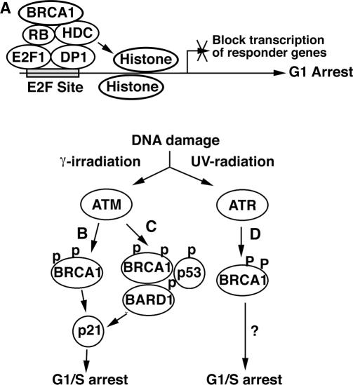
Shortly after the cloning of BRCA1, it was found that inhibition of BRCA1 expression with antisense oligonucleotides enhanced growth of normal and malignant mammary cells (43), and that introduction of wild-type BRCA1 into tumor cells inhibited cell proliferation (44). Interestingly, the growth-inhibitory activity associated with BRCA1 expression was only detected in a subset of cell lines tested, including breast and ovarian but not lung or colon cancer cells (44). Although this observation is consistent with the fact that human BRCA1 mutation carriers mainly develop breast and ovarian tumors, a study attributed this inhibitory activity to the presence of RB protein (45). Aprelikova et al. (45) found that only cells carrying wild-type RB were sensitive to BRCA1 induced G1 arrest, while RB−/− cells were not. To provide a molecular basis for this observation, they further showed that BRCA1 interacts with hypophosphorylated RB. Since hypophosphorylated RB interacts with E2F to prevent transcription of downstream genes and inhibits cell proliferation, it is conceivable that binding to BRCA1 keeps RB in the hypophosphorylated state to achieve growth arrest. BRCA1 C-terminal domains (BRCT) also form a complex with two RB-binding proteins, RbAp46 and RbAp48, and histone deacetylases 1 and 2 (HDAC1 and HDAC2) (46). The RB and histone deacetylase complex (HDC) is thought to suppress transcription of E2F-responsive genes, providing additional evidence for growth inhibition through RB (Figure 1A).
Figure 1.
Current views of BRCA1 functions G1 and G1/S cell cycle checkpoint. (A) A model illustrating a negative role for BRCA1 in G1 arrest. BRCA1 binds to hypophosphorylated RB, which interacts with E2F to form an active complex that blocks transcription. BRCA1 and RB also recruit HDC, which deacetylates histones associated with the promoter, thereby promoting formation of nucleosomes that inhibit transcription. (B and C) Expression of BRCA1 may cause G1/S arrest through a p53-independent (B), and p53 dependent (C) mechanisms. (D) UV radiation may also cause BRCA1-mediated G1/S arrest, although it is not clear whether p21 is involved.
BRCA1-p21 induction
Of note, an earlier investigation attributed the growth-inhibitory effect of BRCA1 to p21WAF1/CIP1, a well-known cyclin-dependent kinase inhibitor (47–49). Somasundaram et al. (50) showed that ectopic over-expression of a wild-type, but not a mutant form, of BRCA1 up-regulated p21 expression and prevented cell cycle progression into the S phase. Such an inhibition effect was not observed in p21−/− cells. Because p53 is a major regulator of p21 expression, they also transfected BRCA1 into p53−/− cells and found that the induction of p21 and inhibition of cell growth by BRCA1 did not require wild-type p53 (Figure 1B).
ATM-BRCA1/BARD1-p53-p21 axis
Contrasted to the above finding, a recent study provided evidence that p53 is involved in the BRCA1 induced G1/S cell cycle checkpoint. It was shown that BRCA1 always forms a heterodimer with BARD1, a BRCA1 associated RING domain protein, in vivo, and the heterodimerization is required to maintain their stability (37,51). Using small interfering RNA (siRNA) to deplete BRCA1 or BARD1, Fabbro et al. (52) demonstrated that the BRCA1–BARD1 complex is required for ATM/ATR (ataxia-telangiectasia-mutated and Rad3-related)-mediated phosphorylation of p53 at Ser-15 following IR or UV radiation-induced DNA damage. This study had several interesting findings. First, the dependence of p53(Ser-15) phosphorylation on the BRCA1–BARD1 complex is quite unique, as phosphorylation of a number of other ATM/ATR targets including H2AX, Chk2, Chk1 and c-jun does not depend on the presence of BRCA1–BARD1 complex. Second, although both IR and UV radiation-induced p53(Ser-15) phosphorylation, only IR radiation could induce G1/S arrest, while UV radiation did not. Because rapid IR-induced phosphorylation of downstream targets is catalyzed by ATM, while ATR mediates rapid UV induced phosphorylation, this observation suggests that the BRCA1–BARD1 complex mediates ATM, but not ATR, catalyzed p53(Ser-15) phosphorylation. Lastly, they demonstrated that the inhibition of p53(Ser-15) phosphorylation by BRCA1-BARD1 acute suppression compromises p21 induction and G1/S checkpoint arrest. Thus, this study reveals an important role of p21 in mediating BRCA1 function in G1/S arrest by IR radiation, and the induction of p21 transcription requires p53 activation (Figure 1C).
A prediction from these observations is that the absence of BRCA1 might decrease p21 expression and impair the G1/S cell cycle checkpoint. However, inconsistently, BRCA1-null embryos exhibited increased levels of p21 and cellular proliferation defects, and died at early postgastrulation stages (28). This is perhaps due to p53 activation triggered by un-repaired DNA damage and genetic instability. Consistently, BRCA1-null embryos survived 1–2 days longer in a p53-null or p21-null genetic background (28,29,53,54). Thus, the activation of p53 triggered by genetic instability in BRCA1−/− embryos obscures the functions of BRCA1 in the G1/S cell cycle checkpoint. It was shown previously that MEF cells carrying double mutations of BRCA1 and p53 were defective at the G1/S cell cycle checkpoint (29). However, this defect cannot be attributed to BRCA1 deficiency, as the absence of p53 alone could impair the G1/S cell cycle checkpoint (49,55).
Interestingly, a recent study using primary fibroblasts from human BRCA1 heterozygotes (BRCA1+/−) and wild-type (BRCA1+/+) indicated that UV radiation could induce G1/S cell cycle arrest in BRCA1+/+ cells, while BRCA1+/− cells displayed an moderate impaired G1/S cell cycle checkpoint (56). Because UV radiation does not activate the p53 mediated G1/S cell cycle checkpoint (52), this study provides an example that reduced dose of BRCA1 could attenuate the G1/S arrest in cells where p53 is not activated. UV induced phosphorylation is primarily mediated by ATR, which has been shown to interact with BRCA1 (57). However, it remains unclear how BRCA1 induces the G1/S arrest upon UV radiation (Figure 1D).
In summary, BRCA1 induced G1/S arrest may occur through a number of distinct pathways that involve many important BRCA1 interacting proteins, including ATM, ATR, BARD1, RB, p53 and p21 and their downstream effectors (Figure 1).
S phase cell cycle checkpoint
Another cell cycle checkpoint induced by ionizing radiation is the S-phase checkpoint, which primarily represents an inhibition of replication initiation upon the DNA damage. A lack of an IR-induced S-phase checkpoint results in persistent DNA synthesis [radioresistant DNA synthesis (RDS)] at early time points after IR. Studies indicated that cells isolated from A–T patients and Nijmegen breakage syndrome (NBS) patients were defective in this checkpoint (58). However, conflicting results were reported regarding whether BRCA1 plays a role in the S-phase checkpoint. One study reported a normal S-phase checkpoint in the BRCA1−/− HCC1937 cells, but no experimental details were provided to support this claim (59). In contrast, Xu et al. (60,61) found that HCC1937 cells were actually defective in S-phase checkpoint, exhibiting a RDS similar to that displayed by AT mutant and NBS mutant cells. Furthermore, their data indicated that the defective IR-induced S-phase checkpoint in HCC1937 cells could be overcome by reconstitution of a wild-type BRCA1, which confirms a role of BRCA1 in this checkpoint.
ATM–BRCA1 interaction
BRCA1 is phosphorylated at several serine sites, including Ser-988, -1387, -1423, -1497 and -1524 upon IR radiation (62,63). To explore whether one of these sites was an important determinant of the IR-induced S-phase checkpoint, mutations of some of these sites were introduced into BRCA1 and tested in HCC1937 cells. Data indicated that wild-type BRCA1 and BRCA1 constructs mutated at either Ser-1423 or both Ser-1423 and Ser-1524 all complemented the defective IR-induced S-phase checkpoint in HCC1937 cells. In contrast, transfection of a construct bearing a mutation at Ser-1387 of BRCA1 failed to complement this defect (61). Because the Ser-1387 in BRCA1 is a known target of ATM phosphorylation, this finding indicates that the phosphorylation of Ser-1387 in BRCA1 is specifically required for the ATM-mediated S-phase checkpoint after ionizing irradiation.
ATR–BRCA1 interaction
Evidence has also been presented that BRCA1 is involved in the S-phase checkpoint activated by stalled replication forks, which can be induced by treatment of cells with UV or hydroxyurea (HU). It has been shown that ATR and ATM phosphorylate BRCA1 on several distinct and overlap Ser/Thr residues, including Ser-1423 (62–64). Tibbetts et al. (64) demonstrated that increased expression of ATR enhanced the phosphorylation of BRCA1 on Ser-1423 following cellular exposure to HU or UV light, whereas doxycycline-induced expression of a kinase-inactive ATR mutant protein inhibited HU- or UV light-induced Ser-1423 phosphorylation. Furthermore, the dramatic relocalization of ATR nuclear foci in response to DNA damage overlaps with the nuclear foci formed by BRCA1. Thus, ATR directly phosphorylates BRCA1 in response to damaged DNA or stalled DNA replication, and both proteins are likely the components of the same genotoxic stress-responsive pathway. These results suggest that ATR activates the intra-S checkpoints in response to stalled replication forks in a manner analogous to that of ATM-dependent induction of these checkpoints after exposure to IR.
BRCA1 also interacts with several other proteins that play an essential role in the S-phase checkpoint. These include the mediator of DNA damage checkpoint protein 1 (MDC1), H2AX, p53 binding protein 1 (53BP1) and MRE11/RAD50/NBS1 (MRN), which form nuclear foci upon IR radiation and arrest cells in the S phase (65). In addition, Durant and Nickoloff (66) have also proposed a cell-cycle-dependent model in which DNA-PK inhibits replication protein A in the S phase of the cell cycle, while BRCA1 inhibits the exonuclease activity of the MRN complex and facilitates DNA repair and S phase arrest.
G2/M cell cycle checkpoint
In addition to the G1/S cell cycle checkpoint and the S phase cell cycle checkpoint, IR radiation also activates the G2/M cell cycle checkpoint, which rapidly delays movement of G2 cells into the mitosis (M) phase. Loss of this checkpoint allows cells with damaged DNA to proceed into the M phase, increasing the likelihood of abnormal chromosomes being passed to the daughter cells. Using MEF cells derived from Brca1Δ11/Δ11 embryos, Xu et al. (15) investigated whether the absence of BRCA1 would affect this cell cycle checkpoint. Their data indicated that BRCA1 wild-type cells exhibited a sharp reduction in mitotic index within 1–4 h after IR radiation. In contrast, Brca1Δ11/Δ11 cells showed no reduction in mitotic index upon IR radiation during the same period of the time. The lack of an immediate mitotic delay following γ-irradiation indicates that elimination of full-length BRCA1 abolishes this checkpoint. They also treated cells with UV radiation and methyl methanesulfonate (MMS). Their data indicated that UV treated Brca1Δ11/Δ11 cells showed a dramatic reduction in mitotic index, while the MMS treated mutant cells were largely defective in the G2/M cell cycle checkpoint. Taken together, these observations suggest that the defect in the G2/M cell cycle checkpoint is specific to certain types of DNA damage.
BRCA1–Chk1 interaction
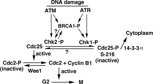
BRCA1 interacts with many proteins that play a role in cell cycle progression (Table 1). It is important to determine whether any BRCA1 interacting proteins are involved in this process, and how they affect the G2/M cell cycle checkpoint in relation to BRCA1. Yarden et al. (67) provided a significant advance on this issue, showing that BRCA1 regulates the expression, phosphorylation and cellular localization of Chk1, a known regulator of the G2-M cell cycle checkpoint. Their data also indicated that BRCA1 affects the expression of both Wee1 kinase, an inhibitor of Cdc2/cyclin B kinase, and the 14-3-3 family of proteins that sequesters phosphorylated Cdc25C and Cdc2/cyclin B kinase in the cytoplasm (68,69). Thus BRCA1 regulates key effectors that control the G2/M checkpoint and is therefore involved in regulating the onset of mitosis (Figure 2).
Figure 2.
A model illustrating a role of BRCA1 in the C2/M cell cycle checkpoint. BRCA1 can be phosphorylated by ATR, ATM and Chk2. BRCA1 regulates expression and cellular localization of Chk1, although it is not clear whether Chk1 can phosphorylates BRCA1. Both Chk1 and Chk2 inactivate Cdc25 by phosphorylate it at Ser-216. The phosphorylation of Cdc25 at Ser-126 not only inactivates this protein but also allows it to bind to 14-3-3α, leading to its exporting from the nucleus. This results in the decrease of the active form of Cdc25, which is a phosphatase involved in dephosphorylation of Cdc2. The dephosphorylated form of Cdc2 is the active form that promotes cell progression from G2 to M phase. Therefore, the reduced amount of Cdc25 results in the decrease of active form of Cdc2, which prevents G2 to M transition. Another two factors are also involved in this pathway. One is Cyclin B1, which forms an active complex with Cdc2 to promote cell progression from G2 to M phase, and the other is Wee1, a kinase that puts phosphate on an inhibitory site of Cdc2, thereby inhibiting function of Cdc2. Thus, expression changes of these genes could theoretically affect G2/M cell cycle checkpoint.
BRCA1–ATM interaction
The chain of the DNA damage response (DDR) starts with upstream kinases, ATM and ATR, which phosphorylate DNA damage sensors, Chk1 and Chk2, and play important roles in the G2/M cell cycle checkpoint (70). ATM, ATR and Chk2 also interact with and phosphorylate BRCA1 (57,62–64). It was revealed that the phosphorylation of BRCA1 by ATM is required for activation of the G2/M checkpoint using the HCC1937 cells, which are defective in both S phase and G2/M cell cycle checkpoints (60,61). In their effort to distinguish the role BRCA1 Ser-1387 and Ser-1423 in these cell cycle checkpoints, they found that mutation of BRCA1 Ser-1423 abolished the ability of BRCA1 to mediate the G2/M checkpoint, while mutation of BRCA1 Ser-1387 affected S phase function of BRCA1 upon IR radiation (60,61). These results clarify the cell cycle checkpoint roles for each of these phosphorylation products.
BRCA1-Chk2 interaction
Chk2, which is an immediate phosphorylation target of ATM, phosphorylates and co-localizes with BRCA1 within discrete nuclear foci prior to DNA damage by γ-irradiation (71). Phosphorylation of BRCA1 at serine 988 is required for the release of BRCA1 from Chk2. This phosphorylation is also important for the ability of BRCA1 to restore survival after DNA damage in the BRCA1-mutated cell line HCC1937. To study the impact of Ser-988 mutation on BRCA1 function, Kim et al. (72) mutated the equivalent serine (Ser-971) in mouse Brca1 using a knock-in approach. The Brca1S971A/S971A mice were developmentally normal, however they suffered a moderately increased risk of spontaneous tumor formation, with a majority of females developing uterus hyperplasia and ovarian abnormalities at two years of age. After treatment with DNA damaging agents, i.e. γ-irradiation and MNNG, Brca1S971A/S971A mice exhibited several abnormalities, including increased body weight, abnormal hair growth pattern, lymphoma, mammary tumor and endometrial tumor. In addition, the onset of tumor formation became accelerated with 80% of mutant mice developing tumors at 1 year of age. These observations suggest that Chk2 phosphorylation of Ser-971 is involved in Brca1 function in modulating DDR and repressing tumor formation. The Brca1S971A/S971A mutant cells also exhibited a partially loss of the G2/M cell cycle checkpoint upon IR radiation. This study suggests that G2/M checkpoint regulation of BRCA is partly modulated by Chk2 phosphorylation in addition to other factors, such as ATM and Chk1 as reviewed earlier (Figure 2).
BRCA1–Aurora interaction
Aurora-A is one of three serine/threonine kinases (A, B and C) and is known to regulate mitotic progression in various organisms [reviewed in (73–76)]. Aurora-A is frequently amplified in several human cancers and over-expression of Aurora-A has been detected in 94% of invasive duct adenocarcinomas of the breast (77–83). It was recently shown that Aurora-A physically binds and phosphorylates BRCA1 at S308, and the phosphorylation is correlated with impaired function of BRCA1 in regulating G2/M transition (84). This finding suggests a link between Aurora-A over-expression and impaired BRCA1 function in genetic instability and tumorigenesis. However, it was recently reported that transgenic mice carrying Aurora-A over-expression in the mammary gland did not develop mammary tumors although they all exhibited mammary hyperplasia (85). In that study, chicken β-actin promoter was linked to the Aurora-A gene through a stopper sequence. The transgene is not expressed unless the stopper is deleted by WAP-Cre, which is activated by pregnancy hormones. Because all the mice only went through one full-cycle of pregnancy, it is possible that the deletion of the stopper, which is required for the expression of the Aurora-A transgene, was incomplete. To test this, we have generated a transgenic mouse strain that over expresses the Aurora-A in the mammary epithelium using the promoter of the mouse mammary tumor virus (MMTV). We found that the MMTV-Aurora transgene is expressed at high levels during the mammary cycle of development and induces mammary tumor formation in about 50% of mice with an average latency of 20 months (X. Wang and C. X. Deng, unpublished data). Further investigations are required to determine whether or not the tumorigenesis in the MMTV-Aurora transgenic mice is entirely or partly due to the impaired BRCA1 function in regulating the G2/M cell cycle checkpoint.
Other BRCA1 interacting proteins
Upon UV radiation, BRCA1 is phosphorylated on Ser-1423 and Ser-1524 by ATR (86). It was shown previously that BRCA1 mutant MEF cells exhibited a normal G2/M cell cycle checkpoint upon UV radiation (15), suggesting that phosphorylation of BRCA1 by ATR may not have a direct impact on this checkpoint. This observation, however, does not rule out a possibility that ATR may play a role in the BRCA1-mediated G2/M checkpoint through other mechanisms. For example, the genetic interplay between BRCA1 and ATR in the G2/M cell cycle checkpoint could occur in their common downstream genes, such as Chk1. In addition, it was shown that BRIT1 (BRCT-repeat inhibitor of hTERT expression), a repressor of human telomerase function that is implicated in cellular immortalization (87,88), is required for the expression of BRCA1, NBS1 and Chk1 (89). Lin et al. (89) found that when BRIT1 expression is depleted by RNAi, cells exhibited defects in both the S phase and G2/M cell cycle checkpoints and become IR radiation sensitive. Thus, the checkpoint defects in the absence of BRIT1 are likely to result from its regulation of Nbs1, BRCA1 and Chk1.
The spindle checkpoint
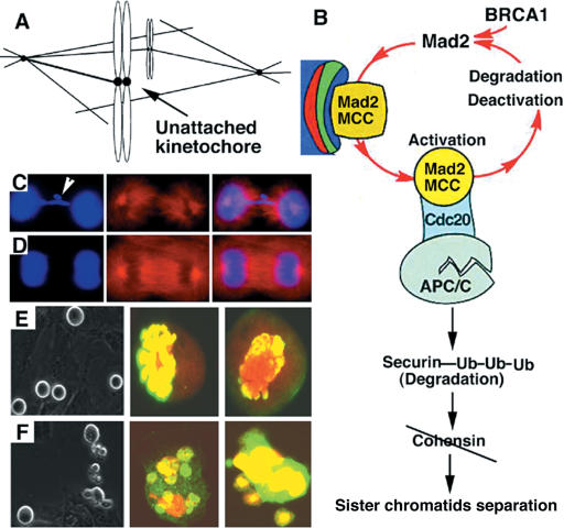
During the mitotic phase, duplicated DNA is first condensed and packed to form sister chromatids, which are then equally separated into newly formed daughter cells. Any premature or mis-separation of sister chromatids will lead to the loss or gain of chromosomes in daughter cells, leading to aneuploidy, which is a prevalent form of genetic instability of human cancers (40). The spindle checkpoint ensures the astonishing accuracy of chromosome segregation by preventing cells with unaligned chromosomes from exiting mitosis. Molecular components of the spindle checkpoint include two evolutionarily conserved protein families, Mad (Mad1, Mad2, Mad3/BubR1) and Bub (Bub1, Bub2, Bub3), as well as other components [reviewed in (90–93)]. In the metaphase, the sister chromatids attach to the mitotic spindle at kinetochores that consist of protein complexes associated with centromeric DNA. After all sister chromatids have attached to the bipolar spindle and their kinetochores are under tension, a large ubiquitin ligase called the anaphase-promoting complex (APC) and its associated substrate-binding co-factor, Cdc20, are activated. The activated APC-Cdc20 tags the securin protein with polyubiquitin chains and promotes its degradation. This, in turn, activates the separase and results in a proteolytic cleavage of the cohesion complex between the sister chromatids and triggers the onset of the anaphase. The spindle checkpoint monitors the attachment of sister chromatids to the spindle and is activated by the lack of microtubule occupancy and tension at the kinetochores (Figure 3A). During the process of spindle checkpoint activation, transient interaction between BubR1-Bub3-Cdc20 and Mad2-Cdc2 complex leads to the formation of the BubR1-Bub3-Mad2-Cdc20 complex (the MCC), which is a more efficient inhibitor of APC (Figure 3B). A number of investigations revealed an essential role of Mad2 in the MCC [reviewed in (94–96)]. Consistently, microinjection of Mad2 antibodies yields premature anaphase onset and chromosome mis-segregation (97). Absence of Mad2 in mouse embryos resulted in accumulation of mitotic errors and apoptosis, leading to early lethality at E5-E6, while haploinsufficiency of Mad2, which results in about 30% less of Mad2 protein, provokes lung tumors after a long latency period (98,99).
Figure 3.
The spindle checkpoint, activation and deficiency. (A) A bipolar spindle showing an unattached kinetochore (arrow). (B) The activation of the spindle checkpoint at the kinetochore leads to the formation of the BubR1-Bub3-Mad2-Cdc20 complex (MCC). The MCC binds and inhibits activity of PPC, preventing sister chromatids from separation. It is shown that Brca1 interacts with Mad2 promoter and positively regulates its expression. (C and D) Images of Brca1Δ11/Δ11 (C) and wild-type (D) MEF cells stained with Dapi and an anti-α-tubulin antibody. Arrows pointed to lagging chromosomes. (E and F) Morphology difference between wild-type (E) and Brca1Δ11/Δ11 MEF (F) cells in responding to nocodazole treatment. Brca1Δ11/Δ11 MEF cells at mitotic phase (as revealed by both round up and positive for phosphorylated histone H3 antibody staining) exhibited significantly more fragmented cells. Phosphorylated histone H3 antibody staining also indicated that many mutant cells contained fragmented chromosomes.
Studying Brca1Δ11/Δ11 cells during the mitotic phase, Wang et al. (31) found that about 30% of these cells displayed abnormal chromosomes, including chromosome bridging, lagging chromosomes in metaphase, anaphase and telophase (Figure 3C and D). It has been shown that a single unattached kinetochore is sufficient to activate the spindle checkpoint and arrest the cell at metaphase (100). Thus, the observations that Brca1Δ11˜Δ11 cells exhibited abnormal chromosome behavior and could advance to the anaphase and telophase suggest that the spindle checkpoint is defective. This notion was confirmed by their further experiment, in which the Brca1Δ11˜Δ11 cells were treated with nocodazole, a reagent that depolymerizes microtubules and activates the spindle checkpoint. Their data indicated that about 60% Brca1 wild-type cells were arrested at metaphase 12 h after the nocodazole treatment (Figure 3E), while Brca1 mutant cells failed to undergo metaphase arrest (Figure 3F), and died due to apoptosis.
Accompanied by these defects, _Brca1_Δ11/Δ11 cells also displayed decreased expression of a number of genes that are involved in the spindle checkpoint, including Mad2, Polo-like-Kinase, Bub1, BubR1 and ZW-10. Given the critical role of Mad2 in the spindle checkpoint (94,97,101), Wang et al. (31) further addressed the possible interaction between Brca1 and Mad2. Using a tetracycline-regulated system to express BRCA1 in UBR60 cells (102), they demonstrated that BRCA1 positively regulates Mad2 by interacting with its promoter. Furthermore, over-expression of Mad2 in mutant cells partially overcame the spindle checkpoint defects. These observations provide strong evidence that Brca1 plays an important role in the spindle checkpoint through maintaining Mad2 expression.
A similar finding was made in BRCA1 wild-type human prostate (DU-145) and breast (MCF-7) cancer cells upon acute suppression of BRCA1 using small interfering RNA that is specific to BRCA1. In these cells, Bae et al. (103) demonstrated that attenuation of the mitotic cell cycle checkpoint was accompanied by down-regulation of multiple genes implicated in the mitotic spindle checkpoint (e.g. Bub1, Bub1b, He, Stk6 and Birc5). Interestingly, their data also revealed expression changes of many other genes that are involved in chromosome segregation (Espl1, Nek2, Pttg1, multiple kinesis and kinesis-like proteins), centrosome function (ASP), cytokinesis (Prc1, Plk, Mphosph1 and Knsl2), and the transition into and progression through mitosis (B-type cyclins, Cdc2 and Cdc20). BRCA1 knockdown also caused the accumulation of binucleated and multinucleated cells, suggesting a defect in the coordination of cytokinesis and karyokinesis. These findings suggest that BRCA1 transcriptionally regulates gene expression for orderly mitotic progression.
DDR AND CANCER EVOLUTION
Early attempts to create animal models for BRCA1 associated breast cancer were not successful, as BRCA1 deficiency invariably results in embryonic lethality, primarily due to elevated cell death and growth retardation (28,29). An animal model carrying mammary specific disruption of BRCA1 (BRCA1Co/Co;MMTV-Cre) also exhibited increased apoptosis and abnormal mammary branch morphogenesis before tumor formation at low frequency with long latency (25). Why do BRCA1 deficient cells die? Mounting experimental evidence indicates that organisms have acquired anti-proliferative and cell death–inducing mechanisms that prevent clonal expansion of mutant cells (104). A key mechanism is the DDR, which can be activated not only by DNA damage but also by some other conditions, such as oncogene expression, loss of tumor suppressors (105). Recently, it was shown that during the early cancerous development of lung or bladder tumors, oncogene induced aberrations in DNA replication activates DDR, as evidenced by the posphorylation of H2AX (γH2AX) followed by the activation of ATM-Chk2-p53 signaling in the tumor tissues (106,107). The progression to malignant transformation requires the inactivation of DDR, which would in turn create genetic instability and accelerate cancer evolution.
Does BRCA1 deficiency activate DDR and thereby cause growth arrest and/or apoptosis? Several lines of evidence indicate that it may be the case. It was shown that the death of BRCA1 mutant cells is directly linked to the activation of p53, as deletion of p53 and/or its downstream mediator p21 partially rescues BRCA1-null embryos (28,29,53,54). Moreover, it was shown that elimination of either one or both wild-type p53 alleles completely overcame embryonic lethality caused by the targeted deletion of full-length Brca1 and allowed Brca1Δ11/Δ11 mutant mice to survive to adulthood (29). Further analysis indicated that haploid loss or complete loss of p53 resulted in attenuated apoptosis and G1-S checkpoint control, allowing Brca1Δ11/Δ11 cells to proliferate (29). Interestingly, most Brca1Δ11/Δ11;p53+/− male mice suffered premature aging, exhibiting higher expression levels of p53 compared with controls (108). This suggests that remaining wild-type p53 is activated, which may be a cause for premature aging.
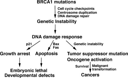
It has been reported that over 90% of human BRCA1 deficient breast cancers also bear p53 mutations, while p53 alterations are only found in about 40% sporadic breast cancers (109). Furthermore, most of mammary tumors developed in the BRCA1Co/Co;MMTV-Cre mouse model also spontaneously mutated their p53 (25), suggesting that the loss of p53 may be responsible for tumorigenesis. To directly test the role of p53 in BRCA1 associated tumorigenesis, a p53-null mutation allele was introduced into BRCA1 conditional mutant model, and the data indicated that introduction of a p53-null mutation into these mice can significantly accelerate mammary tumor formation (25). This data indicated that inactivation of p53 and Brca1 deficiency synergistically induce mammary tumor formation. However the factors responsible for p53 activation in the absence of Brca1 are poorly understood. To investigate this, we employed a genetic test by crossing Brca1Δ11/+ mice with mutant mice carrying targeted mutations of genes in the DDR pathway, including ATM, Chk1, Chk2, p19, Pten, Parp-1, p21 and Gadd45. Our data indicated that ATM or Chk2 inactivation is equivalent to p53 inactivation in that it allows Brca1Δ11/Δ11 embryos to survive to adulthood (L. Cao and C. X. Deng unpublished data). An earlier investigation also revealed that Brca1 deficiency resulted in Chk2 phosphorylation and the Chk2-dependent activation of p53 (110). These observations support a model indicating that BRCA1 deficiency results in genetic instability, leading to the activation of ATM-Chk2-p53 DDR signaling, which, in turn, serves as a natural barrier against malignant transformation of BRCA1 mutant cell (Figure 4).
Figure 4.
A model illustrating connections among cell cycle checkpoints, centrosome duplication, DNA damage repair, genetic instability, DNA damage response, developmental abnormalities and tumorigenesis caused by BRCA1 deficiency.
Experimental data also indicate that the inactivation of DDR is not sufficient for Brca1 deficient cells to undergo malignant transformation. Brca1 mutant mice in either a p53 or Chk2 mutant genetic background developed tumors in a stochastic fashion, suggesting additional factor(s) is needed to tumorigenesis. Consistent with this notion, it has been demonstrated that BRCA1 associated tumors are frequently associated with increased expression of oncogenes, such as cyclin D1, c-Myc and ErbB2, and loss of heterozygosity of tumor suppressor genes (23–27).
CONCLUSIONS AND FUTURE DIRECTIONS
In this review, I have examined experimental evidence regarding the role of BRCA1 in cell cycle checkpoints, genetic stability and tumorigenesis. BRCA1 deficiency results in defective S phase, G2/M and spindle checkpoints. The defects in these cell cycle checkpoints, combined with abnormal centrosome duplication and defective DNA damage repair could cause genetic instability in BRCA1 deficient cells. The genetic instability, in turn, triggers a series of physiological responses, most promptly the DNA damage response, i.e. activation of ATM-Chk2-p53 signals as manifested by G1/S arrest due to up-regulation of p21, and apoptosis due to the activation of pro-apoptotic signals. On the other hand, the absence of BRCA1 allows further genetic alterations, including further tumor suppressor mutations and activation of oncogenes, which overcomes growth defects and ultimately results in breast cancer formation (Figure 4).
The examination of the literature also reveals many unanswered questions. First, the role of BRCA1 in the G1/S cell cycle checkpoint needs further scrutiny, as BRCA1 deficient cells exhibit an intact G1/S cell cycle checkpoint. p53 activation has obscured the role of BRCA1 in this checkpoint and further mechanistic studies should overcome this barrier. Second, it has been shown that expression of many genes that are critical for the spindle checkpoint is down regulated in cells carrying a targeted disruption (31) or RNAi mediated acute suppression of BRCA1 (103). However, it is not clear about the potential relationship between BRCA1 and expression of these genes. Even in the most well studied case, the Mad2 gene, whose expression is regulated by BRCA1 in a number of experimental systems, the published data was ambiguous about whether the interaction of BRCA1 with the promoter of Mad2 was direct or indirect (31). Until this is done, the regulatory role of BRCA1 in the spindle checkpoint remains un-established. Third, acute suppression of BRCA1 also causes altered expression of many genes that are involved in chromosome segregation, centrosome function or cytokinesis (103). Although this observation is interesting, the claim that BRCA1 regulates gene expression for orderly mitotic progression is premature unless solid evidence is provided. Fourth, recent investigations demonstrated that some major functions of BRCA1 could be attributed to the heterodimer formed between BRCA1 and BARD1 (35,37). In addition, germline mutations of BARD1 were also found in breast and ovarian cancers (111,112). Thus potential roles of the BRCA1/BARD1 heterodimer in tumor suppression, DNA damage repair, cell cycle checkpoints and regulation of centrosome duplication should be interesting topics for future studies. Finally, current chemoprevention and therapy are suboptimal (113). The knowledge regarding functions of BRCA1 in cell cycle checkpoints, genome integrity, DDR and cancer evolution may facilitate drug screening and better design of therapeutic approaches. Reagents that cause reactivation of cell cycle checkpoints, cell death, defective DNA damage repair and/or promote mutant cells through an lethal mitosis should be favorable in the treatment of BRCA1 associated breast cancer. Some promising data attacking weakness of BRCA1/2 tumors has been provided by a number of recent publications (114–116).
Acknowledgments
The authors thank Drs Rui-Hong Wang, Thomas Fishler, Joseph De Soto and Cuiying Xiao for critically reading of the manuscript. This work was supported by the intramural Research Program of National Institute of Diabetes, Digestive and Kidney Diseases, National Institutes of Health, USA. The open access publication charge for this paper has been waived by Oxford University Press.
Conflict of interest statement. None declared.
REFERENCES
- 1.Zhang J., Powell S.N. The role of the BRCA1 tumor suppressor in DNA double-strand break repair. Mol. Cancer Res. 2005;3:531–539. doi: 10.1158/1541-7786.MCR-05-0192. [DOI] [PubMed] [Google Scholar]
- 2.Alberg A.J., Lam A.P., Helzlsouer K.J. Epidemiology, prevention, and early detection of breast cancer. Curr. Opin. Oncol. 1999;11:435–441. doi: 10.1097/00001622-199911000-00003. [DOI] [PubMed] [Google Scholar]
- 3.Brody L.C., Biesecker B.B. Breast cancer susceptibility genes. BRCA1 and BRCA2. Medicine (Baltimore) 1998;77:208–226. doi: 10.1097/00005792-199805000-00006. [DOI] [PubMed] [Google Scholar]
- 4.Eccles D.M., Pichert G. Familial non-BRCA1/BRCA2-associated breast cancer. Lancet Oncol. 2005;6:705–711. doi: 10.1016/S1470-2045(05)70318-1. [DOI] [PubMed] [Google Scholar]
- 5.Hall J.M., Lee M.K., Newman B., Morrow J.E., Anderson L.A., Huey B., King M.C. Linkage of early-onset familial breast cancer to chromosome 17q21. Science. 1990;250:1684–1689. doi: 10.1126/science.2270482. [DOI] [PubMed] [Google Scholar]
- 6.Miki T., Bottaro D.P., Fleming T.P., Smith C.L., Burgess W.H., Chan A.M., Aaronson S.A. Determination of ligand-binding specificity by alternative splicing: two distinct growth factor receptors encoded by a single gene. Proc. Natl Acad. Sci. USA. 1992;89:246–250. doi: 10.1073/pnas.89.1.246. [DOI] [PMC free article] [PubMed] [Google Scholar]
- 7.Easton D.F., Ford D., Bishop D.T. Breast and ovarian cancer incidence in BRCA1-mutation carriers. Breast cancer linkage consortium. Am. J. Hum. Genet. 1995;56:265–271. [PMC free article] [PubMed] [Google Scholar]
- 8.Struewing J.P., Hartge P., Wacholder S., Baker S.M., Berlin M., McAdams M., Timmerman M.M., Brody L.C., Tucker M.A. The risk of cancer associated with specific mutations of BRCA1 and BRCA2 among Ashkenazi Jews. N Engl. J. Med. 1997;336:1401–1408. doi: 10.1056/NEJM199705153362001. [DOI] [PubMed] [Google Scholar]
- 9.Ford D., Easton D.F., Stratton M., Narod S., Goldgar D., Devilee P., Bishop D.T., Weber B., Lenoir G., Chang-Claude J., et al. Genetic heterogeneity and penetrance analysis of the BRCA1 and BRCA2 genes in breast cancer families. The breast cancer linkage consortium. Am. J. Hum. Genet. 1998;62:676–689. doi: 10.1086/301749. [DOI] [PMC free article] [PubMed] [Google Scholar]
- 10.Miki Y., Swensen J., Shattuck-Eidens D., Futreal P.A., Harshman K., Tavtigian S., Liu Q., Cochran C., Bennett L.M., Ding W., et al. A strong candidate for the breast and ovarian cancer susceptibility gene BRCA1. Science. 1994;266:66–71. doi: 10.1126/science.7545954. [DOI] [PubMed] [Google Scholar]
- 11.Lane T.F., Deng C., Elson A., Lyu M.S., Kozak C.A., Leder P. Expression of Brca1 is associated with terminal differentiation of ectodermally and mesodermally derived tissues in mice. Genes Dev. 1995;9:2712–2722. doi: 10.1101/gad.9.21.2712. [DOI] [PubMed] [Google Scholar]
- 12.ElShamy W.M., Livingston D.M. Identification of BRCA1-IRIS, a BRCA1 locus product. Nature Cell Biol. 2004;6:954–967. doi: 10.1038/ncb1171. [DOI] [PubMed] [Google Scholar]
- 13.Wilson C.A., Payton M.N., Elliott G.S., Buaas F.W., Cajulis E.E., Grosshans D., Ramos L., Reese D.M., Slamon D.J., Calzone F.J. Differential subcellular localization, expression and biological toxicity of BRCA1 and the splice variant BRCA1-delta11b. Oncogene. 1997;14:1–16. doi: 10.1038/sj.onc.1200924. [DOI] [PubMed] [Google Scholar]
- 14.Thakur S., Zhang H.B., Peng Y., Le H., Carroll B., Ward T., Yao J., Farid L.M., Couch F.J., Wilson R.B., et al. Localization of BRCA1 and a splice variant identifies the nuclear localization signal. Mol. Cell Biol. 1997;17:444–452. doi: 10.1128/mcb.17.1.444. [DOI] [PMC free article] [PubMed] [Google Scholar]
- 15.Xu X., Weaver Z., Linke S.P., Li C., Gotay J., Wang X.W., Harris C.C., Ried T., Deng C.X. Centrosome amplification and a defective G2-M cell cycle checkpoint induce genetic instability in BRCA1 exon 11 isoform-deficient cells. Mol. Cell. 1999;3:389–395. doi: 10.1016/s1097-2765(00)80466-9. [DOI] [PubMed] [Google Scholar]
- 16.Paterson J.W. BRCA1: a review of structure and putative functions. Dis. Markers. 1998;13:261–274. doi: 10.1155/1998/298530. [DOI] [PubMed] [Google Scholar]
- 17.Deng C.X., Brodie S.G. Roles of BRCA1 and its interacting proteins. Bioessays. 2000;22:728–737. doi: 10.1002/1521-1878(200008)22:8<728::AID-BIES6>3.0.CO;2-B. [DOI] [PubMed] [Google Scholar]
- 18.Venkitaraman A.R. Cancer susceptibility and the functions of BRCA1 and BRCA2. Cell. 2002;108:171–182. doi: 10.1016/s0092-8674(02)00615-3. [DOI] [PubMed] [Google Scholar]
- 19.Deng C.X. Tumor formation in Brca1 conditional mutant mice. Environ. Mol. Mutagen. 2002;39:171–177. doi: 10.1002/em.10069. [DOI] [PubMed] [Google Scholar]
- 20.Brodie S.G., Deng C.X. BRCA1-associated tumorigenesis: what have we learned from knockout mice? Trends Genet. 2001;17:S18–S22. doi: 10.1016/s0168-9525(01)02451-9. [DOI] [PubMed] [Google Scholar]
- 21.Deng C.X. Tumorigenesis as a consequence of genetic instability in Brca1 mutant mice. Mutat. Res. 2001;477:183–189. doi: 10.1016/s0027-5107(01)00119-1. [DOI] [PubMed] [Google Scholar]
- 22.Kinzler K.W., Vogelstein B. Cancer-susceptibility genes. Gatekeepers and caretakers. Nature. 1997;386:761–763. doi: 10.1038/386761a0. [DOI] [PubMed] [Google Scholar]
- 23.Tirkkonen M., Johannsson O., Agnarsson B.A., Olsson H., Ingvarsson S., Karhu R., Tanner M., Isola J., Barkardottir R.B., Borg A., et al. Distinct somatic genetic changes associated with tumor progression in carriers of BRCA1 and BRCA2 germ-line mutations. Cancer Res. 1997;57:1222–1227. [PubMed] [Google Scholar]
- 24.Tomlinson G.E., Chen T.T., Stastny V.A., Virmani A.K., Spillman M.A., Tonk V., Blum J.L., Schneider N.R., Wistuba II., Shay J.W., et al. Characterization of a breast cancer cell line derived from a germ-line BRCA1 mutation carrier. Cancer Res. 1998;58:3237–3242. [PubMed] [Google Scholar]
- 25.Xu X., Wagner K.U., Larson D., Weaver Z., Li C., Ried T., Hennighausen L., Wynshaw-Boris A., Deng C.X. Conditional mutation of Brca1 in mammary epithelial cells results in blunted ductal morphogenesis and tumour formation. Nature Genet. 1999;22:37–43. doi: 10.1038/8743. [DOI] [PubMed] [Google Scholar]
- 26.Weaver Z., Montagna C., Xu X., Howard T., Gadina M., Brodie S.G., Deng C.X., Ried T. Mammary tumors in mice conditionally mutant for Brca1 exhibit gross genomic instability and centrosome amplification yet display a recurring distribution of genomic imbalances that is similar to human breast cancer. Oncogene. 2002;21:5097–5107. doi: 10.1038/sj.onc.1205636. [DOI] [PubMed] [Google Scholar]
- 27.Brodie S.G., Xu X., Qiao W., Li W.M., Cao L., Deng C.X. Multiple genetic changes are associated with mammary tumorigenesis in Brca1 conditional knockout mice. Oncogene. 2001;20:7514–7523. doi: 10.1038/sj.onc.1204929. [DOI] [PubMed] [Google Scholar]
- 28.Shen S.X., Weaver Z., Xu X., Li C., Weinstein M., Chen L., Guan X.Y., Ried T., Deng C.X. A targeted disruption of the murine Brca1 gene causes gamma-irradiation hypersensitivity and genetic instability. Oncogene. 1998;17:3115–3124. doi: 10.1038/sj.onc.1202243. [DOI] [PubMed] [Google Scholar]
- 29.Xu X., Qiao W., Linke S.P., Cao L., Li W.M., Furth P.A., Harris C.C., Deng C.X. Genetic interactions between tumor suppressors Brca1 and p53 in apoptosis, cell cycle and tumorigenesis. Nature Genet. 2001;28:266–271. doi: 10.1038/90108. [DOI] [PubMed] [Google Scholar]
- 30.Silver D.P., Livingston D.M. Self-excising retroviral vectors encoding the Cre recombinase overcome Cre-mediated cellular toxicity. Mol. Cell. 2001;8:233–243. doi: 10.1016/s1097-2765(01)00295-7. [DOI] [PubMed] [Google Scholar]
- 31.Wang R.H., Yu H., Deng C.X. A requirement for breast-cancer-associated gene 1 (BRCA1) in the spindle checkpoint. Proc. Natl Acad. Sci. USA. 2004;101:17108–17113. doi: 10.1073/pnas.0407585101. [DOI] [PMC free article] [PubMed] [Google Scholar]
- 32.Deng C.X., Scott F. Role of the tumor suppressor gene Brca1 in genetic stability and mammary gland tumor formation. Oncogene. 2000;19:1059–1064. doi: 10.1038/sj.onc.1203269. [DOI] [PubMed] [Google Scholar]
- 33.Deng C.X. Roles of BRCA1 in centrosome duplication. Oncogene. 2002;21:6222–6227. doi: 10.1038/sj.onc.1205713. [DOI] [PubMed] [Google Scholar]
- 34.Deng C.X., Wang R.H. Roles of BRCA1 in DNA damage repair: a link between development and cancer. Hum. Mol. Genet. 2003;12:R113–R123. doi: 10.1093/hmg/ddg082. [DOI] [PubMed] [Google Scholar]
- 35.Henderson B.R. Regulation of BRCA1, BRCA2 and BARD1 intracellular trafficking. Bioessays. 2005;27:884–893. doi: 10.1002/bies.20277. [DOI] [PubMed] [Google Scholar]
- 36.Cantor S.B., Andreassen P.R. Assessing the link between BACH1 and BRCA1 in the FA pathway. Cell Cycle. 2006;5:164–167. doi: 10.4161/cc.5.2.2338. [DOI] [PubMed] [Google Scholar]
- 37.Baer R., Ludwig T. The BRCA1/BARD1 heterodimer, a tumor suppressor complex with ubiquitin E3 ligase activity. Curr. Opin. Genet. Dev. 2002;12:86–91. doi: 10.1016/s0959-437x(01)00269-6. [DOI] [PubMed] [Google Scholar]
- 38.Elledge S.J. Cell cycle checkpoints: preventing an identity crisis. Science. 1996;274:1664–1672. doi: 10.1126/science.274.5293.1664. [DOI] [PubMed] [Google Scholar]
- 39.Kastan M.B., Bartek J. Cell-cycle checkpoints and cancer. Nature. 2004;432:316–323. doi: 10.1038/nature03097. [DOI] [PubMed] [Google Scholar]
- 40.Kops G.J., Weaver B.A., Cleveland D.W. On the road to cancer: aneuploidy and the mitotic checkpoint. Nature Rev. Cancer. 2005;5:773–785. doi: 10.1038/nrc1714. [DOI] [PubMed] [Google Scholar]
- 41.Vaughn J.P., Davis P.L., Jarboe M.D., Huper G., Evans A.C., Wiseman R.W., Berchuck A., Iglehart J.D., Futreal P.A., Marks J.R. BRCA1 expression is induced before DNA synthesis in both normal and tumor-derived breast cells. Cell Growth Differ. 1996;7:711–715. [PubMed] [Google Scholar]
- 42.Ruffner H., Verma I.M. BRCA1 is a cell cycle-regulated nuclear phosphoprotein. Proc. Natl Acad. Sci. USA. 1997;94:7138–7143. doi: 10.1073/pnas.94.14.7138. [DOI] [PMC free article] [PubMed] [Google Scholar]
- 43.Thompson M.E., Jensen R.A., Obermiller P.S., Page D.L., Holt J.T. Decreased expression of BRCA1 accelerates growth and is often present during sporadic breast cancer progression. Nature Genet. 1995;9:444–450. doi: 10.1038/ng0495-444. [DOI] [PubMed] [Google Scholar]
- 44.Holt J.T., Thompson M.E., Szabo C., Robinson-Benion C., Arteaga C.L., King M.C., Jensen R.A. Growth retardation and tumour inhibition by BRCA1. Nature Genet. 1996;12:298–302. doi: 10.1038/ng0396-298. [DOI] [PubMed] [Google Scholar]
- 45.Aprelikova O.N., Fang B.S., Meissner E.G., Cotter S., Campbell M., Kuthiala A., Bessho M., Jensen R.A., Liu E.T. BRCA1-associated growth arrest is RB-dependent. Proc. Natl Acad. Sci. USA. 1999;96:11866–11871. doi: 10.1073/pnas.96.21.11866. [DOI] [PMC free article] [PubMed] [Google Scholar]
- 46.Yarden R.I., Brody L.C. BRCA1 interacts with components of the histone deacetylase complex. Proc. Natl Acad. Sci. USA. 1999;96:4983–4988. doi: 10.1073/pnas.96.9.4983. [DOI] [PMC free article] [PubMed] [Google Scholar]
- 47.el-Deiry W.S., Tokino T., Velculescu V.E., Levy D.B., Parsons R., Trent J.M., Lin D., Mercer W.E., Kinzler K.W., Vogelstein B. WAF1, a potential mediator of p53 tumor suppression. Cell. 1993;75:817–825. doi: 10.1016/0092-8674(93)90500-p. [DOI] [PubMed] [Google Scholar]
- 48.Harper J.W., Adami G.R., Wei N., Keyomarsi K., Elledge S.J. The p21 Cdk-interacting protein Cip1 is a potent inhibitor of G1 cyclin-dependent kinases. Cell. 1993;75:805–816. doi: 10.1016/0092-8674(93)90499-g. [DOI] [PubMed] [Google Scholar]
- 49.Deng C., Zhang P., Harper J.W., Elledge S.J., Leder P. Mice lacking p21CIP1/WAF1 undergo normal development, but are defective in G1 checkpoint control. Cell. 1995;82:675–684. doi: 10.1016/0092-8674(95)90039-x. [DOI] [PubMed] [Google Scholar]
- 50.Somasundaram K., Zhang H., Zeng Y.X., Houvras Y., Peng Y., Wu G.S., Licht J.D., Weber B.L., El-Deiry W.S. Arrest of the cell cycle by the tumour-suppressor BRCA1 requires the CDK-inhibitor p21WAF1/CiP1. Nature. 1997;389:187–190. doi: 10.1038/38291. [DOI] [PubMed] [Google Scholar]
- 51.Wu L.C., Wang Z.W., Tsan J.T., Spillman M.A., Phung A., Xu X.L., Yang M.C., Hwang L.Y., Bowcock A.M., Baer R. Identification of a RING protein that can interact in vivo with the BRCA1 gene product. Nature Genet. 1996;14:430–440. doi: 10.1038/ng1296-430. [DOI] [PubMed] [Google Scholar]
- 52.Fabbro M., Savage K., Hobson K., Deans A.J., Powell S.N., McArthur G.A., Khanna K.K. BRCA1-BARD1 complexes are required for p53Ser-15 phosphorylation and a G1/S arrest following ionizing radiation-induced DNA damage. J. Biol. Chem. 2004;279:31251–31258. doi: 10.1074/jbc.M405372200. [DOI] [PubMed] [Google Scholar]
- 53.Hakem R., de la Pompa J.L., Elia A., Potter J., Mak T.W. Partial rescue of Brca1 (5–6) early embryonic lethality by p53 or p21 null mutation. Nature Genet. 1997;16:298–302. doi: 10.1038/ng0797-298. [DOI] [PubMed] [Google Scholar]
- 54.Ludwig T., Chapman D.L., Papaioannou V.E., Efstratiadis A. Targeted mutations of breast cancer susceptibility gene homologs in mice: lethal phenotypes of Brca1, Brca2, Brca1/Brca2, Brca1/p53, and Brca2/p53 nullizygous embryos. Genes Dev. 1997;11:1226–1241. doi: 10.1101/gad.11.10.1226. [DOI] [PubMed] [Google Scholar]
- 55.Donehower L.A., Bradley A. The tumor suppressor p53. Biochim. Biophys. Acta. 1993;1155:181–205. doi: 10.1016/0304-419x(93)90004-v. [DOI] [PubMed] [Google Scholar]
- 56.Shorrocks J., Tobi S.E., Latham H., Peacock J.H., Eeles R., Eccles D., McMillan T.J. Primary fibroblasts from BRCA1 heterozygotes display an abnormal G1/S cell cycle checkpoint following UVA irradiation but show normal levels of micronuclei following oxidative stress or mitomycin C treatment. Int. J. Radiat. Oncol. Biol. Phys. 2004;58:470–478. doi: 10.1016/j.ijrobp.2003.09.042. [DOI] [PubMed] [Google Scholar]
- 57.Turner J.M., Aprelikova O., Xu X., Wang R., Kim S., Chandramouli G.V., Barrett J.C., Burgoyne P.S., Deng C.X. BRCA1, histone H2AX phosphorylation, and male meiotic sex chromosome inactivation. Curr. Biol. 2004;14:2135–2142. doi: 10.1016/j.cub.2004.11.032. [DOI] [PubMed] [Google Scholar]
- 58.Shiloh Y. Ataxia-telangiectasia and the Nijmegen breakage syndrome: related disorders but genes apart; pp. 635–662. [DOI] [PubMed] [Google Scholar]
- 59.Scully R., Ganesan S., Vlasakova K., Chen J., Socolovsky M., Livingston D.M. Genetic analysis of BRCA1 function in a defined tumor cell line. Mol. Cell. 1999;4:1093–1099. doi: 10.1016/s1097-2765(00)80238-5. [DOI] [PubMed] [Google Scholar]
- 60.Xu B., Kim S., Kastan M.B. Involvement of Brca1 in S-phase and G(2)-phase checkpoints after ionizing irradiation. Mol. Cell Biol. 2001;21:3445–3450. doi: 10.1128/MCB.21.10.3445-3450.2001. [DOI] [PMC free article] [PubMed] [Google Scholar]
- 61.Xu B., O'Donnell A.H., Kim S.T., Kastan M.B. Phosphorylation of serine 1387 in Brca1 is specifically required for the Atm-mediated S-phase checkpoint after ionizing irradiation. Cancer Res. 2002;62:4588–4591. [PubMed] [Google Scholar]
- 62.Cortez D., Wang Y., Qin J., Elledge S.J. Requirement of ATM-dependent phosphorylation of brca1 in the DNA damage response to double-strand breaks. Science. 1999;286:1162–1166. doi: 10.1126/science.286.5442.1162. [DOI] [PubMed] [Google Scholar]
- 63.Okada S., Ouchi T. Cell cycle differences in DNA damage-induced BRCA1 phosphorylation affect its subcellular localization. J. Biol. Chem. 2003;278:2015–2020. doi: 10.1074/jbc.M208685200. [DOI] [PubMed] [Google Scholar]
- 64.Tibbetts R.S., Cortez D., Brumbaugh K.M., Scully R., Livingston D., Elledge S.J., Abraham R.T. Functional interactions between BRCA1 and the checkpoint kinase ATR during genotoxic stress. Genes Dev. 2000;14:2989–3002. doi: 10.1101/gad.851000. [DOI] [PMC free article] [PubMed] [Google Scholar]
- 65.Stewart G.S., Wang B., Bignell C.R., Taylor A.M., Elledge S.J. MDC1 is a mediator of the mammalian DNA damage checkpoint. Nature. 2003;421:961–966. doi: 10.1038/nature01446. [DOI] [PubMed] [Google Scholar]
- 66.Durant S.T., Nickoloff J.A. Good timing in the cell cycle for precise DNA repair by BRCA1. Cell Cycle. 2005;4:1216–1222. doi: 10.4161/cc.4.9.2027. [DOI] [PubMed] [Google Scholar]
- 67.Yarden R.I., Pardo-Reoyo S., Sgagias M., Cowan K.H., Brody L.C. BRCA1 regulates the G2/M checkpoint by activating Chk1 kinase upon DNA damage. Nature Genet. 2002;30:285–289. doi: 10.1038/ng837. [DOI] [PubMed] [Google Scholar]
- 68.Hutchins J.R., Clarke P.R. Many fingers on the mitotic trigger: post-translational regulation of the Cdc25C phosphatase. Cell Cycle. 2004;3:41–45. [PubMed] [Google Scholar]
- 69.Muslin A.J., Xing H. 14-3-3 proteins: regulation of subcellular localization by molecular interference. Cell Signal. 2000;12:703–709. doi: 10.1016/s0898-6568(00)00131-5. [DOI] [PubMed] [Google Scholar]
- 70.Sancar A., Lindsey-Boltz L.A., Unsal-Kacmaz K., Linn S. Molecular mechanisms of mammalian DNA repair and the DNA damage checkpoints. Annu. Rev. Biochem. 2004;73:39–85. doi: 10.1146/annurev.biochem.73.011303.073723. [DOI] [PubMed] [Google Scholar]
- 71.Lee J.S., Collins K.M., Brown A.L., Lee C.H., Chung J.H. hCds1-mediated phosphorylation of BRCA1 regulates the DNA damage response. Nature. 2000;404:201–204. doi: 10.1038/35004614. [DOI] [PubMed] [Google Scholar]
- 72.Kim S.S., Cao L., Li C., Xu X., Huber L.J., Chodosh L.A., Deng C.X. Uterus hyperplasia and increased carcinogen-induced tumorigenesis in mice carrying a targeted mutation of the Chk2 phosphorylation site in Brca1. Mol. Cell Biol. 2004;24:9498–9507. doi: 10.1128/MCB.24.21.9498-9507.2004. [DOI] [PMC free article] [PubMed] [Google Scholar]
- 73.Meraldi P., Honda R., Nigg E.A. Aurora kinases link chromosome segregation and cell division to cancer susceptibility. Curr. Opin. Genet. Dev. 2004;14:29–36. doi: 10.1016/j.gde.2003.11.006. [DOI] [PubMed] [Google Scholar]
- 74.Katayama H., Brinkley W.R., Sen S. The Aurora kinases: role in cell transformation and tumorigenesis. Cancer Metastasis Rev. 2003;22:451–464. doi: 10.1023/a:1023789416385. [DOI] [PubMed] [Google Scholar]
- 75.Ducat D., Zheng Y. Aurora kinases in spindle assembly and chromosome segregation. Exp. Cell Res. 2004;301:60–67. doi: 10.1016/j.yexcr.2004.08.016. [DOI] [PubMed] [Google Scholar]
- 76.Crane R., Gadea B., Littlepage L., Wu H., Ruderman J.V. Aurora A, meiosis and mitosis. Biol. Cell. 2004;96:215–229. doi: 10.1016/j.biolcel.2003.09.008. [DOI] [PubMed] [Google Scholar]
- 77.Zhou H., Kuang J., Zhong L., Kuo W.L., Gray J.W., Sahin A., Brinkley B.R., Sen S. Tumour amplified kinase STK15/BTAK induces centrosome amplification, aneuploidy and transformation. Nature Genet. 1998;20:189–193. doi: 10.1038/2496. [DOI] [PubMed] [Google Scholar]
- 78.Giet R., Petretti C., Prigent C. Aurora kinases, aneuploidy and cancer, a coincidence or a real link? Trends Cell Biol. 2005;15:241–250. doi: 10.1016/j.tcb.2005.03.004. [DOI] [PubMed] [Google Scholar]
- 79.Bischoff J.R., Anderson L., Zhu Y., Mossie K., Ng L., Souza B., Schryver B., Flanagan P., Clairvoyant F., Ginther C., et al. A homologue of Drosophila aurora kinase is oncogenic and amplified in human colorectal cancers. EMBO J. 1998;17:3052–3065. doi: 10.1093/emboj/17.11.3052. [DOI] [PMC free article] [PubMed] [Google Scholar]
- 80.Hu W., Kavanagh J.J., Deaver M., Johnston D.A., Freedman R.S., Verschraegen C.F., Sen S. Frequent overexpression of STK15/Aurora-A/BTAK and chromosomal instability in tumorigenic cell cultures derived from human ovarian cancer. Oncol. Res. 2005;15:49–57. doi: 10.3727/096504005775082101. [DOI] [PubMed] [Google Scholar]
- 81.Kamada K., Yamada Y., Hirao T., Fujimoto H., Takahama Y., Ueno M., Takayama T., Naito A., Hirao S., Nakajima Y. Amplification/overexpression of Aurora-A in human gastric carcinoma: potential role in differentiated type gastric carcinogenesis. Oncol. Rep. 2004;12:593–599. [PubMed] [Google Scholar]
- 82.Tanaka E., Hashimoto Y., Ito T., Okumura T., Kan T., Watanabe G., Imamura M., Inazawa J., Shimada Y. The clinical significance of Aurora-A/STK15/BTAK expression in human esophageal squamous cell carcinoma. Clin. Cancer Res. 2005;11:1827–1834. doi: 10.1158/1078-0432.CCR-04-1627. [DOI] [PubMed] [Google Scholar]
- 83.Tanaka T., Kimura M., Matsunaga K., Fukada D., Mori H., Okano Y. Centrosomal kinase AIK1 is overexpressed in invasive ductal carcinoma of the breast. Cancer Res. 1999;59:2041–2044. [PubMed] [Google Scholar]
- 84.Ouchi M., Fujiuchi N., Sasai K., Katayama H., Minamishima Y.A., Ongusaha P.P., Deng C., Sen S., Lee S.W., Ouchi T. BRCA1 phosphorylation by Aurora-A in the regulation of G2 to M transition. J. Biol. Chem. 2004;279:19643–19648. doi: 10.1074/jbc.M311780200. [DOI] [PubMed] [Google Scholar]
- 85.Zhang D., Hirota T., Marumoto T., Shimizu M., Kunitoku N., Sasayama T., Arima Y., Feng L., Suzuki M., Takeya M., et al. Cre-loxP-controlled periodic Aurora-A overexpression induces mitotic abnormalities and hyperplasia in mammary glands of mouse models. Oncogene. 2004;23:8720–8730. doi: 10.1038/sj.onc.1208153. [DOI] [PubMed] [Google Scholar]
- 86.Gatei M., Zhou B.B., Hobson K., Scott S., Young D., Khanna K.K. Ataxia telangiectasia mutated (ATM) kinase and ATM and Rad3 related kinase mediate phosphorylation of Brca1 at distinct and overlapping sites. In vivo assessment using phospho-specific antibodies. J. Biol. Chem. 2001;276:17276–17280. doi: 10.1074/jbc.M011681200. [DOI] [PubMed] [Google Scholar]
- 87.Lin S.Y., Elledge S.J. Multiple tumor suppressor pathways negatively regulate telomerase. Cell. 2003;113:881–889. doi: 10.1016/s0092-8674(03)00430-6. [DOI] [PubMed] [Google Scholar]
- 88.Jackson A.P., Eastwood H., Bell S.M., Adu J., Toomes C., Carr I.M., Roberts E., Hampshire D.J., Crow Y.J., Mighell A.J., et al. Identification of microcephalin, a protein implicated in determining the size of the human brain. Am. J. Hum. Genet. 2002;71:136–142. doi: 10.1086/341283. [DOI] [PMC free article] [PubMed] [Google Scholar]
- 89.Lin S.Y., Rai R., Li K., Xu Z.X., Elledge S.J. BRIT1/MCPH1 is a DNA damage responsive protein that regulates the Brca1-Chk1 pathway, implicating checkpoint dysfunction in microcephaly. Proc. Natl Acad. Sci. USA. 2005;102:15105–15109. doi: 10.1073/pnas.0507722102. [DOI] [PMC free article] [PubMed] [Google Scholar]
- 90.Peters J.M. The anaphase-promoting complex: proteolysis in mitosis and beyond. Mol. Cell. 2002;9:931–943. doi: 10.1016/s1097-2765(02)00540-3. [DOI] [PubMed] [Google Scholar]
- 91.Cleveland D.W., Mao Y., Sullivan K.F. Centromeres and kinetochores: from epigenetics to mitotic checkpoint signaling. Cell. 2003;112:407–421. doi: 10.1016/s0092-8674(03)00115-6. [DOI] [PubMed] [Google Scholar]
- 92.Yu H. Regulation of APC-Cdc20 by the spindle checkpoint. Curr. Opin. Cell Biol. 2002;14:706–714. doi: 10.1016/s0955-0674(02)00382-4. [DOI] [PubMed] [Google Scholar]
- 93.Amon A. The spindle checkpoint. Curr. Opin. Genet. Dev. 1999;9:69–75. doi: 10.1016/s0959-437x(99)80010-0. [DOI] [PubMed] [Google Scholar]
- 94.Fang G. Checkpoint protein BubR1 acts synergistically with Mad2 to inhibit anaphase-promoting complex. Mol. Biol. Cell. 2002;13:755–766. doi: 10.1091/mbc.01-09-0437. [DOI] [PMC free article] [PubMed] [Google Scholar]
- 95.Sudakin V., Chan G.K., Yen T.J. Checkpoint inhibition of the APC/C in HeLa cells is mediated by a complex of BUBR1, BUB3, CDC20, and MAD2. J. Cell Biol. 2001;154:925–936. doi: 10.1083/jcb.200102093. [DOI] [PMC free article] [PubMed] [Google Scholar]
- 96.Tang Z., Bharadwaj R., Li B., Yu H. Mad2-Independent inhibition of APCCdc20 by the mitotic checkpoint protein BubR1. Dev. Cell. 2001;1:227–237. doi: 10.1016/s1534-5807(01)00019-3. [DOI] [PubMed] [Google Scholar]
- 97.Gorbsky G.J., Chen R.H., Murray A.W. Microinjection of antibody to Mad2 protein into mammalian cells in mitosis induces premature anaphase. J. Cell Biol. 1998;141:1193–1205. doi: 10.1083/jcb.141.5.1193. [DOI] [PMC free article] [PubMed] [Google Scholar]
- 98.Dobles M., Liberal V., Scott M.L., Benezra R., Sorger P.K. Chromosome missegregation and apoptosis in mice lacking the mitotic checkpoint protein Mad2. Cell. 2000;101:635–645. doi: 10.1016/s0092-8674(00)80875-2. [DOI] [PubMed] [Google Scholar]
- 99.Michel L.S., Liberal V., Chatterjee A., Kirchwegger R., Pasche B., Gerald W., Dobles M., Sorger P.K., Murty V.V., Benezra R. MAD2 haplo-insufficiency causes premature anaphase and chromosome instability in mammalian cells. Nature. 2001;409:355–359. doi: 10.1038/35053094. [DOI] [PubMed] [Google Scholar]
- 100.Nicklas R.B. How cells get the right chromosomes. Science. 1997;275:632–637. doi: 10.1126/science.275.5300.632. [DOI] [PubMed] [Google Scholar]
- 101.Wassmann K., Liberal V., Benezra R. Mad2 phosphorylation regulates its association with Mad1 and the APC/C. EMBO J. 2003;22:797–806. doi: 10.1093/emboj/cdg071. [DOI] [PMC free article] [PubMed] [Google Scholar]
- 102.Harkin D.P., Bean J.M., Miklos D., Song Y.H., Truong V.B., Englert C., Christians F.C., Ellisen L.W., Maheswaran S., Oliner J.D., et al. Induction of GADD45 and JNK/SAPK-dependent apoptosis following inducible expression of BRCA1. Cell. 1999;97:575–586. doi: 10.1016/s0092-8674(00)80769-2. [DOI] [PubMed] [Google Scholar]
- 103.Bae I., Rih J.K., Kim H.J., Kang H.J., Haddad B., Kirilyuk A., Fan S., Avantaggiati M.L., Rosen E.M. BRCA1 regulates gene expression for orderly mitotic progression. Cell Cycle. 2005;4:1641–1666. doi: 10.4161/cc.4.11.2152. [DOI] [PubMed] [Google Scholar]
- 104.Lowe S.W., Cepero E., Evan G. Intrinsic tumour suppression. Nature. 2004;432:307–315. doi: 10.1038/nature03098. [DOI] [PubMed] [Google Scholar]
- 105.Chen Z., Trotman L.C., Shaffer D., Lin H.K., Dotan Z.A., Niki M., Koutcher J.A., Scher H.I., Ludwig T., Gerald W., et al. Crucial role of p53-dependent cellular senescence in suppression of Pten-deficient tumorigenesis. Nature. 2005;436:725–730. doi: 10.1038/nature03918. [DOI] [PMC free article] [PubMed] [Google Scholar]
- 106.Bartkova J., Horejsi Z., Koed K., Kramer A., Tort F., Zieger K., Guldberg P., Sehested M., Nesland J.M., Lukas C., et al. DNA damage response as a candidate anti-cancer barrier in early human tumorigenesis. Nature. 2005;434:864–870. doi: 10.1038/nature03482. [DOI] [PubMed] [Google Scholar]
- 107.Gorgoulis V.G., Vassiliou L.V., Karakaidos P., Zacharatos P., Kotsinas A., Liloglou T., Venere M., Ditullio R.A., Jr, Kastrinakis N.G., Levy B., et al. Activation of the DNA damage checkpoint and genomic instability in human precancerous lesions. Nature. 2005;434:907–913. doi: 10.1038/nature03485. [DOI] [PubMed] [Google Scholar]
- 108.Cao L., Li W., Kim S., Brodie S.G., Deng C.X. Senescence, aging, and malignant transformation mediated by p53 in mice lacking the Brca1 full-length isoform. Genes Dev. 2003;17:201–213. doi: 10.1101/gad.1050003. [DOI] [PMC free article] [PubMed] [Google Scholar]
- 109.Schuyer M., Berns E.M. Is TP53 dysfunction required for BRCA1-associated carcinogenesis? Mol. Cell Endocrinol. 1999;155:143–152. doi: 10.1016/s0303-7207(99)00117-3. [DOI] [PubMed] [Google Scholar]
- 110.McPherson J.P., Lemmers B., Hirao A., Hakem A., Abraham J., Migon E., Matysiak-Zablocki E., Tamblyn L., Sanchez-Sweatman O., Khokha R., et al. Collaboration of Brca1 and Chk2 in tumorigenesis. Genes Dev. 2004;18:1144–1153. doi: 10.1101/gad.1192704. [DOI] [PMC free article] [PubMed] [Google Scholar]
- 111.Ishitobi M., Miyoshi Y., Hasegawa S., Egawa C., Tamaki Y., Monden M., Noguchi S. Mutational analysis of BARD1 in familial breast cancer patients in Japan. Cancer Lett. 2003;200:1–7. doi: 10.1016/s0304-3835(03)00387-2. [DOI] [PubMed] [Google Scholar]
- 112.Ghimenti C., Sensi E., Presciuttini S., Brunetti I.M., Conte P., Bevilacqua G., Caligo M.A. Germline mutations of the BRCA1-associated ring domain (BARD1) gene in breast and breast/ovarian families negative for BRCA1 and BRCA2 alterations. Genes Chromosomes Cancer. 2002;33:235–242. doi: 10.1002/gcc.1223. [DOI] [PubMed] [Google Scholar]
- 113.Senkus-Konefka E., Konefka T., Jassem J. The effects of tamoxifen on the female genital tract. Cancer Treat. Rev. 2004;30:291–301. doi: 10.1016/j.ctrv.2003.09.004. [DOI] [PubMed] [Google Scholar]
- 114.Simeone A.M., Deng C.X., Kelloff G.J., Steele V.E., Johnson M.M., Tari A.M. N-(4-Hydroxyphenyl)retinamide is more potent than other phenylretinamides in inhibiting the growth of BRCA1-mutated breast cancer cells. Carcinogenesis. 2005;26:1000–1007. doi: 10.1093/carcin/bgi038. [DOI] [PubMed] [Google Scholar]
- 115.Farmer H., McCabe N., Lord C.J., Tutt A.N., Johnson D.A., Richardson T.B., Santarosa M., Dillon K.J., Hickson I., Knights C., et al. Targeting the DNA repair defect in BRCA mutant cells as a therapeutic strategy. Nature. 2005;434:917–921. doi: 10.1038/nature03445. [DOI] [PubMed] [Google Scholar]
- 116.Bryant H.E., Schultz N., Thomas H.D., Parker K.M., Flower D., Lopez E., Kyle S., Meuth M., Curtin N.J., Helleday T. Specific killing of BRCA2-deficient tumours with inhibitors of poly(ADP-ribose) polymerase. Nature. 2005;434:913–917. doi: 10.1038/nature03443. [DOI] [PubMed] [Google Scholar]


