Detection of β2-adrenergic receptor dimerization in living cells using bioluminescence resonance energy transfer (BRET) (original) (raw)
Abstract
Heptahelical receptors that interact with heterotrimeric G proteins represent the largest family of proteins involved in signal transduction across biological membranes. Although these receptors generally were believed to be monomeric entities, a growing body of evidence suggests that they may form functionally relevant dimers. However, a definitive demonstration of the existence of G protein-coupled receptor (GPCR) dimers at the surface of living cells is still lacking. Here, using bioluminescence resonance energy transfer (BRET), as a protein–protein interaction assay in whole cells, we unambiguously demonstrate that the human β2-adrenergic receptor (β2AR) forms constitutive homodimers when expressed in HEK-293 cells. Receptor stimulation with the hydrophilic agonist isoproterenol led to an increase in the transfer of energy between β2AR molecules genetically fused to the BRET donor (Renilla luciferase) and acceptor (green fluorescent protein), respectively, indicating that the agonist interacts with receptor dimers at the cell surface. Inhibition of receptor internalization did not prevent agonist-promoted BRET, demonstrating that it did not result from clustering of receptors within endosomes. The notion that receptor dimers exist at the cell surface was confirmed further by the observation that BS3, a cell-impermeable cross-linking agent, increased BRET between β2AR molecules. The selectivity of the constitutive interaction was documented by demonstrating that no BRET occurred between the β2AR and two other unrelated GPCR. In contrast, the well characterized agonist-dependent interaction between the β2AR and the regulatory protein β-arrestin could be monitored by BRET. Taken together, the data demonstrate that GPCR exist as functional dimers in vivo and that BRET-based assays can be used to study both constitutive and hormone-promoted selective protein–protein interactions.
G protein-coupled receptors (GPCR) represent the single largest family of transmembrane receptors involved in cell signaling. Until recently, they were believed, unlike most other membrane receptors, to function as monomeric entities that interact with G proteins once stabilized in their active conformation by agonist binding. However, a growing body of functional and biochemical evidence suggests that they may exist as homo- or heterodimers. The functional evidence is based largely on positive and negative effects that dominant receptor mutants have on wild-type receptor function and on the observation that coexpression of two defective receptors can restore activity (1–6). More recently, coexpression of the type-2b γ-aminobutyric acid receptor GABAb-R2 was found to be essential for the cell surface expression and the function of the GABAb-R1 subtype (7–9), suggesting that heterodimerization between the two receptor molecules is required for function. Biochemically, coimmunoprecipitation of receptors bearing different epitope tags was used to support the notion that GPCR homo- (10–13) and heterodimers (9, 14) can form. However, the consensus models describing GPCR functions still depict them as monomers largely because direct evidence of GPCR dimerization in intact living cells is missing. Indeed, coimmunoprecipitation evidence often is dismissed on the basis that solubilization of hydrophobic proteins could cause artifactual aggregation. Here, we took advantage of a newly developed biophysical approach, known as bioluminescence resonance energy transfer (BRET), to assess whether a prototypical GPCR, the human β2-adrenergic receptor (β2AR), could exist as a homodimer in living cells.
BRET is a naturally occurring phenomenon resulting from the nonradiative transfer of energy between luminescent donor and fluorescent acceptor proteins. In the sea pansy Renilla reniformis, the luminescence resulting from the catalytic degradation of coelenterazine by luciferase [Renilla luciferase (_R_luc)] is transferred to the green fluorescent protein (GFP), which, in turn, emits fluorescence upon dimerization of the two proteins. The strict dependence of the phenomenon on the molecular proximity between energy donors and acceptors makes it a system of choice to monitor protein–protein interactions in living cells. Recently, using a BRET-based approach, Xu et al. (15) took advantage of the emission spectral overlap between the bioluminescent _R_luc and an enhanced red-shifted GFP (YFP) to demonstrate homodimerization of the cyanobacteria clock protein KaiB in Escherichia coli. To apply this approach to the study of β2AR dimerization in intact mammalian cells, fusions β2AR-_R_luc and β2AR-YFP constructs were expressed in HEK-293 cells and the occurrence of BRET was assessed. The potential usefulness of the BRET-based approach to study hormone-promoted interaction between the receptor and the regulatory protein β-arrestin also was investigated.
Materials and Methods
Eucaryotic Expression Vectors.
β2AR-YFP.
The β2AR coding sequence without its stop codon was amplified by using sense and antisense primers harboring unique _Xho_I and _Hin_dIII sites. The fragment then was subcloned in-frame into the _Xho_I/_Hin_dIII site of the yellow variant GFP-topaz vector (pGFP-N1-Topaz; Packard) to give the plasmid pGFP-N1-β2AR-YFP.
β2AR-R_luc._
The pGFP-N1-β2AR-Topaz vector was cut with _Hin_dIII, blunted, and recircularized. This created an additional _Nhe_I restriction site 3′ of the β2AR coding sequence. Digestion with _Nhe_I excised the β2AR coding sequence without its stop codon so that it could be ligated in-frame in the mammalian expression vectors pRL-CMV-_R_luc (Promega). A c-myc-tagged version also was generated by subcloning the _Eco_N1-_Xba_I fragments of pRL-CMV-β2AR-_R_luc into the pCDNA3-myc-β2AR plasmid (10).
β-arrestin-YFP.
The rat β-arrestin 2 coding sequence was amplified out of its original vector (graciously provided by S. G. Fergusson, Robarts Institute, London, ON, Canada) with two _Nhe_I sites containing primers to generate a stop codon-free fragment that then could be subcloned in-frame into the _Nhe_I site of the pGFP-N1-Topaz vector.
YFP-KAIB and _R_luc-KAIB.
pCDNA-3 plasmids encoding these constructs were generously provided by BioSignal (Montreal).
_R_luc-DVED-YFP.
A consensus caspase-3 cleavage site (Asp-Glu-Val-Asp) was introduced into the pT7/Rluc-EYFP (15) by subcloning an appropriate double-stranded DNA oligonucleotide within the linker region joining the two BRET partners using the _Bam_HI and _Kpn_I restriction sites. The _R_luc-caspase-YFP fusion gene then was subcloned from the pT7 plasmid into the mammalian expression vector, pCEP4 (Invitrogen), by using the _Nhe_I and _Not_I restriction sites. Melatonin-1a-_R_luc and CCR5-YFP plasmids were generously provided by R. Jockers and S. Marullo (Institut Cochin, Paris), respectively. DynK44A (16) was a generous gift of J. Benovic (Thomas Jefferson University, Philadelphia).
Cell Culture and Transfection.
HEK293 cells maintained in DMEM supplemented with 10% FBS (Medicorp, Montreal), 100 units/ml penicillin and streptomycin, 2 mM l-glutamine (all from Life Technologies, Gaithersburg, MD) were seeded at a density of 2 × 10⁶ cells per 100-mm dish. Transient transfections were performed the following day by using the calcium phosphate precipitation method (17). For the caspase assay, pCEP4/Rluc-DVED-EYFP DNAs were transfected into HeLa cells by using Lipofectamine (GIBCO/BRL) in 100-mm dishes as described in the manufacturer's protocol.
Spectral Emission Acquisition.
HEK293 cells were transfected with the appropriate plasmids and harvested 48 h later by using PBS/EDTA. The cells then were washed twice in PBS, and 2 × 10⁶ cells were added to 5 μM coelenterazine h (Molecular Probes) in a cuvette. Light-emission (400–600 nm) acquisition was started immediately by using a Spex Fluorolog spectrofluorimeter (Spex Industries, Edison, NJ).
BRET Assay.
Forty-eight hours posttransfection, HEK-293 cells were detached with PBS/EDTA and washed twice in PBS. Approximately 50,000 cells per well were distributed in a 96-well microplate (white Optiplate from Packard) in the presence or absence of isoproterenol (Sigma), propranolol (Sigma), or Mip-1α (Preprotech, Rocky Hill, NJ). The coelenterazine was added at a final concentration of 5 μM, and readings were collected by using a modified topcount apparatus (BRETCount) that allows the sequential integration of the signals detected in the 440- to 500-nm and 510- to 590-nm windows. For the chemical cross-linking experiments, cells were treated with 1 mM bis(sulfosuccinimidyl)suberate (BS3) at 37°C before being detached. For the caspase assay, HeLa cells were harvested 24 h posttransfection and distributed into 96-well microplates at a density of 30,000 cells per well. The following day, apoptosis was induced by adding staurosporine dissolved to a final concentration of 1 μM in Iscove's modified Dulbecco's medium (GIBCO/BRL) without serum and phenol red at 37°C for 5 h in the presence or absence of 2 nM of the specific cell-permeable caspase-3 inhibitor-I (Calbiochem).
Coimmunoprecipitation and Cross-Linking.
Forty-eight hours after transfection, crude membranes were prepared as described elsewhere (18). Proteins (250–500 μg) were solubilized for 1 h at 4°C in a buffer containing 0.5% _n_-dodecyl-β-d-maltoside (Alexis), 25 mM Tris⋅HCl, pH 7.4, 140 mM NaCl, 2 mM EDTA, 0.5 mM PMSF (Sigma), 10 μg/ml benzamidine (Sigma), 5 μg/ml soybean trypsin inhibitor (Sigma), and 5 μg/ml leupeptin (Alexis). The solubilized myc-β2AR-_R_luc receptor was immunoprecipitated by using 40 μl of agarose-conjugated anti-myc mAb (Santa Cruz Biotechnology). Immunopurified receptors subsequently were resolved by SDS/PAGE before being transferred to an Immobilon-P nylon membrane (Millipore) for Western blot analysis. The polyclonal anti-GFP antibody (1:100; CLONTECH) was used to detect the receptors, and the immunoreactivity was revealed by using an horseradish peroxidase-coupled anti-rabbit antibody (1:10,000; Amersham). For cross-linking experiments, whole cells expressing a FLAG-tagged β2AR were incubated with 1 mM BS3 for 30 min at 4°C. Solubilized receptors (500 μg of proteins) then were immunoprecipitated by using 5 μl of the polyclonal anti-β2AR antibody (Santa Cruz Biotechnology) and 40 μl of protein G-Sepharose (Pharmacia). Immunoprecipitated receptors then were resolved by SDS/PAGE and detected with the monoclonal M2-anti-FLAG antibody (Sigma; 1:5,000) after electrophoretic transfer to nitrocellulose.
Results and Discussion
Characterization of the β2AR Fusion Constructs.
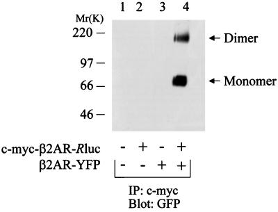
First, in an effort to characterize the proper pharmacological properties of the fusion receptors to be used in the BRET experiments, both radioligand-binding and agonist-promoted adenylyl cyclase activity assays were carried out in HEK-293 cells expressing each of the constructs individually. The binding affinity (_K_d) of the β-adrenergic antagonist [125I]cyanopindolol for the β2AR-YFP (78.5 ± 12 pM) and β2AR-_R_luc (57.5 ± 15 pM) was found to be indistinguishable from that obtained for the wild-type receptor (44.7 ± 13 pM). Similarly, the potency of the agonist isoproterenol to stimulate the adenylyl cyclase activity was the same for the three receptor constructs (β2AR-YFP: 0.3 × 10⁻⁷ ± 0.4 × 10⁻⁷ M; β2AR-_R_luc: 3.0 × 10⁻⁷ ± 3.5 × 10⁻⁷ M; β2AR-WT: 1.5 × 10⁻⁷ ± 1.3 × 10⁻⁷ M). Also, as documented previously for the wild-type β2AR (3, 10), intermolecular interaction between solubilized β2AR-_R_luc and β2AR-YFP could be detected by coimmunoprecipitation. Indeed, as shown in Fig. 1, β2AR-YFP immunoreactivity, as assessed by using an anti-GFP antibody, was found in the myc-β2AR-_R_luc immunoprecipitate only when the two constructs were co-expressed.
Figure 1.
Co-immunoprecipitation of β2AR molecules bearing different immunological epitopes. c-_myc_-β2AR-_R_luc and β2AR-YFP were expressed (lanes 2–4) or not (lane 1) in HEK-293 cells and immunoprecipitated with the agarose-conjugated anti-c-myc mAb (Santa Cruz Biotechnology). The immunoprecipitated proteins then were resolved by SDS/PAGE and immunoblotted with the polyclonal anti-GFP antibody (CLONTECH; this antibody also recognizes YFP). The occurrence of receptor dimerization is revealed by the fact that the YFP-tagged β2AR is coimmunoprecipitated with the c-_myc_-tagged receptor by the anti-c-myc mAb (lane 4).
Assessment of β2AR Dimerization in Vivo.
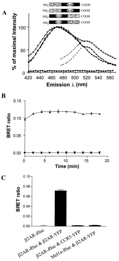
To determine whether the intermolecular interaction detected in vitro, after solubilization of the receptors, could also occur in living cells, light emission spectra were recorded in cells expressing the β2AR-_R_luc and β2AR-YFP constructs simultaneously or individually. Cells expressing both _R_luc-KaiB and YFP-KaiB were used as a positive control. As seen in Fig. 2A, addition of coelenterazine, the cell-permeable substrate for _R_luc, led to a characteristic broad bioluminescence signal with an emission peak at 470 nm in cells expressing either β2AR-_R_luc alone or β2AR-_R_luc and β2AR-YFP. No luminescence was detected in cells expressing β2AR-YFP alone. In addition to the bioluminescence, a fluorescence signal corresponding to the emission wavelength of the Topaz YFP (530 nm) was detected in cells expressing both β2AR-_R_luc and β2AR-YFP, indicating that BRET occurred between the two receptor constructs. Based on the Förster equations (19), it has been estimated that the maximal distance allowing energy transfer between the BRET pair used in the present study is ≈50Å (15). Therefore, the detection of BRET under basal conditions demonstrates a physical proximity between the β2AR-YFP and β2AR-_R_luc that can be explained best by the existence of constitutive receptor dimers or oligomers. For sake of simplicity, the term dimer will be used thereafter as the smallest form of oligomers with the understanding that BRET also could result from the formation of larger complexes. Using a similar approach based on fluorescence resonance energy transfer (FRET), Overton and Blumer (26) recently observed that the α-mating factor receptor also exists as a constitutive dimer in yeast, indicating that constitutive dimerization may be a general feature of GPCR.
Figure 2.
BRET in living HEK-293 cells expressing β2AR-YFP and β2AR-_R_luc. (A) HEK-293 cells expressing β2AR-YFP (asterisks), β2AR-_R_luc (■), or coexpressing β2AR-YFP and β2AR-_R_luc (▴) or KAIB-YFP and KAIB-_R_luc (○) were incubated with 5 μM coelenterazine h (Molecular Probes), and light-emission acquisition was performed immediately. All spectra were normalized as percentage of maximal emission. The spectra shown are representative of four independent experiments. (B) Luminescence and fluorescence signals were quantitated by using a BretCount (Packard), allowing the sequential integration of the signals detected in the 440- to 500-nm and 510- to 590-nm windows. The BRET ratio was defined as [(emission at 510–590) − (emission at 440–500) × Cf]/(emission at 440–500), where Cf corresponded to (emission at 510–590)/(emission at 440–500) for the β2AR-_R_luc expressed alone in the same experiments. Readings were started immediately after coelenterazine addition, and repeated measures were taken for ≈20 min. The data shown represent the mean ± SEM of four independent readings. (C) To assess the specificity of interaction, BRET ratios were measured in cells coexpressing β2AR-_R_luc and β2-YFP together or individually with either CCR5-YFP and Mel1a-_R_luc, respectively.
To quantitate the BRET signal generated, the ratio of the light emitted by the β2AR-YFP (510–590 nm) over that emitted by the β2AR-_R_luc (440–500 nm) was determined by using the equation described in the legend of Fig. 2B. The signal observed in cells coexpressing the two constructs was found to be easily distinguishable from the low background observed in cells expressing β2AR-_R_luc alone and was stable for at least 15 min after the addition of coelenterazine (Fig. 2B). It was also found to be highly reproducible, although differences in the relative expression levels of the receptor constructs led to small variation in the absolute signals observed.
Specificity of β2AR Dimerization.
To control for the specificity of the interaction and to rule out the possibility that the observed dimerization of the β2AR is a consequence of transmembrane protein overexpression, the distantly related chemokine CCR5 receptor fused to YFP (CCR5-YFP) was coexpressed with β2AR-_R_luc. Despite higher levels of receptor expression in these control experiments, no BRET was observed. Similarly, coexpression of β2AR-YFP with a melatonin-1a receptor fused to _R_luc did not lead to BRET (Fig. 2C).
The BRET observed did not result from a spurious interaction between the _R_luc and the YFP moieties of the fusion proteins because no signal could be detected when they were coexpressed as nonfusion proteins (data not shown). This contrasts with the significant BRET signal observed in cells expressing a fusion construct covalently linking _R_luc to YFP (Fig. 3), thus confirming the importance of molecular proximity between the BRET partners for signal detection. The insertion of a consensus cleavage site for caspase-3 within the 17-aa linker region of the fusion protein allowed us to demonstrate that BRET also can be used to monitor dynamic biological processes in living mammalian cells. Indeed, stimulation of caspase-3 activity by a treatment of the cells with staurosporine (20) promoted a marked decrease in the BRET ratio, indicating that a proportion of the fusion protein was cleaved, resulting in the physical separation of the BRET partners. The staurosporine-induced reduction in BRET was blocked by a pretreatment with the caspase-3 inhibitor-1, confirming the specificity of the effect.
Figure 3.
BRET as a sensor for biological processes in vivo. (A) Schematic representation of the _R_luc-YFP fusion construct harboring a consensus cleavage site (DEVD) for caspase-3. (B) The BRET ratio measured in cells expressing the fusion protein treated or not with staurosporine and/or the caspase-3 inhibitor-I. The data shown represent the mean ± SEM of four independent readings.
The absolute level of BRET observed with the _R_luc-YFP fusion protein was significantly higher than those obtained when coexpressing β2AR-_R_luc and β2AR-YFP. This is to be expected given the covalent nature of the construct that ensures a 1:1 interaction ratio. In contrast, even if all receptors were dimeric in the β2AR coexpression experiments, one could not expect more than 50% of the receptor population to generate BRET. Indeed, 50% of the dimers would consist of β2AR-_R_luc/β2AR-_R_luc or β2AR-YFP/β2AR-YFP complexes that cannot generate BRET. However, it is impossible to make direct quantitative comparison between the two systems because the extent of energy transfer not only depends on the number of interactions between energy donor and acceptor but also on the distance between them and on their relative orientation toward each other.
β2AR Dimer Modulation at the Cell Surface.
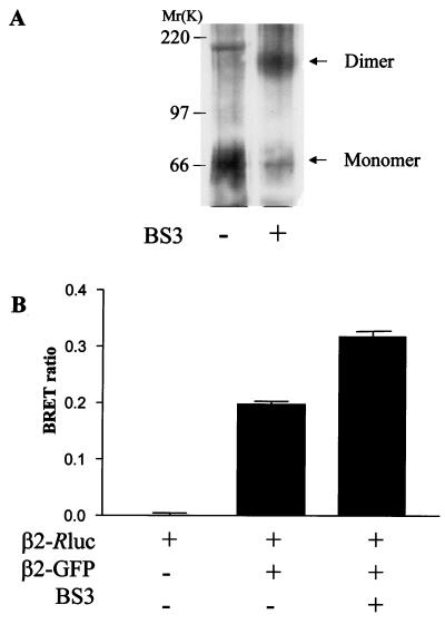
To determine whether modulation of β2AR dimerization at the cell surface could be monitored by BRET, we first investigated the effect of the cell-impermeable cross-linker BS3. As reported previously (10, 11), the chemical cross-linker increased the amount of receptor dimer detected after receptor solubilization and Western blot analysis, most likely reflecting covalent stabilization of the dimer species (Fig. 4A). The BRET signal generated by cells coexpressing β2AR-_R_luc and β2AR-YFP also was increased by the BS3 treatment (Fig. 4B), indicating that covalent cross-linking either increased the proportion of receptor dimers present at the cell surface or changed the conformation of preexisting ones. Indeed, as indicated above, the extent of BRET is affected by both the number of interacting molecules (number of dimers) and by the distance (orientation) between the energy donor and acceptor within each dimer (15). Thus, a change in the conformation of preexisting dimer that would result in closer proximity or more favorable orientation of the BRET partners would be detected as an increase in BRET signal.
Figure 4.
Effect of chemical crosslinking on β2AR dimerization. (A) A FLAG-tagged β2AR was immunoprecipitated from cells treated or not for 30 min with the cell-impermeable chemical crosslinker BS3 by using a polyclonal anti-β2AR antibody (Santa Cruz Biotechnology). Immunoprecipitates then were resolved by SDS/PAGE, and the receptor was detected by Western blot analysis by using the monoclonal anti-FLAG antibody (Sigma). (B) BRET ratio measured in cells expressing the indicated constructs. Cells were treated or not with BS3 for 30 min before the addition of coelenterazine. The data represent the mean ± SEM of four independent readings.
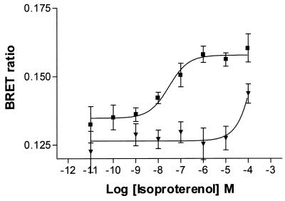
We next examined the effect of biological activation of the receptor on the extent of the energy transfer. The addition of the β-adrenergic agonist, isoproterenol, promoted a dose-dependent increase in the BRET ratio observed with an EC50 of 30 nM (Fig. 5), a value compatible with the high-affinity binding state of the receptor. This effect was competitively blocked by the antagonist propranolol. The agonist-induced increase in BRET was very rapid and was already present at the earliest time point measured (20 sec).
Figure 5.
Effect of agonist treatment. BRET ratio measured in cells expressing β2AR-_R_luc and β2AR-YFP after addition of increasing concentrations of the agonist isoproterenol in the presence (▾) or absence (■) of 10 μM of the β-adrenergic antagonist propranolol. The data represent the mean ± SEM of four independent readings that were analyzed by using a four-parameter logistic equation (GraphPad prism 2.01) and fixing the Hill coefficient to 1 (EC50 = 30 nM). The data also were analyzed, allowing for variable Hill coefficients. In that case, the fitted Hill coefficient was found to be 0.64 and the EC50 was 28 nM.
Given that isoproterenol is a hydrophilic ligand that cannot permeate the plasma membrane, the above results indicate that intermolecular interactions between receptor molecules occur at the cell surface, where they are influenced by receptor activation. In agreement with previous functional evidence (3, 10), these data suggest that β2AR dimers play a role in signal transduction. The effect of the agonist on the BRET ratio is compatible with the idea that agonists promote dimer formation. However, the relatively modest agonist-dependent increase (≈18%) above the constitutive dimerization signal also could suggest that the agonists bind to preformed dimers, leading to conformational changes that favor the transfer of energy within preexisting dimers.
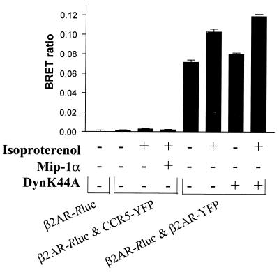
Because agonist stimulation of β2AR is known to promote its clustering and internalization through clathrin-coated pits and vesicles (21–23), the increase in BRET observed upon agonist treatment also could result from elevated local concentration within these delimited structures. To test this hypothesis, the effect of agonist stimulation was assessed in cells coexpressing CCR5-YFP and β2AR-_R_luc. Concomitant stimulation of the β2-adrenergic and CCR-5 receptors by their respective agonists isoproterenol and Mip-1α did not promote any BRET (Fig. 6). Because these two receptors are colocalized in the same clathrin-coated pit (data not shown), it suggests that clustering in these structures is not sufficient to allow transfer of energy between noninteracting receptor molecules. Also consistent with the notion that the agonist-promoted increase in BRET is not a consequence of receptor internalization is the observation that coexpression with a dominant negative mutant of dynamin (K44A) that efficiently blocks receptor internalization (ref. 24 and data not shown) did not affect the elevation of BRET that resulted from receptor activation (Fig. 6).
Figure 6.
Effect of the inhibition of endocytosis on BRET. BRET ratio was measured in cells expressing the indicated constructs in the presence or absence of the β2-adrenergic and CCR5 agonists isoproterenol (10 μM) and MIP-1α (0.2 μM). The effect of coexpressing the dominant negative mutant of dynamin (DynK44A) was also assessed. The data represent the mean ± SEM of four independent readings. Receptor expression levels were as follows: β2AR-_R_luc, 211 fmol/mg protein; β2AR-_R_luc and β2AR-YFP, a total of 398 fmol/mg protein; β2AR-_R_luc and CCR5-YFP, 400 fmol/mg protein for the β2AR and 456 fmol/mg protein for CCR5.
β2AR–β-Arrestin Interaction Measured by BRET.
To determine whether BRET could be used to monitor interactions between GPCR and other interacting molecules, we assessed the association of the β2AR with β-arrestin, which is known to occur exclusively in an agonist-dependent manner (25). In cells coexpressing β2AR_-R_luc and β-arrestin-YFP, the BRET was found to be entirely dependent on the activation of the receptor (Fig. 7). Indeed, the BRET signal observed under basal conditions was indistinguishable from that obtained when expressing β2AR-_R_luc alone but increased in a dose-dependent manner upon addition of the agonist isoproterenol with an EC50 of 0.4 nM. This is entirely consistent with the fact that β-arrestin binds only to the activated, phosphorylated form of the receptor, indicating that BRET is a useful approach to monitor agonist-promoted interactions occurring upon GPCR activation. The relatively low EC50 observed for the isoproterenol-promoted β-arrestin–β2AR interaction compared with that obtained for the β2AR homotropic interaction may be a reflection of different expression levels of the receptor and β-arrestin constructs. However, as indicated before, the extent of BRET observed does not allow one to estimate the stoichiometry of interaction.
Figure 7.
Agonist dependence of β-arrestin/β2AR interactions assessed by BRET. BRET ratio measured in cells coexpressing β2AR-_R_luc and β2-arrestin-YFP in the presence of increasing concentrations of the agonist isoproterenol (■). The open square represents the value observed in the absence of isoproterenol, and the open circle represents the value observed in cells expressing β2AR-_R_luc alone. The data represent the mean ± SEM of four independent readings that were analyzed by using a four-parameter logistic equation (GraphPad prism 2.01) and fixing the Hill coefficient to 1 (EC50 = 0.45 nM). The data also were analyzed allowing for variable Hill coefficients. In this case, the fitted Hill coefficient was found to be 0.74 and the EC50 was 0.43 nM.
Conclusion
Taken together, our results unambiguously demonstrate that the human β2AR forms constitutive homodimers that are expressed at the surface of living mammalian cells, where they interact with agonists. Our study also shows that BRET should prove to be a versatile assay for in vivo assessment of both constitutive and hormone-promoted protein–protein interactions in signal transduction.
Acknowledgments
We are grateful to Louise Cournoyer for her assistance with cell culture and to Claire Normand, Anne Labonté, and Mireille Caron for technical assistance with the BRET assays expertise. This work was supported partly by a grant to M.B and M.D. from the Medical Research Council of Canada (MRCC). S.A. and A.S. hold studentships from the MRCC, and M.B holds an MRCC scientist award.
Abbreviations
GPCR
G protein-coupled receptor(s)
BRET
bioluminescence resonance energy transfer
GFP
green fluorescent protein
_R_luc
_Renilla_-luciferase
YFP
enhanced red-shifted GFP
β2AR
β2-adrenergic receptor
BS3
bis(sulfosuccinimidyl)suberate
Footnotes
Article published online before print: Proc. Natl. Acad. Sci. USA, 10.1073/pnas.060590697.
Article and publication date are at www.pnas.org/cgi/doi/10.1073/pnas.060590697
References
- 1.Maggio R, Vogel Z, Wess J. Proc Natl Acad Sci USA. 1993;90:3103–3107. doi: 10.1073/pnas.90.7.3103. [DOI] [PMC free article] [PubMed] [Google Scholar]
- 2.Monnot C, Bihoreau C, Conchon S, Curnow K M, Corvol P, Clauser E. J Biol Chem. 1996;271:1507–1513. doi: 10.1074/jbc.271.3.1507. [DOI] [PubMed] [Google Scholar]
- 3.Hebert T E, Loisel T P, Adam L, Ethier N, Onge S S, Bouvier M. Biochem J. 1998;330:287–293. doi: 10.1042/bj3300287. [DOI] [PMC free article] [PubMed] [Google Scholar]
- 4.Zhu X, Wess J. Biochemistry. 1998;37:15773–15784. doi: 10.1021/bi981162z. [DOI] [PubMed] [Google Scholar]
- 5.Gouldson P R, Snell C R, Bywater R P, Higgs C, Reynolds C A. Protein Eng. 1998;11:1181–1193. doi: 10.1093/protein/11.12.1181. [DOI] [PubMed] [Google Scholar]
- 6.Blumer K J, Reneke J E, Thorner J. J Biol Chem. 1988;263:10836–10842. [PubMed] [Google Scholar]
- 7.Jones K A, Borowsky B, Tamm J A, Craig D A, Durkin M M, Dai M, Yao W J, Johnson M, Gunwaldsen C, Huang L Y, et al. Nature (London) 1998;396:674–679. doi: 10.1038/25348. [DOI] [PubMed] [Google Scholar]
- 8.Kaupmann K, Malitschek B, Schuler V, Heid J, Froestl W, Beck P, Mosbacher J, Bischoff S, Kulik A, Shigemoto R, et al. Nature (London) 1998;396:683–687. doi: 10.1038/25360. [DOI] [PubMed] [Google Scholar]
- 9.White J H, Wise A, Main M J, Green A, Fraser N J, Disney G H, Barnes A A, Emson P, Foord S M, Marshall F H. Nature (London) 1998;396:679–682. doi: 10.1038/25354. [DOI] [PubMed] [Google Scholar]
- 10.Hebert T E, Moffett S, Morello J P, Loisel T P, Bichet D G, Barret C, Bouvier M. J Biol Chem. 1996;271:16384–16392. doi: 10.1074/jbc.271.27.16384. [DOI] [PubMed] [Google Scholar]
- 11.Cvejic S, Devi L A. J Biol Chem. 1997;272:26959–26964. doi: 10.1074/jbc.272.43.26959. [DOI] [PubMed] [Google Scholar]
- 12.Bai M, Trivedi S, Brown E M. J Biol Chem. 1998;273:23605–23610. doi: 10.1074/jbc.273.36.23605. [DOI] [PubMed] [Google Scholar]
- 13.Romano C, Yang W L, O'Malley K L. J Biol Chem. 1996;271:28612–28616. doi: 10.1074/jbc.271.45.28612. [DOI] [PubMed] [Google Scholar]
- 14.Jordan B A, Devi L A. Nature (London) 1999;399:697–700. doi: 10.1038/21441. [DOI] [PMC free article] [PubMed] [Google Scholar]
- 15.Xu Y, Piston D W, Johnson C H. Proc Natl Acad Sci USA. 1999;96:151–156. doi: 10.1073/pnas.96.1.151. [DOI] [PMC free article] [PubMed] [Google Scholar]
- 16.Gagnon A W, Kallal L, Benovic J L. J Biol Chem. 1998;273:6976–6981. doi: 10.1074/jbc.273.12.6976. [DOI] [PubMed] [Google Scholar]
- 17.Mellon P L, Parker V, Gluzman Y, Maniatis T. Cell. 1981;27:279–288. doi: 10.1016/0092-8674(81)90411-6. [DOI] [PubMed] [Google Scholar]
- 18.Bouvier M, Hnatowich M, Collins S, Kobilka B K, De Blasi A, Lefkowitz R J, Caron M G. Mol Pharmacol. 1988;33:133–139. [PubMed] [Google Scholar]
- 19.Mahajan N P, Linder K, Berry G, Gordon G W, Heim R, Herman B. Nat Biotechnol. 1998;16:547–552. doi: 10.1038/nbt0698-547. [DOI] [PubMed] [Google Scholar]
- 20.Krohn A J, Preis E, Prehn J H. J Neurosci. 1998;18:8186–8197. doi: 10.1523/JNEUROSCI.18-20-08186.1998. [DOI] [PMC free article] [PubMed] [Google Scholar]
- 21.Von Zastrow M, Kobilka B K. J Biol Chem. 1992;267:3530–3538. [PubMed] [Google Scholar]
- 22.Ferguson S S, Downey W E, III, Colapietro A M, Barak L S, Menard L, Caron M G. Science. 1996;271:363–366. doi: 10.1126/science.271.5247.363. [DOI] [PubMed] [Google Scholar]
- 23.Goodman O B, Krupnick J G, Santini F, Gurevich V V, Penn R B, Gagnon A B, Keen J H, Benovic J L. Nature (London) 1996;383:447–450. doi: 10.1038/383447a0. [DOI] [PubMed] [Google Scholar]
- 24.Zhang J, Ferguson S G, Barak L S, Menard L, Caron M G. J Biol Chem. 1996;271:18302–18305. doi: 10.1074/jbc.271.31.18302. [DOI] [PubMed] [Google Scholar]
- 25.Lohse M J, Benovic J L, Codina J, Caron M G, Lefkowitz R J. Science. 1990;248:1547–1550. doi: 10.1126/science.2163110. [DOI] [PubMed] [Google Scholar]
- 26.Overton, M. L. & Blumer, K. J. (2000) Curr. Biol., in press. [DOI] [PubMed]
