Block of C/EBPα function by phosphorylation in acute myeloid leukemia with FLT3 activating mutations (original) (raw)
Abstract
Mutations constitutively activating FLT3 kinase are detected in ∼30% of acute myelogenous leukemia (AML) patients and affect downstream pathways such as extracellular signal–regulated kinase (ERK)1/2. We found that activation of FLT3 in human AML inhibits CCAAT/enhancer binding protein α (C/EBPα**) function by ERK1/2-mediated phosphorylation, which may explain the differentiation block of leukemic blasts. In MV4;11 cells, pharmacological inhibition of either FLT3 or MEK1 leads to granulocytic differentiation. Differentiation of MV4;11 cells was also observed when C/EBPα mutated at serine 21 to alanine (S21A) was stably expressed. In contrast, there was no effect when serine 21 was mutated to aspartate (S21D), which mimics phosphorylation of C/EBPα. Thus, our results suggest that therapies targeting the MEK/ERK cascade or development of protein therapies based on transduction of constitutively active C/EBP**α may prove effective in treatment of FLT3 mutant leukemias resistant to the FLT3 inhibitor therapies.
Acute myelogenous leukemia (AML) can be defined as an accumulation of immature myeloid cells in the bone marrow and blood resulting from dysregulation of normal proliferation, differentiation, and apoptosis. AML is the most common type of leukemia in adults and occurs in approximately one third of newly diagnosed patients. Multiple genetic defects have been implicated in the pathogenesis of AML (1), such as chromosomal deletions or additions, and chromosomal translocations resulting in production of in-frame fusion proteins. Based on current detection techniques, up to 45% of AML cases show normal karyotype (2); thus, in those cases, point mutations or small rearrangements may affect critical genes. One such gene, which is mutated in up to 30% AML cases, is the FLT3 receptor tyrosine kinase gene (3). The most common (20–25% AML patients) form of mutations in FLT3 are small in-frame internal tandem duplications (ITDs) in the juxtamembrane domain (3–5). In ∼7% of AML cases, point mutations in aspartic acid 835 in the kinase domain have been reported as well (6, 7). Both types of mutations result in the constitutive activation of the FLT3 receptor and abnormal activation of the downstream pathways: Stat5, Stat3, Akt, and extracellular signal–regulated kinase (ERK)1/2 (8–11). Because FLT3 is normally expressed in early precursors and plays role in proliferation and differentiation of hematopoietic progenitors (12, 13), it is not surprising that constitutive activation of FLT3 contributes to development of AML. AML patients with FLT3 mutations have poor prognosis (14–19). Therefore, small molecule inhibitors that specifically target FLT3 activity are undergoing clinical trials (20–23), but so far they have produced rather disappointing results. Because FLT3 regulates an intricate signaling network consisting of multiple downstream effectors, identification of the critical FLT3 targets involved in mediating the leukemic phenotype will possibly lead to the identification of novel alternative therapeutical targets for treatment of activated FLT3 leukemias.
Another critical gene involved in the pathogenesis of AML is the CCAAT/enhancer binding protein α (C/EBPα). C/EBPα is a leucine zipper transcription factor that is important for normal myeloid cell differentiation. Within the hematopoietic system, expression of C/EBPα is detectable in early myeloid precursors and is up-regulated as they commit to granulocytic differentiation pathway and mature (24, 25). Consistent with this expression pattern, mice lacking C/EBPα have no mature neutrophils, but rather accumulation of myeloblasts in the bone marrow (26). Conversely, overexpression of C/EBPα in precursor cell lines triggers neutrophilic differentiation (24, 27–29). Several studies from our group and others' showed that expression or function of C/EBPα is inactivated in various types of leukemia (AML and CML) by diverse molecular mechanisms (30–40). Importantly, provision of fully functional C/EBPα into leukemic cells could restore their differentiation program (24, 28, 31).
Recently, we have found that C/EBPα can be directly phosphorylated by ERK1/2 on S21, which affects the ability of C/EBPα to induce differentiation (28). Ectopic expression of the phosphomimetic C/EBPα mutant (S21D) inhibited granulocytic differentiation (28). In the present work, we provide evidence that the activating mutations in FLT3 in AML patients and cell lines inactivate C/EBPα function by ERK1/2-mediated phosphorylation on S21. Either alleviation of ERK1/2 activity or ectopic expression of a functionally active mutant of C/EBPα (S21A) in FLT3 ITD-expressing cells rescues myeloid differentiation. Thus, we provide a new molecular mechanism by which constitutively active FLT3 contributes to the pathogenesis of leukemia.
RESULTS
Activation of FLT3 leads to hyperphosphorylation of C/EBPα on serine 21
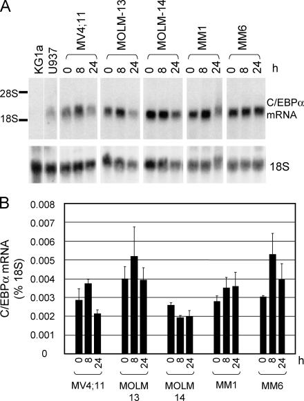
We hypothesized that the differentiation block in AML with FLT3 activating mutations is mediated by compromised expression and/or function of C/EBPα. Therefore, we analyzed C/EBPα mRNA and protein expression in five human FLT3 mutant AML cell lines. Three lines (MOLM-13, MOLM-14 [reference 41], and MV4;11 [references 42, 43]) carry ITD mutations (44) and two (MonoMac1 and MonoMac6 [reference 45]) have an activating point mutation (V592A) in the juxtamembrane domain (46). All cell lines expressed easily detectable C/EBPα mRNA (Fig. 1) and protein (Fig. 2, C and F, and not depicted). During the treatment with a specific FLT3 inhibitor, MLN518 (47), C/EBPα mRNA levels did not change significantly (Fig. 1). Considering that all five cell lines expressed relatively high levels of C/EBPα, which were not up-regulated by MLN518 treatments (Figs. 1 and 2; not depicted), it appears that the suppression of C/EBPα expression by mutated FLT3 is not a general mechanism of the differentiation block in human AML cell lines.
Figure 1.
Human AML cell lines with activating mutations in FLT3 express C/EBPα mRNA. (A) Cell lines (top) were not treated (0) or treated with 1 μM MLN518 for 8 or 24 h (indicated above the lanes). (top) Hybridization of the Northern blot to a human C/EBPα probe. (bottom) A control hybridization to 18S ribosomal RNA. RNA from KG1a and U937 cells served as negative and positive controls, respectively. (B) The same RNA samples were analyzed in triplicate by TaqMan Real-Time PCR and normalized to 18S RNA.
Figure 2.
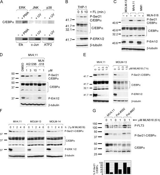
Activation of FLT3 in human AML cells leads to C/EBPα protein phosphorylation on S21 by the ERK1/2 pathway. (A) COS7 cells were untreated or treated with UV-C (60 J/m2; UV) or 100 nM PMA and incubated for 60 min. Endogenous c-Jun NH2-terminal protein kinase (JNK), p38, and ERK were immunoprecipitated and used for in vitro kinase assays. Control tests showed that ERK phosphorylated Elk1, JNK-phosphorylated c-Jun, and p38 MAP kinase phosphorylated ATF2. (B) THP-1 cells expressing wild-type FLT3 were serum starved for 6 h (0), or starved and stimulated with 100 ng/ml FLT3 ligand (FL) for 5 and 10 min before harvest. Western blot of whole cell extracts was sequentially stained with pS21-C/EBPα, C/EBPα, pThr202/Tyr204-ERK1/2, and β-tubulin antibodies. (C) Human AML cell lines with mutant FLT3 have increased phosphorylation of C/EBPα on S21. All cell lines were serum starved for 7 h in the absence (−) or presence (+) of 1 μM MLN518. Western blot was analyzed as in B. (D) MV4;11 cells were serum starved in the absence (−) or presence of the FLT3 inhibitors AG1296 and MLN518 at concentrations marked above the lanes, or with vehicle control (DMSO). Western blot analyses were done as in B. (E) Phosphorylation status of C/EBPα in MV4;11 and MOLM-13 cells as a dose response to MLN518. Cells were serum starved for 7 h in the presence of MLN518 at concentrations indicated above the lanes. Western blot was stained as in B. (F) Time course of FLT3 inhibition and its effect on C/EBPα phosphorylation. At each time point, MV4;11, MOLM-13, or MOLM-14 cells were serum starved for a total period of 8 h. MLN518 (1 μM) was added for the final hours (indicated above the lanes) of the treatment. For example, “1 hr” means that the cells were starved for 7 h in the absence of the inhibitor, and for 1 final hour in its presence. Western blot staining was as described previously. (G) Decrease of pS21-C/EBPα by inhibition of FLT3 activity in AML patient samples. Leukemic blasts from bone marrow (patient no. 592), or peripheral blood (patient no. 667) were cultured in the absence (0), or presence (1) of MLN518 for 6 h. Western blot of whole cell extracts was analyzed with antibodies recognizing activated FLT3 (P-FLT3), phosphorylated C/EBPα (P-Ser21-C/EBPα), or total C/EBPα protein. MV4;11 cells were included for control. Quantification of C/EBPα phosphorylation normalized to the total C/EBPα protein is shown (bottom).
We have recently reported that the function of C/EBPα is inhibited by phosphorylation on S21 by ERK1/2 (28). Interestingly, ERK1/2 is one of several downstream signaling pathways activated by FLT3 (9, 11). Therefore, we sought to determine if C/EBPα protein is phosphorylated upon activation of FLT3. As shown in Fig. 2 A, the NH2-terminal region of C/EBPα (amino acids 1–119) is specifically phosphorylated by ERK1/2 and not by other mitogen-activated protein (MAP) kinases. To determine if activation of FLT3 increases phosphorylation of endogenous C/EBPα, THP-1 cells, which express C/EBPα and wild-type FLT3 receptor, were serum starved and stimulated with FLT3 ligand. Western blot with phospho-specific antibodies (Fig. 2 B) showed that stimulation as short as 5 min resulted in a rapid increase in levels of phosphorylated (activated) ERK1/2 as well as C/EBPα on S21. We also tested whether constitutive activation of mutant FLT3 in AML also results in phosphorylation of C/EBPα. All five cell lines tested (Fig. 2 C and not depicted) demonstrated phosphorylation of C/EBPα on S21, which correlated with activation of ERK1/2. Treatment of these cell lines with MLN518 led to a dramatic reduction in levels of pS21C/EBPα, coincident with a decrease in pERK1/2 levels (Fig. 2 C) and pFLT3 (not depicted). Similarly, treatment of MV4;11 cells with another FLT3 inhibitor, AG1296 (48), showed a dose-dependent decrease in phosphorylation of C/EBPα and ERK1/2 (Fig. 2 D). The absolute levels of C/EBPα protein in all five lines were not significantly altered by the treatments with any of the inhibitors. Fig. 2 E shows that MLN518 reduces pS21-C/EBPα in MV4;11 and MOLM-13 cells in a dose-dependent manner. When normalized for the levels of C/EBPα protein, as little as 0.1 μM MLN518 led to a threefold decline in pS21-C/EBPα levels in both lines, and as great as a 50-fold decrease was noted for treatment of MOLM-13 cells with 1 μM inhibitor. A time course experiment using FLT3 ITD cell lines was performed to test the kinetics of the MLN518 effect on C/EBPα phosphorylation (Fig. 2 F). The inhibition of S21 phosphorylation was apparent within the first 2 h and reached maximum between 4 and 6 h. The treatments had no effect on the overall levels of C/EBPα protein, but correlated with activation of ERK1/2. Activation of ERK1/2 and hyperphosphorylation of C/EBPα were also seen in 293T cells transiently cotransfected with FLT3 ITD and wild-type C/EBPα expression vectors; these effects were abrogated by MLN518 (Fig. S1, available at http://www.jem.org/cgi/content/full/jem.20052242/DC1), indicating a causal role of activated FLT3.
Next, we sought to determine whether constitutive activation of FLT3 affects phosphorylation of C/EBPα in AML patients with FLT3 ITD. Primary cells from BM (patient no. 592) or PBMCs (patient no. 667) were incubated in the absence or presence of MLN518, and whole cell lysates were analyzed by Western blot shown in Fig. 2 G. Staining with pY591-FLT3 antibody, which detects one of the tyrosine residues phosphorylated as a result of activation (unpublished data), showed that treatments with MLN518 led to an ∼50–60% decrease in FLT3 activation. Inhibition of FLT3 in patient samples was also paralleled by a 40–60% decrease in pS21-C/EBPα. Control treatment with DMSO had no effect on phosphorylation of FLT3 or C/EBPα (unpublished data).
Pharmacological inhibition of FLT3 activation by MLN518 triggers myeloid differentiation of MV4;11 cells
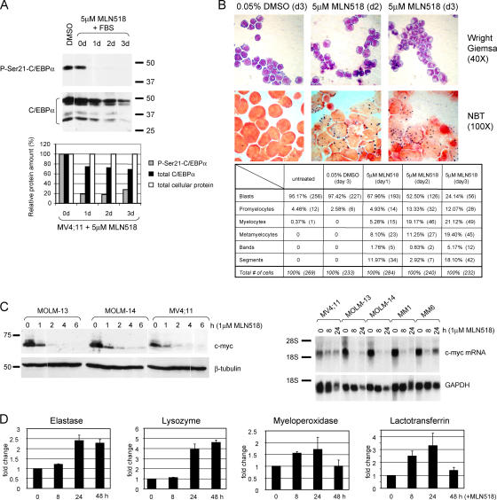
We showed previously that mutating S21 in C/EBPα to D mimics constitutive phosphorylation of S21, inhibits the differentiation-promoting activity of C/EBPα, and can inhibit retinoic acid-mediated granulocytic differentiation of U937 cells (28). Therefore, we hypothesized that constitutive activation of FLT3 may result in differentiation block through phosphorylation and thus, inactivation of C/EBPα. Because MLN518 decreases the level of FLT3 activation and increases the pool of C/EBPα protein not phosphorylated on S21, we expected that the treatment with this inhibitor might result in the onset of differentiation. As shown in Fig. 3 A, the 3-d course of culture of MV4;11 cells with MLN518 in the presence of serum led to a dramatic decrease in phosphorylation of C/EBPα, noted as early as on day 1. In contrast, up to 3 d of DMSO treatment had no effect on C/EBPα phosphorylation. After 2–3 d of MLN518 treatment, MV4;11 cells exhibited indentation and bending of the nuclei and an increase in cytoplasmic/nuclear ratio characteristic of band and metamyelocyte stages of granulocytic differentiation (Fig. 3 B). Cells treated with 0.05% DMSO for the same period of time remained blastic. Morphological changes were accompanied by respiratory burst activity determined by Nitro blue tetrazolium (NBT) reduction assay (Fig. 3 B), another indicator of granulocytic differentiation. Although growth tests indicated that the IC50 of MLN518 for this cell line is between 0.3 and 1 μM (unpublished data), we noted differentiation when MV4;11 cells were cultured in 5 or 10 μM MLN518, but not in 1 μM (unpublished data). To eliminate the possibility that these higher concentrations of MLN518 affects other FLT3-related receptor kinases, such as c-kit or platelet-derived growth factor receptor (PDGFR), we treated MV4;11 and MOLM-13 cells with STI571 (49), and found no effect on phosphorylation of C/EBPα (Fig. S2, available at http://www.jem.org/cgi/content/full/jem.20052242/DC1).
Figure 3.
Inhibition of FLT3 induces granulocytic differentiation of MV4;11 cells. (A) MV4;11 cells were treated with 5 μM MLN518 for 3 d. Samples were withdrawn daily and phosphorylation of C/EBPα on S21 was analyzed by Western blot with phospho-specific (top) and C/EBPα (bottom) antibodies. Quantification of signals with respect to total cellular protein levels (as determined by Ponceau S staining) is shown (bottom). (B) MV4;11 cells were cultured in the presence of 5 μM MLN518 or vehicle control (0.05% DMSO). Cell aliquots were examined daily for morphological changes (top) and for NBT reduction activity (bottom). Differential counts are summarized in the chart. (C) Inhibition of FLT3 leads to down-regulation of c-myc. (top) FLT3 mutant AML cell lines were serum starved for a total of 8 h in the absence or presence of 1 μM MLN518 for the indicated number of hours (at the end of starvation period). Western blot was stained with c-myc or β-tubulin antibodies. (bottom) FLT3 mutant lines, MV4;11, MOLM-13, MOLM-14, MonoMac1 (MM1), and MonoMac6 (MM6) were grown in the absence (0) or presence of 1 μM MLN518 for the indicated times (hours). Total RNA was examined by Northern blot with probes specific for c-myc and GAPDH (to control for RNA integrity). (D) Up-regulation of granulocyte-specific gene mRNA in MLN518-treated cells. Same RNA samples as in C, were examined by Real Time-PCR with primer sets specific for the elastase, lysozyme, myeloperoxidase, and lactoferrin genes. Data were normalized for GAPDH RNA and presented as fold change.
One of the early events essential for granulocytic differentiation is down-regulation of c-myc expression through C/EBPα-mediated inhibition of E2F function (50, 51). Very rapid down-regulation of c-myc protein was seen in all tested FLT3 mutant cell lines in response to MLN518 (Fig. 3 C; not depicted). However, because c-myc protein stability is regulated by ERK1/2-mediated phosphorylation (52), we also investigated c-myc mRNA levels to eliminate the possibility that the decrease in c-myc protein is a result of its dephosphorylation and decrease in its stability. Fig. 3 C shows that the c-myc down-regulation occurs also at the mRNA level in all five cell lines studied. In all cases, the down-regulation of c-myc closely correlated with decrease in pS21-C/EBPα (Figs. 2 F and 3 C show the same blot). In support of the differentiation onset triggered by MLN518 treatment, we also observed up-regulation of elastase, lysozyme, myeloperoxidase, and lactoferrin gene expression in MV4;11 cells (Fig. 3 D). In accordance with earlier published data (47), the MLN518-induced differentiation of MV4;11 cells was accompanied by a notable apoptosis (Table S1, available at http://www.jem.org/cgi/content/full/jem.20052242/DC1).
Inhibition of MEK1 activity decreases phosphorylation of C/EBPα and induces granulocytic differentiation of MV4;11 cells
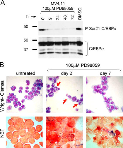
In addition to the ERK1/2 pathway, activation of FLT3 affects other pathways, such as Stat3/5, or Akt, which are also involved in leukemogenesis (8, 9, 11). To determine each pathway's contribution to the differentiation block in FLT3 mutant leukemia, we treated MV4;11 cells with a MEK1 inhibitor, PD98059, as well as a PI3 kinase inhibitor, LY294002, and the JAK-2 protein tyrosine kinase inhibitor, AG490. Cells were monitored daily for morphological changes, NBT reduction activity, cell growth, and apoptosis. LY294002 and AG490 retarded growth and induced apoptosis (Table S1), but did not induce differentiation. In contrast, the MEK1 inhibitor decreased pS21-C/EBPα levels in as early as 9 h (Fig. 4 A) and relieved differentiation block of MV4;11 cells (Fig. 4 B). The first morphological and functional (NBT) signs of differentiation were noted 2 d later (Fig. 4 B). Further incubation of cells with MEK1 inhibitor for up to 7 d resulted in even more pronounced granulocytic morphology (Fig. 4 B). Thus, we conclude that activation of ERK1/2 pathway by FLT3 mutations is the main event responsible for mediating the differentiation block. Because apoptosis, but not differentiation, was noted when cells were treated with inhibitors blocking Stat3/5 or Akt pathways, it appears that activation of those pathways plays a role in cell survival rather than differentiation.
Figure 4.
MV4;11 cells differentiate upon inhibition of the ERK1/2 pathway. (A) Western blot showing decreased pS21-C/EBPα (top) in response to continuous treatment with 100 μM of the MEK1 inhibitor PD98059 or vehicle control (DMSO). For DMSO-treated cells, a 72-h incubation is shown. (bottom) The same blot stained with C/EBPα antibody. (B) (top) MV4;11 cell differentiation was monitored by morphological examination of Wright-Giemsa stained cytospin preparations. Red arrows point to cells with granulocytic morphology. (bottom) Respiratory burst activity as assessed by the NBT assay.
Expression of C/EBPα mutant lacking Ser21 phosphorylation relieves the differentiation block in FLT3-ITD human AML cells
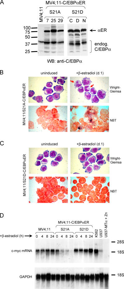
We have demonstrated that C/EBPα with S21 mutated to alanine (S21A), such that it is no longer a substrate for ERK1/2 kinases, retains full ability to induce of granulocytic differentiation of U937 and K562 cells. In contrast, mutation of S21 to aspartate (S21D), which mimics constitutive phosphorylation of S21 (S21D) inhibits granulocytic differentiation (28). We predicted that expressing a C/EBPα protein lacking the ERK1/2 phosphorylation site (S21A mutant) would restore the differentiation of FLT3 mutant AML cells. To test this hypothesis, S21A and S21D C/EBPα mutants were fused to the estrogen receptor (ER) ligand-binding domain and stably introduced into MV4;11 and MOLM-14 cells. In this system, the expression of C/EBPα-ER proteins is constitutive with cytoplasmic localization and their nuclear translocation is induced by treatment with β-estradiol (28, 31, 53). Several independent stable lines of MV4;11 cells were established with high expression of C/EBPα-ER (Fig. 5 A; not depicted). As assessed by examination of Wright-Giemsa stained cells (Fig. 5 B), a 24-h treatment of the MV4;11 stable lines expressing S21A-C/EBPα (MV-S21A) with β-estradiol resulted in morphological changes typical of granulocytic differentiation. These cells also exhibited oxidative burst activity (NBT; Fig. 5 B). In contrast, MV4;11 clones expressing the S21D mutant of C/EBPα (MV-S21D) did not show any of these characteristics and remained immature (Fig. 5 C). Similar results were obtained in MOLM-14 cells (unpublished data). We also tested the effect of induction of the C/EBPα mutant proteins on the expression of c-myc. We found that, whereas β-estradiol–treated MV-S21A lines down-regulated c-myc protein (not depicted) and mRNA (Fig. 5 D), the MV-S21D lines maintained c-myc expression (Fig. 5 D). As down-regulation of c-myc is a critical event in C/EBPα-induced differentiation (50, 51), these results further demonstrate the importance of nonphosphorylated C/EBPα for granulocyte development.
Figure 5.
The dephosphorylated form of C/EBPα is sufficient to mediate granulocytic differentiation of MV4;11 cells. (A) MV4;11 cells were stably transfected with inducible C/EBPα expression vectors. Independent clones were analyzed by Western blot. Parental MV4;11 cells expressing only the endogenous C/EBPα protein are shown (left). Clone nos. 7, 25, and 29 express an ectopic C/EBPα-ER fusion protein in which S21 of C/EBPα was mutated to alanine (S21A). Clones C, D, and N express C/EBPα-ER protein with S21 mutated to aspartate (S21D) to mimic phosphorylation. (B) MV4;11 cells with induced expression of S21A-C/EBPα protein undergo granulocytic differentiation. Granulocyte-specific morphological changes in Wright-Giemsa stained cytospins (indicated by red arrows) were seen (top) as well as an increase in NBT reduction activity (bottom). (C) Induction of S21D-C/EBPα did not induce granulocytic maturation as shown by morphological examinations (top) or NBT reduction assay (bottom). The inductions shown are for 24 h; however, no changes were observed after longer treatments with β-estradiol (not depicted). (D) Parental MV4;11 cells and C/EBP-ER–expressing stable lines (S21A, and S21D) were left untreated (0) or treated with 1 μM β-estradiol for 4, 8, and 24 h. RNA was collected and analyzed by Northern blot with probes specific for c-myc (top) and GAPDH (bottom). For positive controls, K562 and U937 cells were used. The U937 stable line with Zn-inducible expression of ectopic C/EBPα (reference 24) was included as negative control, as they were shown to down-regulate c-myc expression upon induction of C/EBPα expression (reference 51).
DISCUSSION
FLT3 is overexpressed or coexpressed with FLT3 ligand in >90% of AML cases (3, 5), providing another mechanism of constitutive receptor activation. Therefore, specific targeting of activated FLT3 receptor by small molecule inhibitors is an attractive therapeutic approach for AML. Although several chemical compounds have been developed to inhibit FLT3 activity (46–48, 54–56), none of these inhibitors is directed solely to the FLT3 receptor; they can affect activities of other kinases, such as PKC, TrkA, VEGFR, KIT, or PDGFR, thus increasing the possibility of nonspecific toxicity (3, 5). The effectiveness of a given inhibitor may also vary depending on the actual mutation in the FLT3 receptor, raising a possibility of drug resistance. Therefore, elucidating the pathways downstream of FLT3 might lead to the development of better therapeutic approaches in AML.
The major known effect of all FLT3 inhibitors is the induction of apoptosis (47). Ours is the first report demonstrating that treatment of FLT3 ITD cells with MLN518, in addition to apoptosis, can also trigger differentiation. We also showed that inhibition of the ERK1/2 pathway in FLT3 mutant AML line could achieve the same effect, whereas inhibition of other downstream pathways (Akt, Stat3/5) resulted only in apoptosis. Thus, activation of FLT3 affects signaling pathways controlling both differentiation and apoptosis.
Several clinical trials with various FLT3 inhibitors are currently in progress (21–23). In one of them, oral administration of CEP-701 to 14 AML patients expressing FLT3-activating mutations demonstrated sustained FLT3 inhibition and clinical evidence of biologic activity in only 5 patients (22). The same study also showed that two out of eight patients exhibited >90% inhibition in FLT3 receptor activation, but remained resistant to the clinical effect of this compound. The molecular effects of another FLT3 inhibitor, SU11248, were evaluated on patient samples and showed transient or sustained decrease in ERK1/2 activation in 80% of patients. Interestingly, activation of the upstream kinase, MEK1, was seen in only 39% patients. Thus, components of the FLT3–MEK1–ERK1/2 cascade can be dissociated in patients (21).
Previously, we have identified C/EBPα as a transcription factor necessary and sufficient for neutrophilic differentiation (24, 26–29). It was logical to assume that such important molecule might be a target in pathogenesis of leukemia. In fact, the expression or function of C/EBPα are disturbed in various subtypes of leukemia (30–40), providing an explanation for the block in differentiation.
In the course of this work, we show that, in mutant FLT3 AML, the differentiation-promoting function of C/EBPα is inhibited at yet another level: posttranslational modification by phosphorylation. We were able to show, using myeloid and 293T cells, that FLT3 activation induces phosphorylation of C/EBPα on S21, which inactivates C/EBPα function. Consistent with the known role of C/EBPα in promoting granulocytic differentiation, inhibition of FLT3 or introduction into the cells of S21A C/EBPα mutant rescued the differentiation block in AML cells. Furthermore, inhibition of FLT3 activity decreased the levels of S21 phosphorylation in FLT3 mutant cell lines and AML patients. Nonetheless, inhibition of FLT3 was less effective in decreasing the C/EBPα phosphorylation in patient samples when compared with cell lines. One possible explanation for this difference is that the activities of serine/threonine phosphatases, such as protein phosphatase 1 (PP1), or MAP kinase phosphatase-1 (MKP-1) may be lower in patients (57, 58).
Several groups previously generated 32Dcl3 stable lines expressing mutants of FLT3 (59, 60). In those cells, C/EBPα, was shown to be down-regulated at the mRNA level (60). Also, two out of three FLT3 ITD-positive patients had very low levels of C/EBPα mRNA, which increased approximately twofold following the FLT3 inhibition therapy (60). In our own studies with murine 32Dcl3 and 503 (PU.1−/− line expressing endogenous C/EBPα; unpublished data) stable lines, we found that FLT3 ITD mutation did not lead to a strong activation of ERK1/2 and we did not observe an increase in C/EBPα phosphorylation on S21, which is located in a protein region highly conserved among mammalian species. Furthermore, in a mouse transplantation model, FLT3 ITD mutations resulted in development of myeloproliferative disease rather than AML (61). These findings suggest that the signaling pathways activated by FLT3 are different in mice and humans. In fact, intrinsic differences have been reported between mouse and human control of hematopoiesis mediated by C/EBPα (62). We cannot rule out the possibility that both C/EBPα-inactivating mechanisms (transcriptional repression and functional inhibition) may be operational in human FLT3 mutant AML, possibly depending on the differentiation stage of the transformed cell.
Overexpression or constitutive activation of the ERK pathway has been shown to play an important role in the pathogenesis and progression of various cancers (63). Currently, preclinical trials for pancreas, colon, breast cancers, and leukemia are ongoing with compounds specifically inhibiting MEK1/2 component of this pathway (64, 65). Abnormal activation of the ERK pathway also occurs in leukemia because of the activating mutations in FLT3, Ras, as well as genes in other pathways (PI3K, PTEN, Akt) (65). Thus, targeting the Ras–Raf–MEK–ERK pathway in leukemia may offer a potential alternative to standard chemotherapy. It has been shown that the primary effect of down-modulation of MEK–ERK pathway activation in AML primary blasts by a selective inhibitor of MEK1 (PD98059) was a cell cycle arrest followed by apoptosis (66). Our results demonstrate that PD98059 could decrease pS21-C/EBPα levels in the FLT3/ITD AML lines and induce granulocytic differentiation. Our data provide strong indication that inhibitors of MEK–ERK cascade could have significant clinical benefit in the treatment of FLT3 mutant AML, especially in cases of resistance to FLT3 inhibitors. Furthermore, development of protein therapies based on transduction of constitutively active C/EBPα (such as the S21A mutant) may prove effective in treatment of subtypes of leukemia with inadequate expression/function of C/EBPα.
MATERIALS AND METHODS
Cell lines.
MV4;11 (CRL 9591; American Type Culture Collection [references 42, 43]), MOLM13, MOLM14 (67), MonoMac1 (68), U937 (CRL 1593; American Type Culture Collection), and KG1a (CCL 246.1; American Type Culture Collection) were grown in RPMI 1640 with 10% FBS. MonoMac6 (45) were cultured in RPMI 1640/10% FBS supplemented with MEM Non-Essential Amino Acid Solution and OPI Media Supplement (Sigma-Aldrich). THP-1 (TIB 202; American Type Culture Collection) were grown in RPMI 1640/10% FBS with 0.05 mM β-mercaptoethanol. For MV4;11 stable lines, phenol red-free RPMI 1640/10% charcoal dextran stripped FBS and 0.25 μg/ml puromycin were used.
Patient samples.
Patient samples were obtained from bone marrow (patient no. 592) or peripheral blood (patient no. 667) at the Laboratory of Leukemia Diagnostic, Grosshadern. Samples were collected at time of diagnosis before initiation of treatment. The percentage of blasts in the samples was 70% (patient no. 592; male, 71 yr, FAB M2, 46, XY [25], MLL-PTD−, FLT3-LM+, FLT3-TKD−, KIT−, NRAS−) and 95% (patient no. 667; female, 74 yr, FAB M1, 47, XX, +8 [20], MLL-PTD+, FLT3-LM+, FLT3-TKD−, KIT−, NRAS−). Blast cells were collected after patient consent, purified by Ficoll-Hypaque density centrifugation, and frozen in 40% IMDM/50% FCS/10% DMSO. Cells were thawed in IMDM/20% FCS, 400 IE/ml Heparin, 100 U/ml DNase, washed once in IMDM (without serum), and incubated at 37°C for 6 h in IMDM with or without MLN518, or with DMSO (vehicle control).
Reagents.
FLT3 inhibitor MLN518 (CT53518) (47) and FLT3/PDGFR inhibitor AG1296 were reconstituted in DMSO at 10 mM. PD98059 (MEK1 inhibitor), LY294002 (PI3 kinase inhibitor), and tyrphostin AG490 (JAK2 inhibitor) were dissolved in DMSO to 50 mM. The final concentrations were 25 μM or 50 μM for LY294002, and 50 μM or 100 μM for AG490. Recombinant human FLT3 ligand was reconstituted at 10 μg/ml. All reagents, except MLN518 (kept at 4°C) were stored at −20°C.
In vitro kinase assays.
Protein kinase assays were performed as described previously (69). The details are provided in the supplemental Materials and methods section (available at http://www.jem.org/cgi/content/full/jem.20052242/DC1).
Western blot.
5 × 10⁶ cells were spun (1 K, 5 min), washed in PBS, lysed in 400 μl of 1× Laemmli sample buffer (70), and boiled at 100°C for 10 min. 30–40 μl were loaded on 7.5% SDS-PAGE gel. After blocking in 5% milk/TBST (TBST: 25 mM Tris-HCl, pH 7.4, 137 mM NaCl, 2.7 mM KCl, 0.1% Tween 20), membranes were stained with primary antibodies in 5% BSA/TBST/0.1% sodium azide overnight at 4°C and with horseradish peroxidase (HRP)–conjugated secondary antibodies at room temperature for 1 h. Signals were detected by enhanced chemiluminescence and quantified by ImageQuant software (Molecular Dynamics). The primary antibodies were rabbit pS21-C/EBPα (1:1,000; Cell Signaling), goat C/EBPα (1:1,000; Santa Cruz Biotechnology, Inc.), pY591-FLT3 (1:1,000 54H1; Cell Signaling), rabbit p(T202/Y204)-ERK1/2 (1:1,000; Cell Signaling), and β-tubulin (1:1,000 clone 2–28-33; Sigma-Aldrich). All secondary antibodies were HRP-conjugated (Santa Cruz Biotechnology, Inc.) and diluted 1:5,000 for rabbit-HRP, 1:3,000 for mouse-HRP, and 1:2,000 for goat-HRP.
Plasmids.
pBabe-(S21A)C/EBPαER and pBabe-(S21D)C/EBPαER plasmids were described previously (28). They contained coding regions of murine C/EBPα mutated in S21 and fused to the human ER ligand-binding domain. Puromycin gene was included for stable line selection.
Transfections.
Stable lines were made by Nucleofection (Amaxa). Cells were grown in phenol red-free RPMI 1640/10% charcoal-stripped FBS for at least 48 h before transfection. 107 cells were mixed with 100 μl of Nucleofector Solution T and 5 μg of ScaI linearized plasmid. Pulses were delivered with Nucleofector device and program T-05. Cells were resuspended in 10 ml of phenol red-free RPMI 1640/20% charcoal-stripped FBS supplemented with 10% of MV4;11 cells-conditioned medium and plated on 96-well plates. Selection with 0.25 μg/ml puromycin was performed 72 h later.
RNA isolation and analysis.
Total RNA was isolated with TriReagent. For Northern blots, 20 μg of RNA was separated on agarose gels and transferred to Biotrans Plus membranes. The blots were hybridized to the 700-bp EcoRI–HindIII fragment of the human C/EBPα 3′ UTR (24) and a 305-bp XbaI–EcoRI cDNA fragment of human c-myc (50). For loading control, the blots were stripped and rehybridized to GAPDH or 18S ribosomal RNA probes. Quantitation was performed with Image Quant. Details on SYBR green and TaqMan Real-Time PCR assays are provided in supplemental Materials and methods.
Morphological examination.
Approximately 104 cells were spun at 500 revolutions/min for 5 min onto glass slides and Wright-Giemsa stained with DiffQuick solutions (Dade Behring). Differential counts were performed on at least 10 fields from each slide.
Nitroblue tetrazolium reduction assay.
5 × 10⁵ cells were incubated in 0.5 ml solution containing PBS, NBT (1 tablet in 10 ml PBS; Sigma-Aldrich), and 0.33 μM PMA for 20–30 min at 37°C. Cytospin slides were prepared and counterstained with 0.5% safranin in 20% ethanol.
Online supplemental material.
In Fig. S1, FLT3 ITD mutant induces activation of ERK1/2 pathway and phosphorylation of C/EBPα on S21 in transiently transfected 293T cells. Fig. S2 shows that a c-kit and PDGFR inhibitor, STI571, do not affect levels of pS21-C/EBPα in FLT3 ITD cell lines. Table S1 depicts FLT3, PI3K, and Jak2 inhibitors that induce apoptosis of MV4;11 cells. Supplemental Materials and methods contains description of procedures for an in vitro kinase assay and real-time PCR. Online supplemental material is available at http://www.jem.org/cgi/content/full/jem.20052242/DC1.
Supplemental Material
[Supplemental Material Index]
Acknowledgments
We thank C. Sullivan for help with experiments. We are grateful to B. Neel, S. Koschmieder, F. Rosenbauer, G. Huang, and E. Weisberg for useful suggestions; B. Scheijen, and S. Whitman for help in obtaining patient material. We thank Drs. Y. Matsuo for MOLM-13, and S. Heinrichs for MV4;11 and MonoMac1 cell lines. Members of the Tenen and Gilliland laboratories are acknowledged for many useful discussions. We thank K. O'Brien, M. Singleton, and A. Lugay for help in preparation of the manuscript.
This research was supported by grants to D.G. Tenen from the National Institutes of Health (no. P01 CA72009), and to H.S. Radomska from the National Institutes of Health (no. DK62064).
The authors have no conflicting financial interests.
H.S. Radomska and D.S. Bassères contributed equally to this work.
Abbreviations used: AML, acute myelogenous leukemia; C/EBPα, CCAAT/enhancer binding protein α; ER, estrogen receptor; ERK, extracellular signal–regulated kinase; ITD, internal tandem duplication; MAP, mitogen-activated protein; NBT, Nitro blue tetrazolium; PDGFR, platelet-derived growth factor receptor.
References
- 1.Tenen, D.G. 2003. Disruption of differentiation in human cancer: AML shows the way. Nat. Rev. Cancer. 3:89–101. [DOI] [PubMed] [Google Scholar]
- 2.Mrozek, K., K. Heinonen, and C.D. Bloomfield. 2001. Clinical importance of cytogenetics in acute myeloid leukaemia. Best. Pract. Res. Clin. Haematol. 14:19–47. [DOI] [PubMed] [Google Scholar]
- 3.Gilliland, D.G., and J.D. Griffin. 2002. The roles of FLT3 in hematopoiesis and leukemia. Blood. 100:1532–1542. [DOI] [PubMed] [Google Scholar]
- 4.Nakao, M., S. Yokota, T. Iwai, H. Kaneko, S. Horiike, K. Kashima, Y. Sonoda, T. Fujimoto, and S. Misawa. 1996. Internal tandem duplication of the flt3 gene found in acute myeloid leukemia. Leukemia. 10:1911–1918. [PubMed] [Google Scholar]
- 5.Brown, P., and D. Small. 2004. FLT3 inhibitors: a paradigm for the development of targeted therapeutics for paediatric cancer. Eur. J. Cancer. 40:707–721. [DOI] [PubMed] [Google Scholar]
- 6.Yamamoto, Y., H. Kiyoi, Y. Nakano, R. Suzuki, Y. Kodera, S. Miyawaki, N. Asou, K. Kuriyama, F. Yagasaki, C. Shimazaki, et al. 2001. Activating mutation of D835 within the activation loop of FLT3 in human hematologic malignancies. Blood. 97:2434–2439. [DOI] [PubMed] [Google Scholar]
- 7.Abu-Duhier, F.M., A.C. Goodeve, G.A. Wilson, R.S. Care, I.R. Peake, and J.T. Reilly. 2001. Identification of novel FLT-3 Asp835 mutations in adult acute myeloid leukaemia. Br. J. Haematol. 113:983–988. [DOI] [PubMed] [Google Scholar]
- 8.Tse, K.F., G. Mukherjee, and D. Small. 2000. Constitutive activation of FLT3 stimulates multiple intracellular signal transducers and results in transformation. Leukemia. 14:1766–1776. [DOI] [PubMed] [Google Scholar]
- 9.Hayakawa, F., M. Towatari, H. Kiyoi, M. Tanimoto, T. Kitamura, H. Saito, and T. Naoe. 2000. Tandem-duplicated Flt3 constitutively activates STAT5 and MAP kinase and introduces autonomous cell growth in IL-3-dependent cell lines. Oncogene. 19:624–631. [DOI] [PubMed] [Google Scholar]
- 10.Mizuki, M., R. Fenski, H. Halfter, I. Matsumura, R. Schmidt, C. Muller, W. Gruning, K. Kratz-Albers, S. Serve, C. Steur, et al. 2000. Flt3 mutations from patients with acute myeloid leukemia induce transformation of 32D cells mediated by the Ras and STAT5 pathways. Blood. 96:3907–3914. [PubMed] [Google Scholar]
- 11.Spiekermann, K., K. Bagrintseva, R. Schwab, K. Schmieja, and W. Hiddemann. 2003. Overexpression and constitutive activation of FLT3 induces STAT5 activation in primary acute myeloid leukemia blast cells. Clin. Cancer Res. 9:2140–2150. [PubMed] [Google Scholar]
- 12.Rosnet, O., H.J. Buhring, S. Marchetto, I. Rappold, C. Lavagna, D. Sainty, C. Arnoulet, C. Chabannon, L. Kanz, C. Hannum, and D. Birnbaum. 1996. Human FLT3/FLK2 receptor tyrosine kinase is expressed at the surface of normal and malignant hematopoietic cells. Leukemia. 10:238–248. [PubMed] [Google Scholar]
- 13.Rasko, J.E., D. Metcalf, M.T. Rossner, C.G. Begley, and N.A. Nicola. 1995. The flt3/flk-2 ligand: receptor distribution and action on murine haemopoietic cell survival and proliferation. Leukemia. 9:2058–2066. [PubMed] [Google Scholar]
- 14.Abu-Duhier, F.M., A.C. Goodeve, G.A. Wilson, M.A. Gari, I.R. Peake, D.C. Rees, E.A. Vandenberghe, P.R. Winship, and J.T. Reilly. 2000. FLT3 internal tandem duplication mutations in adult acute myeloid leukaemia define a high-risk group. Br. J. Haematol. 111:190–195. [DOI] [PubMed] [Google Scholar]
- 15.Kiyoi, H., T. Naoe, Y. Nakano, S. Yokota, S. Minami, S. Miyawaki, N. Asou, K. Kuriyama, I. Jinnai, C. Shimazaki, et al. 1999. Prognostic implication of FLT3 and N-RAS gene mutations in acute myeloid leukemia. Blood. 93:3074–3080. [PubMed] [Google Scholar]
- 16.Kottaridis, P.D., R.E. Gale, M.E. Frew, G. Harrison, S.E. Langabeer, A.A. Belton, H. Walker, K. Wheatley, D.T. Bowen, A.K. Burnett, et al. 2001. The presence of a FLT3 internal tandem duplication in patients with acute myeloid leukemia (AML) adds important prognostic information to cytogenetic risk group and response to the first cycle of chemotherapy: analysis of 854 patients from the UK Medical Research Council AML 10 and 12 trials. Blood. 98:1752–1759. [DOI] [PubMed] [Google Scholar]
- 17.Rombouts, W.J., I. Blokland, B. Lowenberg, and R.E. Ploemacher. 2000. Biological characteristics and prognosis of adult acute myeloid leukemia with internal tandem duplications in the Flt3 gene. Leukemia. 14:675–683. [DOI] [PubMed] [Google Scholar]
- 18.Whitman, S.P., K.J. Archer, L. Feng, C. Baldus, B. Becknell, B.D. Carlson, A.J. Carroll, K. Mrozek, J.W. Vardiman, S.L. George, et al. 2001. Absence of the wild-type allele predicts poor prognosis in adult de novo acute myeloid leukemia with normal cytogenetics and the internal tandem duplication of FLT3: a cancer and leukemia group B study. Cancer Res. 61:7233–7239. [PubMed] [Google Scholar]
- 19.Frohling, S., R.F. Schlenk, J. Breitruck, A. Benner, S. Kreitmeier, K. Tobis, H. Dohner, and K. Dohner. 2002. Prognostic significance of activating FLT3 mutations in younger adults (16 to 60 years) with acute myeloid leukemia and normal cytogenetics: a study of the AML Study Group Ulm. Blood. 100:4372–4380. [DOI] [PubMed] [Google Scholar]
- 20.Fiedler, W., R. Mesters, H. Tinnefeld, S. Loges, P. Staib, U. Duhrsen, M. Flasshove, O.G. Ottmann, W. Jung, F. Cavalli, et al. 2003. A phase 2 clinical study of SU5416 in patients with refractory acute myeloid leukemia. Blood. 102:2763–2767. [DOI] [PubMed] [Google Scholar]
- 21.O'Farrell, A.M., J.M. Foran, W. Fiedler, H. Serve, R.L. Paquette, M.A. Cooper, H.A. Yuen, S.G. Louie, H. Kim, S. Nicholas, et al. 2003. An innovative phase I clinical study demonstrates inhibition of FLT3 phosphorylation by SU11248 in acute myeloid leukemia patients. Clin. Cancer Res. 9:5465–5476. [PubMed] [Google Scholar]
- 22.Smith, B.D., M. Levis, M. Beran, F. Giles, H. Kantarjian, K. Berg, K.M. Murphy, T. Dauses, J. Allebach, and D. Small. 2004. Single-agent CEP-701, a novel FLT3 inhibitor, shows biologic and clinical activity in patients with relapsed or refractory acute myeloid leukemia. Blood. 103:3669–3676. [DOI] [PubMed] [Google Scholar]
- 23.Stone, R.M., J. De Angelo, I. Galinsky, E. Estey, V. Klimek, W. Grandin, D. Lebwohl, A. Yap, P. Cohen, E. Fox, et al. 2004. PKC 412 FLT3 inhibitor therapy in AML: results of a phase II trial. Ann. Hematol. 83:S89–S90. [DOI] [PubMed] [Google Scholar]
- 24.Radomska, H.S., C.S. Huettner, P. Zhang, T. Cheng, D.T. Scadden, and D.G. Tenen. 1998. CCAAT/enhancer binding protein α is a regulatory switch sufficient for induction of granulocytic development from bipotential myeloid progenitors. Mol. Cell. Biol. 18:4301–4314. [DOI] [PMC free article] [PubMed] [Google Scholar]
- 25.Akashi, K., D. Traver, T. Miyamoto, and I.L. Weissman. 2000. A clonogenic common myeloid progenitor that gives rise to all myeloid lineages. Nature. 404:193–197. [DOI] [PubMed] [Google Scholar]
- 26.Zhang, D.-E., P. Zhang, N.D. Wang, C.J. Hetherington, G.J. Darlington, and D.G. Tenen. 1997. Absence of granulocyte colony-stimulating factor signaling and neutrophil development in CCAAT enhancer binding protein α-deficient mice. Proc. Natl. Acad. Sci. USA. 94:569–574. [DOI] [PMC free article] [PubMed] [Google Scholar]
- 27.Wang, X., E.W. Scott, C.L. Sawyers, and A.D. Friedman. 1999. C/EBPα bypasses granulocyte colony-stimulating factor signals to rapidly induce PU.1 gene expression, stimulate granulocytic differentiation, and limit proliferation in 32D cl3 myeloblasts. Blood. 94:560–571. [PubMed] [Google Scholar]
- 28.Ross, S.E., H.S. Radomska, B. Wu, P. Zhang, J.N. Winnay, L. Bajnok, W.S. Wright, F. Schaufele, D.G. Tenen, and O.A. MacDougald. 2004. Phosphorylation of C/EBPα inhibits granulopoiesis. Mol. Cell. Biol. 24:675–686. [DOI] [PMC free article] [PubMed] [Google Scholar]
- 29.Zhang, P., E.A. Nelson, H.S. Radomska, J. Iwasaki-Arai, K. Akashi, A.D. Friedman, and D.G. Tenen. 2002. Induction of granulocytic differentiation by two pathways. Blood. 99:4406–4412. [DOI] [PubMed] [Google Scholar]
- 30.Pabst, T., B.U. Mueller, P. Zhang, H.S. Radomska, S. Narravula, S. Schnittger, G. Behre, W. Hiddemann, and D.G. Tenen. 2001. Dominant negative mutations of CEBPA, encoding CCAAT/enhancer binding protein-α (C/EBPα), in acute myeloid leukemia. Nat. Genet. 27:263–270. [DOI] [PubMed] [Google Scholar]
- 31.Pabst, T., B.U. Mueller, N. Harakawa, D.-E. Zhang, and D.G. Tenen. 2001. AML1-ETO downregulates the granulocytic differentiation factor C/EBPα in t(8;21) myeloid leukemia. Nat. Med. 7:444–451. [DOI] [PubMed] [Google Scholar]
- 32.Perrotti, D., V. Cesi, R. Trotta, C. Guerzoni, G. Santilli, K. Campbell, A. Iervolino, F. Condorelli, C. Gambacorti-Passerini, M.A. Caligiuri, and B. Calabretta. 2001. BCR/ABL suppresses C/EBPα expression through inhibitory activity of hnRNP E 2. Nat. Genet. 30:48–58. [DOI] [PubMed] [Google Scholar]
- 33.Helbling, D., B. Mueller, N.A. Timchenko, M.F. Fey, and T. Pabst. 2003. The myeloid transcription factor C/EBPα is a key target gene of the leukaemic fusion gene AML1/MDS1/EVI 1. Proc. Natl. Acad. Sci. USA. 101:13312–13317. [DOI] [PMC free article] [PubMed] [Google Scholar]
- 34.Westendorf, J.J., C.M. Yamamoto, N. Lenny, J.R. Downing, M.E. Selsted, and S.W. Hiebert. 1998. The t(8:21) fusion product, AML-1-ETO, associates with C/EBP-α, inhibits C/EBP-α-dependent transcription, and blocks granulocytic differentiation. Mol. Cell. Biol. 18:322–333. [DOI] [PMC free article] [PubMed] [Google Scholar]
- 35.Gombart, A.F., W.K. Hofmann, S. Kawano, S. Takeuchi, U. Krug, S.W. Kwok, R.J. Larson, H. Asou, C.W. Miller, D. Hoelzer, and H.P. Koeffler. 2002. Mutations in the gene encoding the transcription factor CCAAT/enhancer binding protein α in myelodysplastic syndromes and acute myeloid leukemias. Blood. 99:1332–1340. [DOI] [PubMed] [Google Scholar]
- 36.Preudhomme, C., C. Sagot, N. Boissel, J.M. Cayuela, I. Tigaud, S. de Botton, X. Thomas, E. Rafffoux, C. Lamandin, S. Castaigne, et al. 2002. Favorable prognostic significance of CEBPA mutations in patients with de novo acute myeloid leukemia: a study from the Acute Leukemia French Association (ALFA). Blood. 100:2717–2723. [DOI] [PubMed] [Google Scholar]
- 37.Barjesteh van Waalwijk van Doorn-Khosrovani, S., C. Erpelinck, J. Meijer, S. van Oosterhoud, W.L. van Putten, P.J. Valk, H. Berna Beverloo, D.G. Tenen, B. Lowenberg, and R. Delwel. 2003. Biallelic mutations in the CEBPA gene and low CEBPA expression levels as prognostic markers in intermediate-risk AML. Hematol. J. 4:31–40. [DOI] [PubMed] [Google Scholar]
- 38.Kaeferstein, A., U. Krug, J. Tiesmeier, M. Aivado, M. Faulhaber, M. Stadler, J. Krauter, U. Germing, W.K. Hofmann, H.P. Koeffler, et al. 2003. The emergence of a C/EBPα mutation in the clonal evolution of MDS towards secondary AML. Leukemia. 17:343–349. [DOI] [PubMed] [Google Scholar]
- 39.Snaddon, J., M.L. Smith, M. Neat, M. Cambal-Parrales, A. Dixon-McIver, R. Arch, J.A. Amess, A.Z. Rohatiner, T.A. Lister, and J. Fitzgibbon. 2003. Mutations of CEBPA in acute myeloid leukemia FAB types M1 and M 2. Genes Chromosomes Cancer. 37:72–78. [DOI] [PubMed] [Google Scholar]
- 40.Frohling, S., R.F. Schlenk, I. Stolze, J. Bihlmayr, A. Benner, S. Kreitmeier, K. Tobis, H. Dohner, and K. Dohner. 2004. CEBPA mutations in younger adults with acute myeloid leukemia and normal cytogenetics: prognostic relevance and analysis of cooperating mutations. J. Clin. Oncol. 22:624–633. [DOI] [PubMed] [Google Scholar]
- 41.Yokota, S., H. Kiyoi, M. Nakao, T. Iwai, S. Misawa, T. Okuda, Y. Sonoda, T. Abe, K. Kahsima, Y. Matsuo, and T. Naoe. 1997. Internal tandem duplication of the FLT3 gene is preferentially seen in acute myeloid leukemia and myelodysplastic syndrome among various hematological malignancies. A study on a large series of patients and cell lines. Leukemia. 11:1605–1609. [DOI] [PubMed] [Google Scholar]
- 42.Lange, B., M. Valtieri, D. Santoli, D. Caracciolo, F. Mavilio, I. Gemperlein, C. Griffin, B. Emanuel, J. Finan, and P. Nowell. 1987. Growth factor requirements of childhood acute leukemia: establishment of GM-CSF-dependent cell lines. Blood. 70:192–199. [PubMed] [Google Scholar]
- 43.Santoli, D., Y.C. Yang, S.C. Clark, B.L. Kreider, D. Caracciolo, and G. Rovera. 1987. Synergistic and antagonistic effects of recombinant human interleukin (IL) 3, IL-1 α, granulocyte and macrophage colony-stimulating factors (G-CSF and M-CSF) on the growth of GM-CSF-dependent leukemic cell lines. J. Immunol. 139:3348–3354. [PubMed] [Google Scholar]
- 44.Quentmeier, H., J. Reinhardt, M. Zaborski, and H.G. Drexler. 2003. FLT3 mutations in acute myeloid leukemia cell lines. Leukemia. 17:120–124. [DOI] [PubMed] [Google Scholar]
- 45.Ziegler-Heitbrock, H.W., E. Thiel, A. Futterer, V. Herzog, A. Wirtz, and G. Riethmuller. 1988. Establishment of a human cell line (Mono Mac 6) with characteristics of mature monocytes. Int. J. Cancer. 41:456–461. [DOI] [PubMed] [Google Scholar]
- 46.Spiekermann, K., R.J. Dirschinger, R. Schwab, K. Bagrintseva, F. Faber, C. Buske, S. Schnittger, L.M. Kelly, D.G. Gilliland, and W. Hiddemann. 2003. The protein tyrosine kinase inhibitor SU5614 inhibits FLT3 and induces growth arrest and apoptosis in AML-derived cell lines expressing a constitutively activated FLT 3. Blood. 101:1494–1504. [DOI] [PubMed] [Google Scholar]
- 47.Kelly, L.M., J.C. Yu, C.L. Boulton, M. Apatira, J. Li, C.M. Sullivan, I. Williams, S.M. Amaral, D.P. Curley, N. Duclos, et al. 2002. CT53518, a novel selective FLT3 antagonist for the treatment of acute myelogenous leukemia (AML). Cancer Cell. 1:421–432. [DOI] [PubMed] [Google Scholar]
- 48.Tse, K.F., E. Novelli, C.I. Civin, F.D. Bohmer, and D. Small. 2001. Inhibition of FLT3-mediated transformation by use of a tyrosine kinase inhibitor. Leukemia. 15:1001–1010. [DOI] [PubMed] [Google Scholar]
- 49.Buchdunger, E., C.L. Cioffi, N. Law, D. Stover, S. Ohno-Jones, B.J. Druker, and N.B. Lydon. 2000. Abl protein-tyrosine kinase inhibitor STI571 inhibits in vitro signal transduction mediated by c-kit and platelet-derived growth factor receptors. J. Pharmacol. Exp. Ther. 295:139–145. [PubMed] [Google Scholar]
- 50.Johansen, L.M., A. Iwama, T.A. Lodie, K. Sasaki, D.W. Felsher, T.R. Golub, and D.G. Tenen. 2001. C-myc is a critical target for C/EBPα in granulopoiesis. Mol. Cell. Biol. 21:3789–3806. [DOI] [PMC free article] [PubMed] [Google Scholar]
- 51.D'Alo', F., L.M. Johansen, E.A. Nelson, H.S. Radomska, E.K. Evans, P. Zhang, C. Nerlov, and D.G. Tenen. 2003. The amino terminal and E2F interaction domains are critical for C/EBPα-mediated induction of granulopoietic development of hematopoietic cells. Blood. 102:3163–3171. [DOI] [PubMed] [Google Scholar]
- 52.Sears, R., F. Nuckolls, E. Haura, Y. Taya, K. Tamai, and J.R. Nevins. 2000. Multiple Ras-dependent phosphorylation pathways regulate Myc protein stability. Genes Dev. 14:2501–2514. [DOI] [PMC free article] [PubMed] [Google Scholar]
- 53.Umek, R.M., A.D. Friedman, and S.L. McKnight. 1991. CCAAT-enhancer binding protein: a component of a differentiation switch. Science. 251:288–292. [DOI] [PubMed] [Google Scholar]
- 54.Levis, M., K.F. Tse, B.D. Smith, E. Garrett, and D. Small. 2001. A FLT3 tyrosine kinase inhibitor is selectively cytotoxic to acute myeloid leukemia blasts harboring FLT3 internal tandem duplication mutations. Blood. 98:885–887. [DOI] [PubMed] [Google Scholar]
- 55.Levis, M., J. Allebach, K.F. Tse, R. Zheng, B.R. Baldwin, B.D. Smith, S. Jones-Bolin, B. Ruggeri, C. Dionne, and D. Small. 2002. A FLT3-targeted tyrosine kinase inhibitor is cytotoxic to leukemia cells in vitro and in vivo. Blood. 99:3885–3891. [DOI] [PubMed] [Google Scholar]
- 56.Weisberg, E., C. Boulton, L.M. Kelly, P. Manley, D. Fabbro, T. Meyer, D.G. Gilliland, and J.D. Griffin. 2002. Inhibition of mutant FLT3 receptors in leukemia cells by the small molecule tyrosine kinase inhibitor PKC412. Cancer Cell. 1:433–443. [DOI] [PubMed] [Google Scholar]
- 57.Nishikawa, M., M. Yamamoto, Y. Watanabe, K. Kita, and H. Shiku. 2001. Clinical significance of low protein phosphatase-1 activity of blasts in acute myelogenous leukemia with high white cell counts. Int. J. Oncol. 18:559–565. [DOI] [PubMed] [Google Scholar]
- 58.Franklin, C.C., and A.S. Kraft. 1995. Constitutively active MAP kinase kinase (MEK1) stimulates SAP kinase and c-Jun transcriptional activity in U937 human leukemic cells. Oncogene. 11:2365–2374. [PubMed] [Google Scholar]
- 59.Mizuki, M., J. Schwable, C. Steur, C. Choudhary, S. Agrawal, B. Sargin, B. Steffen, I. Matsumura, Y. Kanakura, F.D. Bohmer, et al. 2003. Suppression of myeloid transcription factors and induction of STAT response genes by AML-specific Flt3 mutations. Blood. 101:3164–3173. [DOI] [PubMed] [Google Scholar]
- 60.Zheng, R., A.D. Friedman, M. Levis, L. Li, E.G. Weir, and D. Small. 2004. Internal tandem duplication mutation of FLT3 blocks myeloid differentiation through suppression of C/EBPα expression. Blood. 103:1883–1890. [DOI] [PubMed] [Google Scholar]
- 61.Kelly, L.M., Q. Liu, J.L. Kutok, I.R. Williams, C.L. Boulton, and D.G. Gilliland. 2002. FLT3 internal tandem duplication mutations associated with human acute myeloid leukemias induce myeloproliferative disease in a murine bone marrow transplant model. Blood. 99:310–318. [DOI] [PubMed] [Google Scholar]
- 62.Schwieger, M., J. Lohler, M. Fischer, U. Herwig, D.G. Tenen, and C. Stocking. 2004. A dominant-negative mutant of C/EBPα, associated with acute myeloid leukemias, inhibits differentiation of myeloid and erythroid progenitors of man but not mouse. Blood. 103:2744–2752. [DOI] [PubMed] [Google Scholar]
- 63.Platanias, L.C. 2003. Map kinase signaling pathways and hematologic malignancies. Blood. 101:4667–4679. [DOI] [PubMed] [Google Scholar]
- 64.Allen, L.F., J. Sebolt-Leopold, and M.B. Meyer. 2003. CI-1040 (PD184352), a targeted signal transduction inhibitor of MEK (MAPKK). Semin. Oncol. 30:105–116. [DOI] [PubMed] [Google Scholar]
- 65.Chang, F., L.S. Steelman, J.T. Lee, J.G. Shelton, P.M. Navolanic, W.L. Blalock, R.A. Franklin, and J.A. McCubrey. 2003. Signal transduction mediated by the Ras/Raf/MEK/ERK pathway from cytokine receptors to transcription factors: potential targeting for therapeutic intervention. Leukemia. 17:1263–1293. [DOI] [PubMed] [Google Scholar]
- 66.Lunghi, P., A. Tabilio, P.P. Dall'Aglio, E. Ridolo, C. Carlo-Stella, P.G. Pelicci, and A. Bonati. 2003. Downmodulation of ERK activity inhibits the proliferation and induces the apoptosis of primary acute myelogenous leukemia blasts. Leukemia. 17:1783–1793. [DOI] [PubMed] [Google Scholar]
- 67.Matsuo, Y., R.A. MacLeod, C.C. Uphoff, H.G. Drexler, C. Nishizaki, Y. Katayama, G. Kimura, N. Fujii, E. Omoto, M. Harada, and K. Orita. 1997. Two acute monocytic leukemia (AML-M5a) cell lines (MOLM-13 and MOLM-14) with interclonal phenotypic heterogeneity showing MLL-AF9 fusion resulting from an occult chromosome insertion, ins(11;9)(q23;p22p23). Leukemia. 11:1469–1477. [DOI] [PubMed] [Google Scholar]
- 68.Steube, K.G., D. Teepe, C. Meyer, M. Zaborski, and H.G. Drexler. 1997. A model system in haematology and immunology: the human monocytic cell line MONO-MAC- 1. Leuk. Res. 21:327–335. [DOI] [PubMed] [Google Scholar]
- 69.Whitmarsh, A.J., and R.J. Davis. 2001. Analyzing JNK and p38 mitogen-activated protein kinase activity. Methods Enzymol. 332:319–336. [DOI] [PubMed] [Google Scholar]
- 70.Laemmli, U.K. 1970. Cleavage of structural proteins during the assembly of the head of bacteriophage T 4. Nature. 227:680–685. [DOI] [PubMed] [Google Scholar]
Associated Data
This section collects any data citations, data availability statements, or supplementary materials included in this article.
Supplementary Materials
[Supplemental Material Index]
