Ets-1 is a negative regulator of Th17 differentiation (original) (raw)
Abstract
IL-17 is a proinflammatory cytokine that plays a role in the clearance of extracellular bacteria and contributes to the pathology of many autoimmune and allergic conditions. IL-17 is produced mainly by a newly characterized subset of T helper (Th) cells termed Th17. Although the role of Th17 cells in the pathology of autoimmune diseases is well established, the transcription factors regulating the differentiation of Th17 cells remain poorly characterized. We report that Ets-1–deficient Th cells differentiated more efficiently to Th17 cells than wild-type cells. This was attributed to both low IL-2 production and increased resistance to the inhibitory effect of IL-2 on Th17 differentiation. The resistance to IL-2 suppression was caused by a defect downstream of STAT5 phosphorylation, but was not caused by a difference in the level of RORγt. Furthermore, Ets-1–deficient mice contained an abnormally high level of IL-17 transcripts in their lungs and exhibited increased mucus production by airway epithelial cells in an IL-17–dependent manner. Based on these observations, we report that Ets-1 is a negative regulator of Th17 differentiation.
The IL-17 family of cytokines is composed of six members (IL-17A–F) that share little homology to other cytokine families. The first described member, IL-17A (CTLA-8; referred to hereafter as IL-17), has been described as a proinflammatory cytokine acting on epithelial and endothelial cells (1). Increased production of IL-17 has been described in various human autoimmune and allergic diseases, such as rheumatoid arthritis (2), multiple sclerosis (3), and asthma (4). Animal models have mostly confirmed the crucial pathological role of IL-17 in these diseases (5). IL-17 is also indispensable for eradicating extracellular microorganisms, partly because of its ability to recruit neutrophils to the infected organs/tissues (6, 7).
Intense focus has been put on defining the cell types responsible for IL-17 production over the past few years. Initial animal studies looking at the role of IL-12 or -23 in experimental autoimmune encephalomyelitis models identified the IL-23/-17 axis as a requirement for disease establishment (8, 9). The major cell type responsible for IL-17 production in this model was found to be CD4+ Th cells. Th cell subsets have been classically divided into Th1 or Th2 based on their cytokine production profile, as well as expression of transcription factors (5, 10, 11). Th1 cells are crucial for resistance to intracellular pathogens, secrete IFN-γ, and require the transcription factor T-bet. Th2 cells are characterized by the expression of GATA-3 and c-Maf, secrete IL-4, -5, and -13, and play a role in parasite clearance and allergic diseases. Initially, it was hypothesized that IL-17–producing Th cells might be derived from a progenitor that also gives rise to Th1 cells (12). However, recent findings have defined the IL-17–producing cells as a new Th cell lineage, renamed Th17, which are characterized by their ability to secrete IL-17F and -22, in addition to IL-17 (13–16). Differentiation of Th17 cells requires TGFβ1 and IL-6 both in vivo and in vitro. Although IL-23 is required for the maintenance of Th17 cells in vivo, it cannot drive de novo differentiation of naive Th cells (13, 14, 16). Recently, the nuclear orphan receptor RORγt has been found to be indispensable for the differentiation of Th17 cells (17). However, it is still unclear whether RORγt directly controls IL-17 expression and what other transcription factors may be involved in regulating the differentiation of this subset of Th cells. The differentiation of Th17 cells is also subject to negative regulation by non-Th17 cytokines, such as IL-4, IFN-γ, IL-27, and IL-2 (15, 18–20). It is believed that these cytokines inhibit the differentiation of Th17 cells by suppressing the expression of RORγt.
Ets-1, which is the prototype member of the Ets family of transcription factors, recognizes the conserved GGAA/T motif and binds DNA through the conserved Ets domain. Ets-1 has been shown to play a role in hematopoietic development, angiogenesis, and tumor progression (21). We have also previously demonstrated that deficiency in Ets-1 has a profound impact on Th1 immune responses (22). Ets-1–deficient (Ets-1 KO) Th1 cells produced abnormally low levels of IFN-γ, IL-2, and TNF-α, but, unexpectedly, expressed a very high level of IL-10. However, the role of Ets-1 in regulating the differentiation and function of Th17 cells remains unknown.
We report that Ets-1 is a negative regulator of Th17 differentiation. Ets-1 KO Th cells produced an increased level of IL-17 after differentiation in the presence of TGFβ1 and IL-6. Ets-1–deficient cells produced less IL-2 than WT cells. At the same time, they were resistant to IL-2 because of a defect downstream of STAT5 phosphorylation. However, the increased resistance to IL-2 did not lead to increased levels of RORγt in differentiating Ets-1 KO Th17 cells. Finally, Ets-1 KO mice spontaneously expressed an abnormally high level of IL-17 in their lungs and exhibit mucus overproduction by their airway epithelial cells. This overproduction of mucus can be attenuated by an antibody against IL-17.
RESULTS
Expression and phosphorylation of Ets-1 in Th17 cells
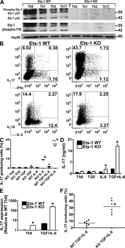
Ets-1 is expressed in two forms (p55 and p42) in Th cells because of alternative splicing, and it can undergo phosphorylation in response to stimulation through T cell receptors (23–25). The known phosphorylation sites of Ets-1 are Ser251, Ser257, Ser282, and Ser285 (collectively termed 4S) and Thr38. The p42 isoform of Ets-1 lacks exon 7, and therefore does not contain the 4S phosphorylation site. Previous in vitro studies have indicated that these phosphorylation events substantially influence the DNA-binding and -activating properties of Ets-1 (25–29). To examine the role of Ets-1 in regulating the differentiation of Th17 cells, we first set out to determine whether Ets-1 is expressed in Th17 and, if it is, whether Ets-1 can also undergo phosphorylation in activated Th17 cells. Th0, Th1, Th2, and Th17 cells were generated in vitro and restimulated with PMA/ionomycin for 4 h. The cytokine profile of each Th subset was confirmed by intracellular cytokine staining (ICS; Fig. S1, available at http://www.jem.org/cgi/content/full/jem.20070994/DC1). The level and phosphorylation of Ets-1 proteins was then examined by Western analysis. As shown in Fig. 1 A, all Th subsets expressed comparable levels of both the full-length p55 and the alternatively spliced p42 Ets-1 isoforms. In addition, phosphorylation of Thr38 (as detected by anti-phosphoT38) and 4S (as measured by a shift in the molecular weight of p55) was observed in all Th subsets. As expected, expression of Ets-1 was not detected in any of the Ets-1 KO Th subsets.
Figure 1.
Ets-1 KO Th cells produce increased levels of IL-17. Total Th cells derived from WT or Ets-1 KO mice were cultured as described in the Materials and methods. (A) After 5 d in culture under Th0, Th1, Th2, or Th17 conditions, cells were restimulated with PMA/ionomycin for 10 min and lysed. Whole-cell extract was analyzed by Western blotting using antibodies against Ets-1 or phospho-T38 Ets-1. An antibody against Hsp90 was used as loading control. (B) Total Th cells from WT and Ets-1 KO mice were cultured under Th17 conditions for 5 d and restimulated with PMA/ionomycin. Cytokine production was assessed by ICS. The numbers represent the percentages of cells stained positive for the indicated cytokines. (C) Total Ets-1 KO and WT Th cells were differentiated for 5 d in the presence of WT irradiated splenocytes and indicated cytokines, and the production of IL-17 in response to PMA/ionomycin stimulation was analyzed by ICS. The percentages of IL-17–positive cells from five independent experiments are shown. (D) Some of the differentiated Th cells (at 1 million cells/ml) generated in C were restimulated with 0.1 μg/ml of plate-bound anti-CD3 for 24 h, and the level of IL-17 protein in the supernatant was measured by ELISA. (E) A fraction of the PMA/ionomycin-restimulated cells from C were analyzed for IL-17 expression by real-time PCR analysis. (F) Naive WT and Ets-1 KO Th cells were differentiated under Th17 conditions, and the production of IL-17 by the differentiated cells was analyzed by ICS after 5 d. Cumulative results of six independent experiments are shown. Data are presented as the mean ± the SD. *, P < 0.05.
Increased IL-17 production by Ets-1 KO Th cells
To determine whether Ets-1 plays a role in regulating production of cytokines by Th17 cells, we used intracellular cytokine staining to compare the cytokine profiles between WT and Ets-1 KO Th17 cells. Surprisingly, we found that Ets-1 KO Th17 cells produced an abnormally high level of IL-17 compared with WT cells. When differentiated under Th17 conditions, ∼6–10% of the differentiated WT cells stained positive for IL-17, whereas >35% of Ets-1 KO Th17 cells produced IL-17 (Fig. 1, B and C). We also observed a substantial reduction in the level of IL-2 in Ets-1 KO Th17 cells, a result that is consistent with our previous findings in Ets-1 KO Th1 cells (22). However, the levels of IL-10 and IFN-γ were relatively low, but comparable between WT and Ets-1 KO Th17 cells, suggesting that the role of Ets-1 in regulating the expression of these two cytokines is Th subset dependent. Although deficiency of Ets-1 leads to overproduction of IL-17, Ets-1 KO Th cells are not “preprogrammed” to become IL-17–producing cells. When differentiated in the absence of polarizing cytokines (Th0 conditions), very little IL-17 was expressed by either WT or Ets-1 KO Th cells (Fig. 1 C). Interestingly, polarization with IL-6 alone, but not TGFβ1, already led to a significant increase in IL-17 production by Ets-1 KO cells, which was probably caused by the presence of a low level of TGFβ1 in fetal calf serum (Fig. 1 C). The increase in IL-17 staining of Ets-1 KO Th17 cells correlated very well with elevated levels of IL-17 protein in the supernatant of anti-CD3–stimulated Ets-1 KO Th17 cells, and with a marked increase in the level of IL-17 transcripts (Fig. 1, D and E). Thus, deficiency of Ets-1 leads to increased IL-17 expression by Th17 cells, and this effect of Ets-1 deficiency is mediated at the transcriptional level.
Ets-1 KO mice have previously been reported to contain an increased number of memory T cells (30). It is possible that Ets-1 KO APC preferentially drive Th cells into the Th17 pathway. Thus, the enhanced IL-17 production may simply reflect the presence of more memory Th17 cells in the starting bulk Ets-1 KO Th population. We found that WT and Ets-1 KO APCs were equally competent in supporting the in vitro differentiation of WT Th17 cells (unpublished data). In addition, when naive CD4+ CD62Lhi CD25− Th cells were sorted and cultured under Th17 conditions, the differentiated Ets-1 KO Th17 cells still exhibited overproduction of IL-17 compared with their WT counterparts (Fig. 1 F).
Increased IL-17 expression by Ets-1 KO Th17 cells is reversible and is caused by enhanced Th17 differentiation
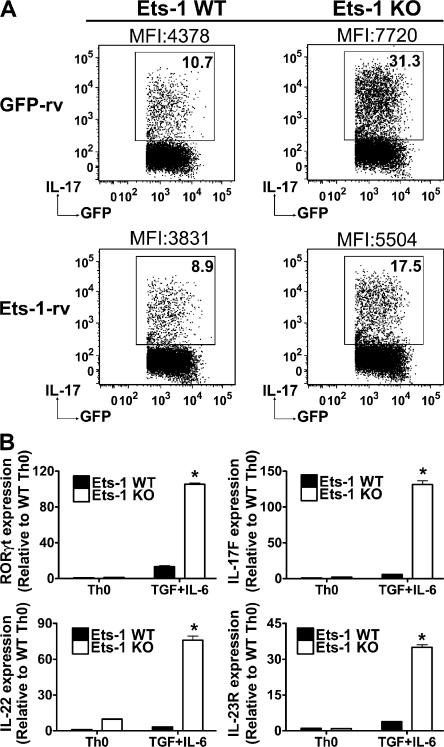
Deficiency of Ets-1 also significantly alters the development of thymocytes (30–32). To eliminate the possibility that increased IL-17 production by Ets-1 KO Th17 cells is caused by aberrant lymphocyte development, we retrovirally transduced differentiating WT and Ets-1 KO Th17 cells with either a retrovirus expressing GFP alone (GFP-rv) or the full-length p55 Ets-1 isoform (Ets-1-rv). The expression of IL-17 by the transduced (GFP-positive) Th17 cells was analyzed by ICS. Expression of retroviral Ets-1 in WT Th17 cells had only a minimal effect on the number of IL-17–producing cells compared with GFP-rv. The already high level of Ets-1 protein in WT Th17 cells probably explains the modest effect of the Ets-1-rv transduction on IL-17 production. In contrast, transduction with the Ets-1-rv retrovirus led to a 50% reduction in the number of IL-17–producing cells from differentiating Ets-1 KO Th17 cells (Fig. 2 A). Mean fluorescence intensity in the Ets-1-rv–transduced Ets-1 KO cells still expressing IL-17 was also lower than in the GFP-rv–transduced group. Incomplete normalization of IL-17 production is probably caused by the kinetics of infection. Because retroviral transduction requires proliferating cells, it is conceivable that at the time of infection on day 2, a population of cells is already committed to IL-17 production. These data demonstrate that the increase in IL-17 production by Ets-1 KO Th17 cells can be reversed by restoring the expression of Ets-1, and is thus unlikely to be caused by altered thymic development.
Figure 2.
Increased IL-17 production by Ets-1 KO Th cells is reversible, and is associated with increased expression of Th17 markers. (A) Total Th cells from WT and Ets-1 KO mice were infected 2 d after activation under Th17 skewing conditions with either empty virus (GFP-rv) or virus expressing full-length Ets-1 isoform (Ets-1-rv). The panels are gated on GFP-positive cells. The production of IL-17 by the transduced (GFP-positive) cells was examined by ICS. Mean fluorescence intensity (MFI) of the IL-17-positive cells is indicated. Results are representative of three independent experiments. (B) After 5 d in culture in the indicated conditions, differentiated WT and Ets-1 KO Th cells were restimulated for 4 h with PMA/ionomycin, and the transcript levels of IL-17F, IL-22, IL-23R, and RORγt were measured by real-time PCR. Data are presented as the mean ± the SD and are representative of three independent experiments. *, P < 0.05.
Although IL-17 is the prototypical cytokine of Th17 cells, other factors, such as IL-17F, IL-22, RORγt, and IL-23R have been found to be associated with cells committed to this subset. Deficiency of Ets-1 could specifically enhance the transcription of the Il17a gene. Alternatively, lack of Ets-1 could lead to augmented differentiation of Th17 cells. In the latter scenario, Ets-1 KO Th17 cells should also overexpress the aforementioned Th17-specific genes. We therefore used quantitative real-time PCR to analyze the expression of other Th17 marker genes in restimulated WT and Ets-1 KO Th17 cells (Fig. 2 B). In agreement with published reports, these Th17 marker genes were detected in WT Th17 cells, but not Th0 cells. Deficiency of Ets-1 resulted in a striking increase in the levels of all the known Th17 markers. This result clearly indicates that Ets-1 inhibits the differentiation of Th17 cells, and not merely the transcription of the Il17a gene.
Ets-1 does not bind to the IL-17 gene or interfere with early signaling events during Th17 differentiation
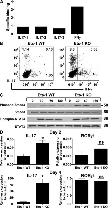
We next tried to identify possible mechanisms mediating the enhanced Th17 differentiation of Ets-1 KO Th cells. We first determined whether Ets-1 could regulate the expression of IL-17 by directly binding to the Il17a gene. Several conserved potential Ets-binding sites in the promoter and first intron of the IL-17 genetic region were tested for Ets-1 binding by chromatin immunoprecipitation (CHIP). However, we could not demonstrate any binding of Ets-1 (Fig. 3 A). As a positive control, substantial binding of Ets-1 to the IFN-γ promoter was detected, a result that is consistent with our previously published data (22). In our hands, optimal differentiation of Th17 cells requires APCs (irradiated WT splenocytes), IL-6, and TGFβ1. Ets-1 KO Th cells may be more sensitive than WT cells to signals derived from APCs, thereby preferentially differentiating into Th17 cells. To test this hypothesis, we cultured WT and Ets-1 KO Th cells under Th17 conditions, but in the absence of APC. Although the number of IL-17–producing cells was lower in both WT and Ets-1 KO populations compared with those cultured in the presence of APC, Ets-1 KO Th17 cells still produced more IL-17 than their WT counterparts (Fig. 3 B). Thus, the predilection for Th17 differentiation of Ets-1 KO Th cells is not dependent on APC-derived signals.
Figure 3.
Ets-1 does not bind to the IL-17 promoter or interfere with early signaling events during Th17 differentiation. (A) CHIP was performed on WT Th17 cells using anti–Ets-1 antibody. Binding to conserved Ets sites in the IL-17 and IFN-γ genetic region was analyzed by real-time PCR. Relative binding was calculated as described in the Materials and methods. (B) Total WT and Ets-1 KO Th cells were plated on anti-CD3–coated plates and given anti-CD28, but in the absence of APC, and skewed to the Th17 lineage with IL-6 and TGFβ1 for 5 d. The production of indicated cytokines by the differentiated Th cells was measured by ICS. (C) Freshly isolated total WT and Ets-1 KO Th cells were stimulated with TGFβ1 and IL-6 for the indicated number of minutes. The levels of phospho-Smad3, total Smad2/3, phospho-STAT3, and total STAT3 were examined with Western blotting. (D) Naive Th cells were cultured under Th17 conditions, and RNA was harvested on day 2 or 4 and analyzed for expression of IL-17 and RORγt by real-time PCR. Data are representative of three independent experiments and are presented as the mean ± the SD. *, P < 0.05.
Another possible explanation for increased Th17 differentiation is augmented signal transduction from TGFβR and/or IL-6R. To address this possibility, freshly isolated Th cells were stimulated with TGFβ1 and IL-6, and the activation of proximal signal transduction components were examined by Western analysis. No phosphorylated STAT3 was detected before the addition of IL-6. Treatment with IL-6 quickly induced the phosphorylation of STAT3 within 15 min, and the levels and kinetics of phosphorylation of STAT3 were comparable between WT and Ets-1 KO Th cells up to 3–6 h after IL-6 stimulation (Fig. 3 C and not depicted). A low level of phosphorylated Smad3 was detected even in the absence of exogenous TGFβ1 probably caused by preexisting TGFβ1 in fetal calf serum. The addition of TGFβ1 further boosted the level of phosphorylated Smad3, which remained relatively constant for 60 min before returning to baseline. In most, but not all, of the experiments, we detected a subtle increase in the level of phosphorylated Smad3 in Ets-1 KO Th cells after the addition of exogenous TGFβ1 (Fig. 3 C). We therefore conclude that early signaling events downstream of TGFβR and IL-6R occur normally in Ets-1 KO Th cells.
Increased Th17 differentiation in Ets-1 KO Th cells despite normal expression of RORγt
RORγt is essential and sufficient for the differentiation of Th17 cells (17). The data shown in Fig. 2 B indicate a high level of RORγt in the differentiated Ets-1 KO Th17 cells. This finding might explain why Ets-1 KO Th17 cells produce more IL-17 than WT cells. On the other hand, the elevated RORγt level may simply reflect the fact that there are more IL-17–producing cells in the differentiated Ets-1 KO population and that this is not the cause of enhanced Th17 differentiation. In support of the latter scenario, when naive Th cells were examined 2 d into the Th17 differentiation protocol, an abnormally high level of IL-17 transcripts was already detected in the differentiating Ets-1 KO Th17 cells (Fig. 3 D). However, there was no statistically significant difference in the level of RORγt between WT and Ets-1 KO populations. The overproduction of IL-17 by differentiating Ets-1 KO Th17 cells was even more obvious on the fourth day of the differentiation protocol, but the levels of RORγt were still very comparable (Fig. 3 D). These data demonstrate that naive Ets-1 KO Th cells differentiate more readily than WT into Th17 cells, and that the enhanced early IL-17 production is not associated with an increase in the expression of RORγt.
Deficiency of Ets-1 enhances Th17 differentiation by an IL-2–dependent mechanism
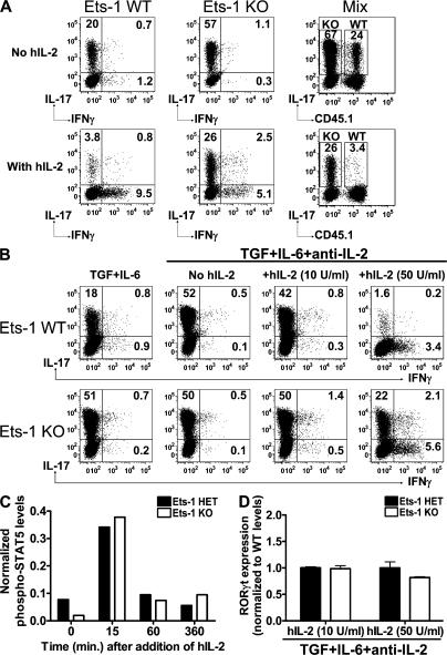
IL-2 was recently shown to be a potent inhibitor of Th17 differentiation (19). Exogenous human IL-2 (hIL-2 at 50 U/ml) was added to our Th17 culture conditions without knowing the negative effect of IL-2. Our intent had been to equalize the level of active IL-2 because Ets-1 KO Th cells, regardless of functional subsets, have a substantial defect in IL-2 production. The difference in IL-2 production was readily detected even at 48 h after the initial stimulation (22). Despite the effort, exogenous hIL-2, even added in the beginning of in vitro differentiation and whenever Th cells were expanded, may not be sufficient to equalize the level of IL-2. Such a difference in the level of IL-2 may result in the abnormal Th17 differentiation of Ets-1 KO cells. To test this hypothesis, congenic CD45.1 C57BL/6 and CD45.2 Ets-1 KO cells were separately cultured or cocultured under Th17 skewing conditions in the absence of exogenous hIL-2. In this coculture system, WT and Ets-1 KO Th cells were allowed to differentiate into Th17 cells in the same cytokine milieu, thereby avoiding any confounding effects caused by aberrant production of endogenous IL-2. We found that the coculture system did not alter the production of IL-17 by either Ets-1 KO or WT cells (Fig. 4 A). Thus, the defect in IL-2 production cannot fully explain the aberrant Th17 differentiation of Ets-1 KO cells.
Figure 4.
Increased Th17 differentiation in Ets-1 KO Th cells is dependent on IL-2. (A) Total Th cells from congenic CD45.1 C57BL/6 and CD45.2 Ets-1 KO mice were cultured separately or cocultured (Mix) at a 1:1 ratio under Th17 skewing conditions in the presence (added at 0 h) or absence of exogenous IL-2. The cells were stained for the CD45.1 isoform, and the production of indicated cytokines was analyzed by ICS after restimulation with PMA/ionomycin. (B) Total Th cells from WT and Ets-1 KO mice were cultured under Th17 conditions in the presence of anti–mouse IL-2 and various concentrations of hIL-2. Cytokine production was assessed by ICS. The numbers represent the percentages of cells stained positive for indicated cytokines. (C) Total Th cells from WT and Ets-1 KO mice were cultured for 3 d with TGFβ1, IL-6, anti–mouse IL-2, and hIL-2 (10 U/ml). Cells were subsequently rested for 2 h in the presence of anti–mouse IL-2 before being stimulated with 50 U/ml hIL-2 for the indicated amount of time. Cell lysates were harvested and subjected to Western blot analyses using antibody against phosphorylated or total STAT5. The blots were scanned and the levels of phosphorylated STAT5 were normalized to the total amount of STAT5. (D) After 5 d in culture in the indicated conditions, RNA from differentiated WT and Ets-1 KO Th17 cells was collected, and the transcript level of RORγt was measured by real-time PCR. Data are presented as the mean ± the SD and are from two independent experiments. *, P < 0.05.
In parallel experiments, exogenous hIL-2 was added to the coculture system. Again, the coculture system failed to equalize the differentiation of Th17 cells in the presence of exogenous hIL-2 (Fig. 4 A). But exogenous hIL-2 strongly inhibited the differentiation of WT Th17 cells, a result that is consistent with a recent publication (19). Interestingly, the negative effect of IL-2 on Ets-1 KO Th17 cells was much less dramatic than that on WT cells. This intriguing observation raised the possibility that Ets-1 KO–differentiating Th17 cells were more resistant to IL-2 suppression. To test this hypothesis, we first determined whether the effect of Ets-1 deficiency was dependent on IL-2. We set up Th17 differentiation in the absence of IL-2/STAT5 signaling. WT and Ets-1 KO Th cells were activated in vitro under Th17 conditions, but in the absence of exogenous hIL-2. In addition, anti–murine IL-2 antibody was added in a separate set of samples to further neutralize endogenous IL-2. The production of IL-17 by the differentiated Th17 cells was examined by ICS. Similar to what was shown in Fig. 4 A, Ets-1 KO Th17 cells produced a much higher level of IL-17 than WT cells when differentiated in the absence of exogenous hIL-2 (Fig. 4 B). Remarkably, neutralization of endogenous IL-2 markedly enhanced IL-17 production by WT Th17 cells to a level comparable to that of Ets-1 KO Th17 cells. In contrast, anti–IL-2 had a negligible effect on the production of IL-17 by Ets-1 KO Th17 cells. Thus, the Th17-promoting effect of Ets-1 deficiency is only apparent in the presence of IL-2 signaling, and is therefore dependent on IL-2.
Ets-1 KO-differentiating Th17 cells are more resistant to IL-2 suppression than WT cells
To further examine the response of differentiating Th17 cells to IL-2 in a dose-dependent manner, we then added incremental doses of hIL-2 to the culture shown in Fig. 4 B. As hIL-2 is not recognized by the anti–murine IL-2 antibody, IL-2 activity in this culture system came solely from exogenous hIL-2. The differentiation of WT Th17 cells started to be suppressed by exogenous hIL-2 at a concentration of 10 U/ml and was almost completely inhibited at 50 U/ml. Surprisingly, the differentiation of Ets-1 KO Th17 cells was only reduced by 50% at 50 U/ml of hIL-2. Collectively, our data clearly demonstrate that Ets-1 KO Th17 cells not only have a defect in IL-2 production, but also are more resistant than WT cells to IL-2 suppression.
IL-2 is known to induce the phosphorylation of STAT5, which in turn inhibits the expression of RORγt in differentiating Th17 cells. Deficiency of Ets-1 may affect the level and/or kinetics of STAT5 phosphorylation, explaining the increased resistance to the negative effects of IL-2. To address this question, on day 3 we harvested differentiating WT and Ets-1 KO Th17 cells from anti–mouse IL-2– and hIL-2–treated (10 U/ml) cultures. The cells were washed thoroughly and treated with 50 U/ml of fresh hIL-2. The level of phospho-STAT5 was then examined at different time points. We found that the level and kinetics of STAT5 phosphorylation was very comparable between WT and Ets-1 KO Th cells (Fig. 4 C). Furthermore, the levels of RORγt in differentiating WT and Ets-1 KO Th17 cells that were cultivated with either 10 or 50 U/ml of hIL-2 were also comparable (Fig. 4 D). Thus, the resistance to IL-2 is not caused by a defect in STAT5 phosphorylation or induction of RORγt.
Ets-1 KO mice display increased expression of Th17 markers in vivo
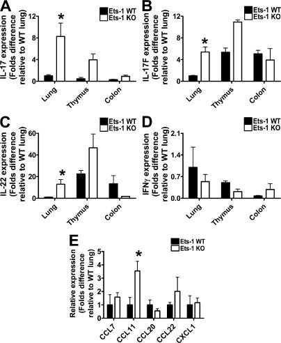
To determine whether the aberrant Th17 differentiation in vitro can also be observed in vivo, we performed expression analyses on various organs from WT and Ets-1 KO mice. We detected significant increases in Th17-associated cytokines (IL-17, -17F, and -22) in the lungs of Ets-1 KO mice (Fig. 5, A–C). In contrast, we did not observe any difference in the expression of the Th1 cytokine IFN-γ (Fig. 5 D). IL-13 is also a potent mediator of airway inflammation (33), but we found that the transcript level of IL-13 was normal in Ets-1 KO lung tissue (Fig. S2, available at http://www.jem.org/cgi/content/full/jem.20070994/DC1). There was also a strong trend toward higher expression of Th17 markers in Ets-1 KO thymus. Interestingly, no significant differences in the level of Th17 markers were observed in the colon of WT and Ets-1 KO mice, suggesting that the overexpression of Th17 cytokines is restricted to certain organs/tissues. Ectopic expression of an IL-17 transgene has been shown to induce the expression of several chemokines in the lung tissue (15). We also detected an abnormally high level of CCL11, but not CCL7, CCL20, CCL22, or CXCL1 in the lung of Ets-1 KO mice (Fig. 5 E).
Figure 5.
In vivo evidence of increased Th17 differentiation in Ets-1 KO mice. Total RNA of indicated tissues from three WT and Ets-1 KO mice was extracted, and the transcript levels of IL-17 (A), IL-17F (B), IL-22 (C), and IFNγ (D) were quantified by real-time PCR. (E) Expression of various chemokines was analyzed in the lungs of WT and Ets-1 KO mice. Data are presented as the mean ± the SD. *, P < 0.05.
IL-17–dependent overproduction of mucus by Ets-1 KO airway epithelial cells
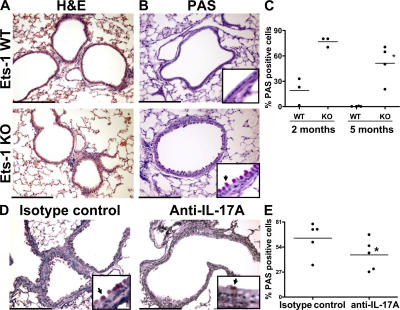
Ectopic expression of IL-17 by lung epithelial cells has been shown to induce striking airway inflammation (15). The observation that Ets-1 KO mice expressed a high level of IL-17 in the lungs prompted us to perform thorough histological analysis of lungs from WT and Ets-1 KO mice to determine whether increased expression of Th17 mediators had any pathological consequences. We did not observe any significant differences in cellular infiltration, fibrosis, or bronchoalveolar lavage composition (Fig. 6 A and not depicted). Interestingly, Ets-1 KO lungs showed a striking increase in the number of mucus-producing cells (Fig. 6 B). Approximately 60–80% of airway epithelial cells of Ets-1 KO mice were stained positive with periodic acid Schiff (PAS), whereas only 0–20% of WT epithelial cells produced mucus. This marked increase in mucus production was observed as early as 2 mo after birth, and it was still present in 5-mo-old Ets-1 KO mice (Fig. 6 C). This observation is consistent with a previous study showing that IL-17 is a potent inducer of mucin production (34). To confirm that the increased mucus production in Ets-1 KO lungs was caused by overproduction of IL-17, 2-mo-old Ets-1 KO mice were treated every other day for 2 wk with either an anti–IL-17 antibody or isotype control. Neutralization of IL-17 led to a statistically significant, although modest, reduction in the number of mucus-producing cells (Fig. 6, D and E). In addition, the overall staining in the remaining PAS-positive cells was less intense in the anti–IL-17–treated mice than in control IgG-treated mice. The modest effect of anti–IL-17 treatment shown in Fig. 6 is in line with that reported in animal models of colitis and arthritis (35, 36). These data collectively indicate that the overproduction of mucus in Ets-1 KO mice can be partially attributed to excessive IL-17.
Figure 6.
IL-17–dependent mucus overproduction by Ets-1 KO airway epithelial cells. Histological sections of inflated lungs from WT and Ets-1 KO mice were stained for hematoxylin and eosin (A) and PAS (B). Bars, 1 mm. The arrow in B points out PAS-positive epithelial cells. The percentages of PAS-positive epithelial cells observed in WT and KO mice (n = 3–4) at the indicated ages are shown in C. 2-mo-old Ets-1 KO mice were injected intraperitoneally with anti–IL-17 or control IgG every other day for 2 wk (6 injections in total) before killing. The histological sections of inflated lungs were stained with PAS (D). The percentages of PAS-positive epithelial cells are shown in F. Data are presented as the mean ± the SD. *, P < 0.05.
DISCUSSION
In this study, we report an inhibitory role for the transcription factor Ets-1 in Th17 differentiation. Besides STAT molecules (37), there are only a handful of transcription factors that have been shown to control the differentiation and function of this new subset of Th cells. RORγt is a well-known promoter of Th17 cells, whereas Foxp3 and T-bet suppress the differentiation of Th17 cells (12, 17). Differentiated Th17 cells maintain a high level of RORγt (17), but shut off the expression of Foxp3 (13) and T-bet (38). In this regard, Ets-1 is quite unique because it is expressed at a substantial level in Th17 population, despite its function as a negative regulator.
The differentiation of Th17 cells is regulated by several signaling pathways. APC-derived signals, such as signals downstream of ICOS/ICOSL (15), critically influence the differentiation of Th17 cells. But the preferential Th17 differentiation of Ets-1 KO Th cells was still observed in the absence of APC. In addition, the strength of early signal transduction events downstream of TGFβR and IL-6R was apparently normal in Ets-1 KO cells. Non-Th17 cytokines, such as IFN-γ and IL-4, can also inhibit the differentiation of Th17 cells. Deficiency of Ets-1 attenuates the production of IFN-γ and IL-4 by Th1 and Th2 cells, respectively, and may therefore lead to enhanced Th17 differentiation. However, Ets-1 KO and WT Th17 cells produced low, but comparable, levels of these two cytokines (Fig. 1 B and not depicted). In addition, anti–IL-4 and –IFN-γ, when added into Th17 culture conditions, were unable to equalize Th17 differentiation of WT and Ets-1KO cells (Fig. S3, available at http://www.jem.org/cgi/content/full/jem.20070994/DC1). During the revision of this manuscript, it was reported that IL-21, induced by IL-6 via STAT3, facilitated Th17 differentiation in a STAT3-dependent positive feedback manner (39–41). We found that the transcript levels of IL-21 in differentiating WT and Ets-1 KO Th17 cells were comparable (unpublished data). This result is consistent with the observation that the level and kinetics of IL-6–induced STAT3 phosphorylation were normal in Ets-1 KO Th cells. It is also unlikely that Ets-1 controls the expression of the known Th17 regulators, thereby indirectly suppressing the differentiation of Th17 cells. We did not detect reduced expression of T-bet in differentiating WT and Ets-1 KO Th17 cells (unpublished data). Foxp3 was transiently induced during the differentiation of Th17 cells, but the level and kinetics of induction of Foxp3 were normal in Ets-1 KO Th cells (Fig. S4).
Our data instead indicate that the effect of Ets-1 deficiency on Th17 differentiation is only apparent in the presence of IL-2. IL-2 is known to inhibit Th17 differentiation by suppressing the expression of RORγt (19). Ets-1 KO Th cells have a profound defect in the production of IL-2. Such a defect surely facilitates Th17 differentiation because eliminating IL-2 from the culture markedly enhanced the differentiation of WT Th17 cells. But our data further demonstrate that differentiating Ets-1 KO Th cells are also more resistant to the negative effect of IL-2. This resistance was clearly revealed when the level of bioactive IL-2 in cytokine milieu was normalized either by the coculture system or by the combination of anti–murine IL-2 and hIL-2. More importantly, we found that the resistance was not caused by a defect in the phosphorylation of STAT5 or in the induction of RORγt. This observation strongly suggests that IL-2 can also inhibit the differentiation of Th17 cells by acting on a molecular event that takes place after the induction of RORγt. To the best of our knowledge, this is the first evidence showing that IL-2 can suppress the differentiation of Th17 cells by a mechanism different from suppressing the induction of RORγt.
It is intriguing to know that an early study has demonstrated physical interactions between Ets-1 and STAT5 in human T cells (42). STAT5 has been shown to directly bind to the promoter of IL-17 (19). Ets-1 may form a protein complex with STAT5, and then bind to the IL-17 promoter. However, we could not find any evolutionarily conserved STAT5–Ets-1 composite sites in the IL-17 locus, nor could we demonstrate any direct binding of Ets-1 to the IL-17 gene (Fig. 3 A). This may suggest that the target of the STAT5–Ets-1 complex, if indeed present, is not IL-17. Although Ets-1 KO Th17 cells were resistant to inhibition by IL-2, other IL-2–induced responses, such as induction of CD25, were intact in Ets-1 KO Th cells (unpublished data). Thus, deficiency of Ets-1 selectively interferes with a novel IL-2–dependent molecular event that is critical for the differentiation of Th17 cells. Identifying the target genes of Ets-1 will help us understand this novel molecular event.
IL-17 is a highly inflammatory cytokine, and it is the main effector molecule in several organ-specific autoimmune diseases, such as experimental autoimmune encephalomyelitis (43), colitis (36), and arthritis (44), in addition to airway inflammation (15). But why do Ets-1 KO mice not exhibit pathological changes in organs other than the lungs? The answer may rest in the observation that the overproduction of IL-17 in Ets-1 KO mice is mainly confined to the lungs. This observation also strongly suggests that the differentiation of Ets-1 KO Th cells is still subject to regulation by local environmental factors. Indeed, Ets-1 KO Th cells, despite their propensity for Th17 differentiation, still require APC-derived signals, IL-6, and TGFβ1 to optimally differentiate into IL-17–producing cells. In addition, it was recently shown that engagement of dectin-1 by yeast β glucan–activated dendritic cells favored the production of IL-23, but not IL-12 (45). The dectin-activated dendritic cells then “instructed” Th cells to become IL-17–producing cells. This observation further highlights the critical influence of environmental factors in determining the fate of differentiating Th cells. Thus, the interplays between Ets-1 KO Th cells and “permissive” environmental factors that are present in the lungs, but not the colon, lead to lung-restricted overproduction of IL-17.
Given the observation that anti–IL-17 treatment attenuated mucus production in Ets-1 KO mice, we conclude that the overproduction of mucus is at least partally attributed to excessive IL-17. It is not unexpected that the effect of anti–IL-17 treatment is only modest for the following reasons. First, the dose, timing, and duration of treatment may not be optimal. Second, the lungs of Ets-1 KO mice also contain high levels of other Th17 cytokines, such as IL-17F and -22, which may also directly or indirectly promote mucus production. The anti–IL-17 antibody used in this study will not block the biological function of Th17-cytokines other than IL-17.
Our findings further support a pathogenic role of Th17 cells in airway inflammation. IL-17–deficient mice were resistant to airway inflammation in one animal model of acute allergic asthma (46). In addition, constitutively forced expression of IL-17 by airway epithelial cells induced marked lung inflammation that was characterized by airway remodeling and mucus production (15). But the pathological consequence of excessive IL-17 derived from bona fide IL-17–producing cells, namely Th17 cells, in response to physiological stimuli remains unclear. Our data indicate that chronic exposure to Th17 cell–derived cytokines can, indeed, lead to mucus overproduction. However, we did not detect any abnormal cellular infiltration or increased fibrosis, which is a feature of airway remodeling, in the lungs of 5-mo-old Ets-1 KO mice. The development of cellular infiltration and fibrosis may require a longer exposure to a higher level of Th17-derived cytokines than that was detected in the relatively young Ets-1 KO lungs. It is also possible that lung tissue may respond differently to Th17-derived cytokines compared with ectopically expressed IL-17. The differences in the level and source of IL-17 between Ets-1 KO mice and the IL-17 transgenic mice may also explain why we only observed aberrant expression of CCL11, but not other chemokines in the lung of Ets-1 KO mice.
Elevated levels of IL-17 have been reproducibly detected in bronchial lavage obtained from patients with allergic asthma (2, 47), and the contribution of Th17 cells to the pathogenesis of this disease has begun to be elucidated. Our data raise the possibility that loss of Ets-1 function may indirectly lead to exacerbation of asthma in a subset of patients by boosting the production of IL-17 by pulmonary Th cells. The chromosomal locus of the ets1 gene is not known to be associated with susceptibility to allergic asthma. But it has been demonstrated in vitro that the transcriptional activity of Ets-1 is subject to regulation by posttranslational modifications, including phosphorylation and sumoylation, in response to various signals (25, 27–29, 48). It is foreseeable that the activity of Ets-1 can be manipulated by altering the posttranslational modifications with pharmacological and/or biological reagents. Such reagents carry the potential of becoming effective treatments for allergic asthma.
MATERIALS AND METHODS
Mice.
The Ets-1 KO mice have been previously described (49). Mice used in these experiments have been backcrossed to the C57BL/6 background for six generations. All experiments were performed using 6–8-wk-old male or female littermate pairs. Heterozygous mice were used as WT controls. Congenic CD45.1 C57BL/6 mice were purchased from Taconic. The animals were housed under specific pathogen-free conditions, and experiments were performed in accordance with the institutional guidelines for animal care at the Dana-Farber Cancer Institute under approved protocols.
Antibodies and cytokines.
Anti-CD3 (2C11) and -IFNγ (XMJ 1.2) were purchased from BD Biosciences. Recombinant hIL-2 and anti–IL-4 (11B11) were provided by the National Cancer Institute. IL-12 and -4 were obtained from Peprotech. TGFβ1, IL-6, and anti–mouse IL-2 were purchased from R&D Systems. The following antibodies for surface and intracellular cytokine staining were purchased from BD Biosciences: CD4 (L3T4), CD25 (PC61), CD44 (IM7), CD62L (MEL-14), IFNγ (XMJ 1.2), IL-2 (JES6-5H4), IL-4 (11B11), IL-10 (JES5-16E3), and IL-17 (TC11-18H10). For in vivo IL-17 neutralization, anti–IL-17 (50104) and control rat IgG2a were purchased from R&D Systems.
Cell purification and in vitro differentiation.
Spleen and peripheral lymph nodes were harvested from WT and Ets-1 KO mice. Total CD4+ cells were purified using magnetic cell separation according to the manufacturer's protocol (Miltenyi Biotech). To obtain naive Th cells, CD4+ cells were stained for CD4, CD25, and CD62L and sorted on a High-Speed MoFlo sorter (DakoCytomation). The CD4+CD25−CD62Lhi population was used for in vitro Th cell differentiation. Total or sorted CD4+ naive cells were cultured in the presence of WT irradiated splenocytes (1:3 ratio) and 2 μg/ml soluble anti-CD3 in the presence of cytokines to obtain either Th0 (no additional cytokines or antibodies), Th1 (3 ng/ml IL-12 and 10 μg/ml anti-IL-4), Th2 (10 ng/ml IL-4 and 10 μg/ml anti-IFN-γ), or Th17 (3 ng/ml TGFβ1 and 20 ng/ml IL-6) cells. 50 U/ml IL-2 was added to the cultures after 24 h. Analysis of cytokine production was performed 5 d later. Retroviral transduction was performed 2 d after activation. The protocol for retroviral transduction and the GFP-rv and Ets-1-rv construct have been previously described (22).
Analysis of cytokine production.
Intracellular cytokine staining was performed as previously described (50). After staining, the samples were run on a FACSCanto flow cytometer (BD Biosciences) and analyzed using the FlowJo software package. IL-17 Duoset ELISA kit was purchased from R&D Systems and used according to the manufacturer's protocol.
Histology.
Immediately after the killing of the mouse, the lungs were removed, inflated with 4% paraformaldehyde, dehydrated, mounted in paraffin, and sectioned. Deparaffinized and hydrated sections were stained with hematoxylin and eosin and PAS stains following standard procedure. For quantitation, 300 airway epithelial cells in the lungs were counted in a blinded fashion, and the number of PAS-positive cells was quantified.
Western blotting.
2 × 10⁶ CD4+ cells were lysed in SDS-PAGE sample buffer containing 2.5% 2-mercaptoethanol. The lysates were subsequently sheared using a 26-gauge needle. Samples were loaded onto 8% polyacrylamide gels and transferred onto PVDF membrane (Polyscreen; Perkin Elmer). The membrane was subsequently blocked in 5% milk and probed with antibodies according to the manufacturer's protocol. The Ets-1 (C-20) and HSP90 antibodies were purchased from Santa Cruz Biotechnology. Anti-phospho T38 was purchased from Biosource. Phospho-Smad3, total Smad3, phospho-STAT3, total STAT3, phospho-STAT5, and total STAT5 antibodies were obtained from Cell Signaling Technology.
RNA analysis.
Total RNA was purified using a Trizol Plus kit (Invitrogen). First-strand cDNA synthesis was performed on 1 μg of total RNA using the QuantiTect Reverse Transcription kit (QIAGEN). Gene expression levels were determined by real-time PCR analysis performed using the Brilliant SYBR Green QPCR kit according to the manufacturer's protocol (Stratagene) on a MX-3000P apparatus (Stratagene) using the following cycling conditions: denaturation at 95°C for 30 s, annealing at 56°C for 60 s, and extension at 72°C for 30 s. Primer sets were designed using the Primer3 web utility or using the Primer Bank database (51). Sequences used in this study are presented in Table I and were confirmed to generate only one product with a minimum of 90% efficiency. Levels of cytokine mRNA were adjusted for differences in β-actin expression and normalized to the levels found in the Ets-1 WT group, where indicated.
Table I.
Sequences used in this study
| Gene | Sense (5′–3′) | Antisense (5′–3′) | Primer bank ID |
|---|---|---|---|
| IL-13 | GCAACATCACACAAGACCAGA | GTCAGGGAATCCAGGGCTAC | 6680403a2 |
| IL-17A | actttcagggtcgagaaga | ttctgaatctgcctctgaat | |
| IL-17F | TGCTACTGTTGATGTTGGGAC | AATGCCCTGGTTTTGGTTGAA | 22003916a1 |
| IL-22 | GTGAGAAGCTAACGTCCATC | GTCTACCTCTGGTCTCATGG | |
| IL-23R | TTCAGATGGGCATGAATGTTTCT | CCAAATCCGAGCTGTTGTTCTAT | 21362353a1 |
| IFNγ | tgaacgctacacactgcatct | cgactccttttccgcttcctg | |
| RORγt | AGCTTTGTGCAGATCTAAGG | TGTCCTCCTCAGTAGGGTAG | |
| CCL7 | GCTGCTTTCAGCATCCAAGTG | CCAGGGACACCGACTACTG | 7305463a1 |
| CCL11 | GAATCACCAACAACAGATGCAC | ATCCTGGACCCACTTCTTCTT | 6755418a1 |
| CCL20 | GCCTCTCGTACATACAGACGC | CCAGTTCTGCTTTGGATCAGC | 8394248a1 |
| CCL22 | AGGTCCCTATGGTGCCAATGT | CGGCAGGATTTTGAGGTCCA | 6677879a1 |
| CXCL1 | CTGGGATTCACCTCAAGAACATC | CAGGGTCAAGGCAAGCCTC | 6680109a1 |
| β-actin | AGAGGGAAATCGTGCGTGAC | CAATAGTGATGACCTGGCCGT |
CHIP.
CHIP was performed as previously described (22). Precipitated DNA fragments were amplified by quantitative PCR. The antibodies used for immunoprecipitation were anti-Ets1 (C-20) and rabbit control IgG (both from Santa Cruz Biotechnology). Identification of conserved Ets-binding site was done using the rVista 2.0 web utility (52). IL-17 Ets sites were located at site 1 (5 kb upstream of the transcriptional start site), site 2 (within the first intron), and site 3 (1 kb upstream of the transcriptional start site). All sites were conserved between mouse and rat genomes. Additionally, site 1 was conserved in human and dog genomes. Specific binding was calculated as follows: specific binding = 2^ − (CtEts1-Ctinput)/2^ − (CtrbIg − Ctinput). The following primer pairs were used: IL17-site1, 5′-CGTGTGGTTTGGTTTACTTA-3′ and 5′-GCTGACTTCATCTGATACCC-3′; IL17-site2, 5′-TATGCTCTGCACTCGTATTC-3′ and 5′-GTACACCAGCTATCCTCCAG-3′; IL17-site3, 5′-GGGATGTTAATTCAAACTGC-3′ and 5′-CTCACACACACCTCTGATTG-3′; and IFN-γ promoter, 5′-CTTTCAGAGAATCCCACAAG-3′ and 5′-TTAAGATGGTGACAGATAGGTG-3′.
Statistical analysis.
A Mann-Whitney nonparametric test was performed to calculate statistical significance for all experiments, except for the in vivo IL-17 neutralization, where an unpaired t test using Welch correction was performed (Prism 4; GraphPad Softwares). Differences among treatments are considered significant if P ≤ 0.05.
Online supplemental material.
Fig. S1 shows ICS staining of various Th subsets derived from WT cells after restimulation with PMA/ionomycin. Fig. S2 shows expression of IL-13 in the lungs of WT and Ets-1 KO mice. Fig. S3 shows WT and Ets-1 KO Th17 differentiation in the presence of neutralizing antibodies to IL-4 and IFNγ. Fig. S4 represents FoxP3 expression in differentiating WT and Ets-1 KO Th17 cells. The online version of this article is available at http://www.jem.org/cgi/content/full/jem.20070994/DC1.
Supplemental Material
[Supplemental Material Index]
Acknowledgments
The authors would like to thank Dr. S.Y. Pai for critical review of the manuscript and T. Bowman for help in preparing and staining histological samples.
This work was supported by a National Institutes of Health (NIH) R03 (AI0678801; I.-C. Ho), NIH R01 (AI073542-01; M. Oukka), and a National Multiple Sclerosis Society (RG-3882-A-1; M. Oukka) grant, as well as a Senior Research Award from the Crohn's and Colitis Foundation of America (I.-C. Ho).
The authors have no conflicting financial interests.
Abbreviations used: CHIP, chromatin immunoprecipitation; ICS, intracellular cytokine staining; PAS, periodic acid Schiff.
References
- 1.Weaver, C.T., R.D. Hatton, P.R. Mangan, and L.E. Harrington. 2007. IL-17 family cytokines and the expanding diversity of effector t cell lineages. Annu. Rev. Immunol. 25:821–852. [DOI] [PubMed] [Google Scholar]
- 2.Ziolkowska, M., A. Koc, G. Luszczykiewicz, K. Ksiezopolska-Pietrzak, E. Klimczak, H. Chwalinska-Sadowska, and W. Maslinski. 2000. High levels of IL-17 in rheumatoid arthritis patients: IL-15 triggers in vitro IL-17 production via cyclosporin A-sensitive mechanism. J. Immunol. 164:2832–2838. [DOI] [PubMed] [Google Scholar]
- 3.Matusevicius, D., P. Kivisakk, B. He, N. Kostulas, V. Ozenci, S. Fredrikson, and H. Link. 1999. Interleukin-17 mRNA expression in blood and CSF mononuclear cells is augmented in multiple sclerosis. Mult. Scler. 5:101–104. [DOI] [PubMed] [Google Scholar]
- 4.Molet, S., Q. Hamid, F. Davoine, E. Nutku, R. Taha, N. Page, R. Olivenstein, J. Elias, and J. Chakir. 2001. IL-17 is increased in asthmatic airways and induces human bronchial fibroblasts to produce cytokines. J. Allergy Clin. Immunol. 108:430–438. [DOI] [PubMed] [Google Scholar]
- 5.Bettelli, E., M. Oukka, and V.K. Kuchroo. 2007. T(H)-17 cells in the circle of immunity and autoimmunity. Nat. Immunol. 8:345–350. [DOI] [PubMed] [Google Scholar]
- 6.Ye, P., F.H. Rodriguez, S. Kanaly, K.L. Stocking, J. Schurr, P. Schwarzenberger, P. Oliver, W. Huang, P. Zhang, J. Zhang, et al. 2001. Requirement of interleukin 17 receptor signaling for lung CXC chemokine and granulocyte colony-stimulating factor expression, neutrophil recruitment, and host defense. J. Exp. Med. 194:519–527. [DOI] [PMC free article] [PubMed] [Google Scholar]
- 7.Ye, P., P.B. Garvey, P. Zhang, S. Nelson, G. Bagby, W.R. Summer, P. Schwarzenberger, J.E. Shellito, and J.K. Kolls. 2001. Interleukin-17 and lung host defense against Klebsiella pneumoniae infection. Am. J. Respir. Cell Mol. Biol. 25:335–340. [DOI] [PubMed] [Google Scholar]
- 8.Cua, D.J., J. Sherlock, Y. Chen, C.A. Murphy, B. Joyce, B. Seymour, L. Lucian, W. To, S. Kwan, T. Churakova, et al. 2003. Interleukin-23 rather than interleukin-12 is the critical cytokine for autoimmune inflammation of the brain. Nature. 421:744–748. [DOI] [PubMed] [Google Scholar]
- 9.Langrish, C.L., Y. Chen, W.M. Blumenschein, J. Mattson, B. Basham, J.D. Sedgwick, T. McClanahan, R.A. Kastelein, and D.J. Cua. 2005. IL-23 drives a pathogenic T cell population that induces autoimmune inflammation. J. Exp. Med. 201:233–240. [DOI] [PMC free article] [PubMed] [Google Scholar]
- 10.Reiner, S.L. 2007. Development in motion: helper T cells at work. Cell. 129:33–36. [DOI] [PubMed] [Google Scholar]
- 11.Steinman, L. 2007. A brief history of T(H)17, the first major revision in the T(H)1/T(H)2 hypothesis of T cell-mediated tissue damage. Nat. Med. 13:139–145. [DOI] [PubMed] [Google Scholar]
- 12.Mathur, A.N., H.C. Chang, D.G. Zisoulis, R. Kapur, M.L. Belladonna, G.S. Kansas, and M.H. Kaplan. 2006. T-bet is a critical determinant in the instability of the IL-17-secreting T-helper phenotype. Blood. 108:1595–1601. [DOI] [PMC free article] [PubMed] [Google Scholar]
- 13.Bettelli, E., Y. Carrier, W. Gao, T. Korn, T.B. Strom, M. Oukka, H.L. Weiner, and V.K. Kuchroo. 2006. Reciprocal developmental pathways for the generation of pathogenic effector TH17 and regulatory T cells. Nature. 441:235–238. [DOI] [PubMed] [Google Scholar]
- 14.Mangan, P.R., L.E. Harrington, D.B. O'Quinn, W.S. Helms, D.C. Bullard, C.O. Elson, R.D. Hatton, S.M. Wahl, T.R. Schoeb, and C.T. Weaver. 2006. Transforming growth factor-beta induces development of the T(H)17 lineage. Nature. 441:231–234. [DOI] [PubMed] [Google Scholar]
- 15.Park, H., Z. Li, X.O. Yang, S.H. Chang, R. Nurieva, Y.H. Wang, Y. Wang, L. Hood, Z. Zhu, Q. Tian, and C. Dong. 2005. A distinct lineage of CD4 T cells regulates tissue inflammation by producing interleukin 17. Nat. Immunol. 6:1133–1141. [DOI] [PMC free article] [PubMed] [Google Scholar]
- 16.Veldhoen, M., R.J. Hocking, C.J. Atkins, R.M. Locksley, and B. Stockinger. 2006. TGFbeta in the context of an inflammatory cytokine milieu supports de novo differentiation of IL-17-producing T cells. Immunity. 24:179–189. [DOI] [PubMed] [Google Scholar]
- 17.Ivanov, I.I., B.S. McKenzie, L. Zhou, C.E. Tadokoro, A. Lepelley, J.J. Lafaille, D.J. Cua, and D.R. Littman. 2006. The orphan nuclear receptor RORgammat directs the differentiation program of proinflammatory IL-17+ T helper cells. Cell. 126:1121–1133. [DOI] [PubMed] [Google Scholar]
- 18.Batten, M., J. Li, S. Yi, N.M. Kljavin, D.M. Danilenko, S. Lucas, J. Lee, F. J. de Sauvage, and N. Ghilardi. 2006. Interleukin 27 limits autoimmune encephalomyelitis by suppressing the development of interleukin 17-producing T cells. Nat. Immunol. 7:929–936. [DOI] [PubMed] [Google Scholar]
- 19.Laurence, A., C.M. Tato, T.S. Davidson, Y. Kanno, Z. Chen, Z. Yao, R.B. Blank, F. Meylan, R. Siegel, L. Hennighausen, et al. 2007. Interleukin-2 signaling via STAT5 constrains T helper 17 cell generation. Immunity. 26:371–381. [DOI] [PubMed] [Google Scholar]
- 20.Stumhofer, J.S., A. Laurence, E.H. Wilson, E. Huang, C.M. Tato, L.M. Johnson, A.V. Villarino, Q. Huang, A. Yoshimura, D. Sehy, et al. 2006. Interleukin 27 negatively regulates the development of interleukin 17-producing T helper cells during chronic inflammation of the central nervous system. Nat. Immunol. 7:937–945. [DOI] [PubMed] [Google Scholar]
- 21.Dittmer, J. 2003. The biology of the Ets1 proto-oncogene. Mol. Cancer. 2:1–21. [DOI] [PMC free article] [PubMed] [Google Scholar]
- 22.Grenningloh, R., B.Y. Kang, and I.C. Ho. 2005. Ets-1, a functional cofactor of T-bet, is essential for Th1 inflammatory responses. J. Exp. Med. 201:615–626. [DOI] [PMC free article] [PubMed] [Google Scholar]
- 23.Koizumi, S., R.J. Fisher, S. Fujiwara, C. Jorcyk, N.K. Bhat, A. Seth, and T.S. Papas. 1990. Isoforms of the human ets-1 protein: generation by alternative splicing and differential phosphorylation. Oncogene. 5:675–681. [PubMed] [Google Scholar]
- 24.Lionneton, F., E. Lelievre, D. Baillat, D. Stehelin, and F. Soncin. 2003. Characterization and functional analysis of the p42Ets-1 variant of the mouse Ets-1 transcription factor. Oncogene. 22:9156–9164. [DOI] [PubMed] [Google Scholar]
- 25.Rabault, B., and J. Ghysdael. 1994. Calcium-induced phosphorylation of ETS1 inhibits its specific DNA binding activity. J. Biol. Chem. 269:28143–28151. [PubMed] [Google Scholar]
- 26.Yang, B.S., C.A. Hauser, G. Henkel, M.S. Colman, B.C. Van, K.J. Stacey, D.A. Hume, R.A. Maki, and M.C. Ostrowski. 1996. Ras-mediated phosphorylation of a conserved threonine residue enhances the transactivation activities of c-Ets1 and c-Ets2. Mol. Cell. Biol. 16:538–547. [DOI] [PMC free article] [PubMed] [Google Scholar]
- 27.Cowley, D.O., and B.J. Graves. 2000. Phosphorylation represses Ets-1 DNA binding by reinforcing autoinhibition. Genes Dev. 14:366–376. [PMC free article] [PubMed] [Google Scholar]
- 28.Goetz, T.L., T.L. Gu, N.A. Speck, and B.J. Graves. 2000. Auto-inhibition of Ets-1 is counteracted by DNA binding cooperativity with core-binding factor alpha2. Mol. Cell. Biol. 20:81–90. [DOI] [PMC free article] [PubMed] [Google Scholar]
- 29.Kim, W.Y., M. Sieweke, E. Ogawa, H.J. Wee, U. Englmeier, T. Graf, and Y. Ito. 1999. Mutual activation of Ets-1 and AML1 DNA binding by direct interaction of their autoinhibitory domains. EMBO J. 18:1609–1620. [DOI] [PMC free article] [PubMed] [Google Scholar]
- 30.Clements, J.L., S.A. John, and L.A. Garrett-Sinha. 2006. Impaired generation of CD8+ thymocytes in Ets-1-deficient mice. J. Immunol. 177:905–912. [DOI] [PubMed] [Google Scholar]
- 31.Bories, J.C., D.M. Willerford, D. Grevin, L. Davidson, A. Camus, P. Martin, D. Stehelin, and F.W. Alt. 1995. Increased T-cell apoptosis and terminal B-cell differentiation induced by inactivation of the Ets-1 proto-oncogene. Nature. 377:635–638. [DOI] [PubMed] [Google Scholar]
- 32.Muthusamy, N., K. Barton, and J.M. Leiden. 1995. Defective activation and survival of T cells lacking the Ets-1 transcription factor. Nature. 377:639–642. [DOI] [PubMed] [Google Scholar]
- 33.Wills-Karp, M. 2004. Interleukin-13 in asthma pathogenesis. Immunol. Rev. 202:175–190. [DOI] [PubMed] [Google Scholar]
- 34.Chen, Y., P. Thai, Y.H. Zhao, Y.S. Ho, M.M. DeSouza, and R. Wu. 2003. Stimulation of airway mucin gene expression by interleukin (IL)-17 through IL-6 paracrine/autocrine loop. J. Biol. Chem. 278:17036–17043. [DOI] [PubMed] [Google Scholar]
- 35.Lubberts, E., M.I. Koenders, B. Oppers-Walgreen, L. van den Bersselaar, C.J. Coenen-de Roo, L.A. Joosten, and W.B. van den Berg. 2004. Treatment with a neutralizing anti-murine interleukin-17 antibody after the onset of collagen-induced arthritis reduces joint inflammation, cartilage destruction, and bone erosion. Arthritis Rheum. 50:650–659. [DOI] [PubMed] [Google Scholar]
- 36.Yen, D., J. Cheung, H. Scheerens, F. Poulet, T. McClanahan, B. McKenzie, M.A. Kleinschek, A. Owyang, J. Mattson, W. Blumenschein, et al. 2006. IL-23 is essential for T cell-mediated colitis and promotes inflammation via IL-17 and IL-6. J. Clin. Invest. 116:1310–1316. [DOI] [PMC free article] [PubMed] [Google Scholar]
- 37.Mathur, A.N., H.C. Chang, D.G. Zisoulis, G.L. Stritesky, Q. Yu, J.T. O'Malley, R. Kapur, D.E. Levy, G.S. Kansas, and M.H. Kaplan. 2007. Stat3 and Stat4 direct development of IL-17-secreting Th cells. J. Immunol. 178:4901–4907. [DOI] [PubMed] [Google Scholar]
- 38.Harrington, L.E., R.D. Hatton, P.R. Mangan, H. Turner, T.L. Murphy, K.M. Murphy, and C.T. Weaver. 2005. Interleukin 17-producing CD4+ effector T cells develop via a lineage distinct from the T helper type 1 and 2 lineages. Nat. Immunol. 6:1123–1132. [DOI] [PubMed] [Google Scholar]
- 39.Korn, T., E. Bettelli, W. Gao, A. Awasthi, A. Jager, T.B. Strom, M. Oukka, and V.K. Kuchroo. 2007. IL-21 initiates an alternative pathway to induce proinflammatory T(H)17 cells. Nature. 441:235–238. [DOI] [PMC free article] [PubMed] [Google Scholar]
- 40.Nurieva, R., X.O. Yang, G. Martinez, Y. Zhang, A.D. Panopoulos, L. Ma, K. Schluns, Q. Tian, S.S. Watowich, A.M. Jetten, and C. Dong. 2007. Essential autocrine regulation by IL-21 in the generation of inflammatory T cells. Nature. 448:480–483. [DOI] [PubMed] [Google Scholar]
- 41.Zhou, L., I.I. Ivanov, R. Spolski, R. Min, K. Shenderov, T. Egawa, D.E. Levy, W.J. Leonard, and D.R. Littman. 2007. IL-6 programs T(H)-17 cell differentiation by promoting sequential engagement of the IL-21 and IL-23 pathways. Nat. Immunol. 8:967–974. [DOI] [PubMed] [Google Scholar]
- 42.Rameil, P., P. Lecine, J. Ghysdael, F. Gouilleux, B. Kahn-Perles, and J. Imbert. 2000. IL-2 and long-term T cell activation induce physical and functional interaction between STAT5 and ETS transcription factors in human T cells. Oncogene. 19:2086–2097. [DOI] [PubMed] [Google Scholar]
- 43.Komiyama, Y., S. Nakae, T. Matsuki, A. Nambu, H. Ishigame, S. Kakuta, K. Sudo, and Y. Iwakura. 2006. IL-17 plays an important role in the development of experimental autoimmune encephalomyelitis. J. Immunol. 177:566–573. [DOI] [PubMed] [Google Scholar]
- 44.Lubberts, E., L.A. Joosten, B. Oppers, L. van den Bersselaar, C.J. Coenen-de Roo, J.K. Kolls, P. Schwarzenberger, F.A. van de Loo, and W.B. van den Berg. 2001. IL-1-independent role of IL-17 in synovial inflammation and joint destruction during collagen-induced arthritis. J. Immunol. 167:1004–1013. [DOI] [PubMed] [Google Scholar]
- 45.Leibundgut-Landmann, S., O. Gross, M.J. Robinson, F. Osorio, E.C. Slack, S.V. Tsoni, E. Schweighoffer, V. Tybulewicz, G.D. Brown, J. Ruland, and C. Reis e Sousa. 2007. Syk- and CARD9-dependent coupling of innate immunity to the induction of T helper cells that produce interleukin 17. Nat. Immunol. 8:630–638. [DOI] [PubMed] [Google Scholar]
- 46.Schnyder-Candrian, S., D. Togbe, I. Couillin, I. Mercier, F. Brombacher, V. Quesniaux, F. Fossiez, B. Ryffel, and B. Schnyder. 2006. Interleukin-17 is a negative regulator of established allergic asthma. J. Exp. Med. 203:2715–2725. [DOI] [PMC free article] [PubMed] [Google Scholar]
- 47.Bullens, D.M., E. Truyen, L. Coteur, E. Dilissen, P.W. Hellings, L.J. Dupont, and J.L. Ceuppens. 2006. IL-17 mRNA in sputum of asthmatic patients: linking T cell driven inflammation and granulocytic influx? Respir. Res. 7:135. [DOI] [PMC free article] [PubMed] [Google Scholar]
- 48.Ji, Z., C. Degerny, N. Vintonenko, J. Deheuninck, B. Foveau, C. Leroy, J. Coll, D. Tulasne, J.L. Baert, and V. Fafeur. 2007. Regulation of the Ets-1 transcription factor by sumoylation and ubiquitinylation. Oncogene. 26:395–406. [DOI] [PubMed] [Google Scholar]
- 49.Barton, K., N. Muthusamy, C. Fischer, C.N. Ting, T.L. Walunas, L.L. Lanier, and J.M. Leiden. 1998. The Ets-1 transcription factor is required for the development of natural killer cells in mice. Immunity. 9:555–563. [DOI] [PubMed] [Google Scholar]
- 50.Ouyang, W., S.H. Ranganath, K. Weindel, D. Bhattacharya, T.L. Murphy, W.C. Sha, and K.M. Murphy. 1998. Inhibition of Th1 development mediated by GATA-3 through an IL-4-independent mechanism. Immunity. 9:745–755. [DOI] [PubMed] [Google Scholar]
- 51.Wang, X., and B. Seed. 2003. A PCR primer bank for quantitative gene expression analysis. Nucleic Acids Res. 31:e154–e161. [DOI] [PMC free article] [PubMed] [Google Scholar]
- 52.Loots, G.G., and I. Ovcharenko. 2004. rVISTA 2.0: evolutionary analysis of transcription factor binding sites. Nucleic Acids Res. 32:W217–W221. [DOI] [PMC free article] [PubMed] [Google Scholar]
Associated Data
This section collects any data citations, data availability statements, or supplementary materials included in this article.
Supplementary Materials
[Supplemental Material Index]
