Anaphase-Promoting Complex/Cyclosome–Dependent Proteolysis of Human Cyclin a Starts at the Beginning of Mitosis and Is Not Subject to the Spindle Assembly Checkpoint (original) (raw)
Abstract
Cyclin A is a stable protein in S and G2 phases, but is destabilized when cells enter mitosis and is almost completely degraded before the metaphase to anaphase transition. Microinjection of antibodies against subunits of the anaphase-promoting complex/cyclosome (APC/C) or against human Cdc20 (fizzy) arrested cells at metaphase and stabilized both cyclins A and B1. Cyclin A was efficiently polyubiquitylated by Cdc20 or Cdh1-activated APC/C in vitro, but in contrast to cyclin B1, the proteolysis of cyclin A was not delayed by the spindle assembly checkpoint. The degradation of cyclin B1 was accelerated by inhibition of the spindle assembly checkpoint. These data suggest that the APC/C is activated as cells enter mitosis and immediately targets cyclin A for degradation, whereas the spindle assembly checkpoint delays the degradation of cyclin B1 until the metaphase to anaphase transition. The “destruction box” (D-box) of cyclin A is 10–20 residues longer than that of cyclin B. Overexpression of wild-type cyclin A delayed the metaphase to anaphase transition, whereas expression of cyclin A mutants lacking a D-box arrested cells in anaphase.
Keywords: cyclin A, ubiquitin, APC/C, spindle assembly checkpoint, mitosis
Introduction
Cyclin A is an essential gene in Drosophila and mice (Lehner and O'Farrell 1989; Murphy et al. 1997). In Drosophila, the single cyclin A activates Cdk1 and is essential for entry into mitosis (Lehner and O'Farrell 1989; Knoblich and Lehner 1993). In Xenopus (Minshull et al. 1990; Howe et al. 1995), mice (Sweeney et al. 1996), and humans (Yang et al. 1997), two versions of cyclin A exist, both of which can form complexes with either Cdk1 or Cdk2. Cyclin A1 is only expressed in meiosis and very early embryos, whereas cyclin A2 is found in proliferating somatic cells. The only essential function of cyclin A1 in mice occurs in spermatogenesis (Liu et al. 1998), whereas deletion of the somatic form of cyclin A causes embryonic lethality (Murphy et al. 1997). The precise function of somatic cyclin A is not well defined, and roles in both S phase and mitosis have been suggested (Girard et al. 1991; Pagano et al. 1992; Cardoso et al. 1993; Krude et al. 1997; Furuno et al. 1999; Jiang et al. 1999; Petersen et al. 1999). In cultured cells, cyclin A starts to be detectable as cells enter S phase, and its level increases throughout S and G2 phase, peaking in early mitosis (Pines and Hunter 1990; Poon et al. 1994; Erlandsson et al. 2000). It is rapidly degraded during mitosis, undergoing proteolytic destruction before the B-type cyclins (Minshull et al. 1990; Whitfield et al. 1990; Hunt et al. 1992; Sigrist et al. 1995; Kramer et al. 2000). In addition to cyclins, securins must be degraded to allow the onset of anaphase (Cohen-Fix et al. 1996; Funabiki et al. 1996; Zou et al. 1999). All of these proteins are degraded by ubiquitin-mediated delivery to the proteasome, which requires a dedicated ubiquitin ligase called the anaphase-promoting complex/cyclosome (APC/C) (for review see Peters 1999). Substrates for the APC/C carry one or more short sequences in their NH2 terminus called a destruction box (D-box) (Glotzer et al. 1991). The degradation of Xenopus laevis cyclin A1 not only requires a functional D-box but also correct folding to allow binding to Cdk1 or 2, and unlike that of cyclin B1, the D-box of frog cyclin A1 cannot act as an independent destruction module when grafted onto heterologous proteins (King et al. 1996; Klotzbücher et al. 1996).
The activation of the APC/C in mitosis involves phosphorylation (Rudner and Murray 2000) and also requires Cdc20/fizzy (for review see Zachariae and Nasmyth 1999). Inhibition of or inactivating mutations in the APC/C arrest cells at metaphase and stabilize both securin and cyclin (Holloway et al. 1993; Irniger et al. 1995; King et al. 1995; Tugendreich et al. 1995; Cohen-Fix et al. 1996). Because premature activation of the APC/C may cause unequal chromosome segregation, its activation is further regulated by the spindle assembly checkpoint which monitors the attachment of microtubules to kinetochores and delays the activation of the APC/C until all chromosomes are properly attached to the mitotic spindle (Rudner and Murray 1996). When activated, this checkpoint mediates the inhibition of the APC/C by the action of Mad2 (Li et al. 1997; Fang et al. 1998a).
In Drosophila, the Cdc20 homologue fizzy (fzy) is required for the degradation of both cyclins A and B (Dawson et al. 1995; Sigrist et al. 1995), indicating that the same destruction machinery acts on both cyclins. Nevertheless, their degradation is differentially regulated as cyclin A disappears before cyclin B. Furthermore, when embryos are exposed to colchicine, cyclin B but not cyclin A is stabilized (Whitfield et al. 1990). In Drosophila, the proteolysis of cyclin A is essential for progression through mitosis, since expression of stable cyclin A arrests cells (transiently) at metaphase (Sigrist et al. 1995), whereas expression of stable cyclin B arrests cells at later stages of mitosis (Holloway et al. 1993; Surana et al. 1993; Rimmington et al. 1994; Sigrist et al. 1995; Yamano et al. 1996).
In this paper, we show that human cyclin A is a relatively stable protein during S and G2 phase, but becomes extremely short lived when cells enter mitosis. We show that microinjection of antibodies against the APC/C stabilizes cyclin A and arrests cells in metaphase. We define a novel D-box for cyclin A and find that expression of wild-type cyclin A delays the onset of anaphase by a mechanism which requires a functional D-box but not associated kinase activity. By contrast, expression of indestructible mutants of cyclin A with high kinase activity arrests cells in anaphase. We also show that, unlike the proteolysis of the B-type cyclins, that of cyclin A is not sensitive to the spindle assembly checkpoint.
Materials and Methods
Cell Culture and Transfections
Cells were grown in E4 medium supplemented with 10% bovine calf serum, 100 U/ml penicillin, 100 μg/ml streptomycin, and 2 μg/ml butyl-_p_-hydroxybenzoate at 37°C, 6% CO2, and saturated humidity. HeLa (clone Ohio) and HTB96 (U2OS) cells were synchronized by double thymidine block or mitotic shake-off after nocodazole treatment as described by Krek and DeCaprio 1995. For microinjection experiments, cells were injected 6–8 h after release from the second thymidine block. For transient transfections (efficiency 30–50%), unsynchronized cells were transfected using Superfect (QIAGEN) following the manufacturer's instructions. For stable transfections, HTB96 cells were transfected with 10 μg plasmid using a modified CaPO4 coprecipitation method (Pear et al. 1993). The tetracycline-inducible cell line ONK2 was generated by transfecting HTB96 cells with pUHG17-1 (Gossen et al. 1995) (a gift from H. Bujard, Zentrum für Molekulare Biologie, Heidelberg, Germany) and pEFblas-tetKRAB. Stable cell lines were established by selection against 5 μg/ml Blasticidin S (Calbiochem) and characterized by transiently transfected pUHD10-3–green fluorescent protein (GFP) and induction with 200 ng/ml doxycycline for 24 h. Cyclin ANΔ170–inducible cell lines were obtained by transfecting ONK2 cells with pUHD10-3-ANΔ170 and pKJ-1 (a gift from J. Penninger, Amgen, Toronto, Canada), selected against G-418 (1 mg/ml) and Blasticidin S (5 μg/ml), and characterized by immunoblotting 24 h after induction.
Plasmids
Cyclin A was amplified by PCR from pKS-myc-Cyclin A (a gift from J. Pines, University of Cambridge, Cambridge, UK) to remove the 9E10 epitope and introduce unique NH2- and COOH-terminal restriction sites. Mouse cyclin B1 and a D-box mutant thereof were amplified from plasmids 1213 and 1213dm, respectively (a gift from M. Brandeis, Hebrew University, Jerusalem, Isreal), and human Mad2 was amplified from HeLa cell cDNA. All cDNA fragments were subcloned into the mammalian expression vector pEFT7MCS (Pepperkok et al. 1999). Point mutations were generated either by oligonucleotide-directed second strand synthesis of phage R408-generated single stranded DNA or standard PCR-based protocols (Ausubel et al. 1999) and verified by restriction digests or sequencing. NH2- or COOH-terminal deletions were generated by PCR. The cyclin A-GFP fusion was constructed by subcloning either a GFP or cyan fluorescent protein (CFP) (provided by R. Tsien, University of California at San Diego, La Jolla, CA) encoding cDNA fragment into the COOH terminus of cyclin A. Cyclin B–yellow fluorescent protein (YFP) fusions have been described (Pepperkok et al. 1999). GFP leaks out of methanol-fixed cells, so we generated GFPNeo as a transfection marker. GFPNeo was constructed by amplifying GFP and neomycin phosphotransferase from plasmids pEFTT7-GFP and pCDNA3.1 (Invitrogen), respectively, with primers containing overlapping sequences and subcloned into pEFT7MCS. We generated the bicistronic expression vector pTSIGN in which GFPNeo was expressed from the encephalomyocarditis virus (EMCV) interal ribosome entry site (IRES) in pEFT7MCS. The Mad2 cDNA was cloned upstream of the EMCV-IRES and is expressed under the control of the EF1 α-promoter. DnBub1 was generated by amplifying the NH2-terminal 334 residues from the mouse Bub1 coding region (provided by S. Taylor, School of Biological Sciences, Manchester, UK) and subcloning it into pEFT7MCS. The expression vector for the tetracycline repressible transcriptional repressor tet-KRAB was constructed by subcloning the coding sequence of tet-KRAB (Deuschle et al. 1995) into the expression vector pEFblas (a gift from G. Wahl, Salk Institute, La Jolla, CA). The expression vector for hsMad1 was a gift from T. Jeang (National Institute of Allergy and Infectious Diseases, Bethesda, MD) (Jin et al. 1998).
Antibodies
Polyclonal (JG39) and monoclonal antibodies (E23) were raised against a fragment of bovine cyclin A. For indirect immunofluorescence, JG39 was used at a 1:100 dilution. Anti-cyclin B1 antibodies (V152) were used at 10 μg/ml for immunostaining and 5 μg/ml for immunoblotting. E23 was used at 2 μg/ml for immunoblotting. Anti-Cdc27 and anti-Cdc20 antibodies have been described (Kramer et al. 1998; Gieffers et al. 1999) and were used at 1 mg/ml in microinjection experiments.
Microinjection and Time-lapse Videomicroscopy
HeLa and HTBH2B cells were grown in glass-bottomed 3-cm dishes (MaTek Corp.) and microinjected using a semiautomatic Eppendorf microinjection system mounted on a ZEISS Axiovert 35 microscope. For microinjection experiments, proteins were diluted in sterile filtered PBS to 1 mg/ml and plasmid DNA in sterile filtered water to 25–100 ng/μl. Fluorescent dextran (M r > 2 MD) (Sigma-Aldrich) was used at a final concentration of 0.5 mg/ml. All samples were centrifuged for 20 min at 14,000 rpm at 4°C before 2 μl was loaded into a borosilicate capillary pulled on a P-97 micropipette puller (Sutter Instrument Co.). For live cell observations, cells were cultured in phenol red–free medium and placed into small workshop-made microincubation chambers fitted onto the stage of ZEISS Axiovert 135TV or Nikon inverted microscopes. The microscopes were heated to 37°C and equipped with a CO2 supply. Phase and fluorescence shutters were controlled by Lambda 10-2 (Sutter Instrument Co.) or Ludl filter wheels. The ZEISS microscopes were equipped with MicroMax 1300 (Roper Scientific) mounted at the bottom port, whereas the Nikon microscopes had Orca I (Hamamatsu) cameras mounted at the side port. The cooled CCD cameras were driven by IPLabSpectrum P (Scanalytics), Openlab (Improvision), or AQM2000 (Kinetic Imaging) software packages. For dual color GFP imaging, we used the commercial filter set XF135 (Omega Optical, Inc.). To minimize light damage to the cells, neutral density filters were put into the light path. Time-lapse series were generated by taking images every 5–6 min that were then converted to 8-bit images, assembled into QuickTime™ movies, and quantified using NIH Image 1.62.
Immunofluorescence
Cells grown on glass coverslips were washed in PBS and fixed with methanol at −20°C for 4 min or in 3.7% paraformaldehyde for 20 min at room temperature and permeabilized with 0.1% Triton X-100 for 2 min. After incubation with the primary antibodies (diluted in PBS/1% FCS/0.2% Tween 20) for 20 min at room temperature, cells were washed twice with PBS and incubated with secondary FITC- (Dako), Alexa 488– (Molecular Probes), Cy3- (Sigma-Aldrich), or Alexa 546–conjugated secondary antibodies. For direct immunofluorescence, affinity-purified anti-cyclin A polyclonal rabbit serum JG39 was conjugated with Cy3 (Amersham Pharmacia Biotech). For DNA staining, living or fixed cells were incubated with Hoechst 33342 (Sigma-Aldrich) at 1 μg/ml for 10 min and then washed in PBS. Cells were mounted using Mowiol 4-88 (Calbiochem) and analyzed using standard FITC, rhodamine, and DAPI filter sets on a ZEISS Axioskop microscope. Pictures were taken using an Orca I camera (Hamamatsu) and Openlab image software (Improvision).
Metabolic Labeling, Immunoprecipitation, and Immunoblotting
Logarithmically growing HeLa cells (105/ml) were washed twice in medium lacking methionine and cysteine and cultured for 30 min in this medium supplemented with 10% dialyzed FCS. To label newly synthesized proteins, 100 μCi [35S]methionine and [35S]cysteine (Promix; Amersham Pharmacia Biotech) were added for 4 h. Cells were washed three times with PBS and then incubated in complete medium containing 2 mM cold methionine and cysteine. For immunoprecipitation, dishes were put on ice, rinsed with ice-cold PBS, and the cells were scraped into Eppendorf tubes, pelleted at 1,000 g for 2 min, and frozen. Extracts were prepared by NP-40 lysis as described below. After preclearing, 2 μl of rabbit polyclonal antiserum JG39 was added to 1 mg of protein, incubated for 2 h at 4°C, and centrifuged at 14,000 rpm before adding 10 μl of Affi-Prep protein A beads (Bio-Rad Laboratories). After incubation for 1 h at 4°C, beads were pelleted and washed three times in lysis buffer, transferred to new tubes, and washed in lysis buffer containing 500 mM NaCl. The pellet was finally resuspended in 20 μl SDS sample buffer and analyzed by SDS-PAGE and autoradiography using a PhosphorImager (Molecular Dynamics).
For immunoblotting, cell pellets were incubated for 30 min on ice in NP-40 lysis buffer (50 mM Tris-HCl, pH 8.0, 250 mM NaCl, 20 mM EGTA, 50 mM NaF, 100 μM Na3VO4, and 0.1% NP-40) and spun at 10,000 g for 10 min. The soluble extracts (10–20 μg protein) were analyzed by PAGE, transferred to nitrocellulose membranes, and processed by standard immunoblotting procedures (Ausubel et al. 1999). The bands were visualized by ECL (Amersham Pharmacia Biotech).
Destruction and Ubiquitylation Assays
Substrates for in vitro degradation assays were prepared by translating 1 μg of in vitro T7 RNA polymerase transcribed and capped mRNA in 10 μl of a 50:50 (vol/vol) mixture of ribonuclease-treated rabbit reticulocyte lysate and frog egg extract (Murray 1991) containing 5 μCi [35S]methionine and [35S]cysteine (Promix; Amersham Pharmacia Biotech) for 90 min at 23°C. For degradation assays, 1 μl of substrate was added to 14 μl egg extract, activated by addition of 0.4 mM CaCl2, and incubated at 23°C. 1-μl aliquots were diluted in 20 μl SDS sample buffer and analyzed by SDS-PAGE and autoradiography.
Substrates for in vitro ubiquitylation reactions were prepared using the Promega TnT kit and [35S]Promix. Recombinant human Cdh1 or Cdc20 was purified from baculovirus-infected insect cells and added to APC/C immunopurified from interphase Xenopus egg extracts. The ubiquitylation reactions were performed as described by Kramer et al. 2000.
Online Supplemental Material
The online version of this paper contains two video clips (available at http://www.jcb.org/cgi/content/full/153/1/137/DC1) corresponding to Fig. 8A and Fig. B. The first video, shows fluorescence images of HTB96 cells expressing H2B-GFP undergoing mitosis. The second video shows H2B-GFP–expressing cells that have been injected with an expression plasmid for stable cyclin A (Cyclin AΔ47-83) which arrest in anaphase with continuing chromosome movements. Fig. 8A and Fig. B, show selected frames from the videos with the times indicated in minutes. Time point “0” is arbitrarily set to the first image, which does not correspond with the first frame of the video.
Figure 8.
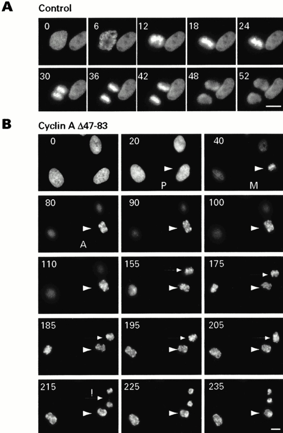
Stable cyclin A arrests cells in anaphase. (A) Time-lapse analysis of an H2B-GFP–expressing HTB96 cell going through mitosis. (B) H2B-GFP–expressing cells were injected in G2 phase with DNA encoding cyclin AΔ44-83 and TRITC-dextran as an injection marker. The arrowhead marks a cell that was arrested in anaphase for >2 h. The arrow marks a cell that underwent an abortive cytokinesis at 215 min but failed to decondense its chromosomes. Bars, 20 μm.
Results
Cyclin A Is Stable in Interphase and Becomes Unstable in Mitosis
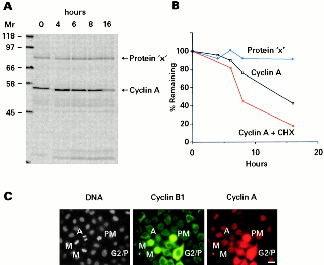
Cyclin A expression starts at the beginning of S phase, and its levels rise steadily until late G2 phase, but it is absent in late mitosis and G1 phase (Pines and Hunter 1991; Erlandsson et al. 2000). To understand the regulation of cyclin A protein levels, we measured its half-life in proliferating cells. An asynchronously growing population of HeLa cells was labeled with 35S-amino acids and transferred to nonradioactive growth medium after 4 h. Cells were harvested at the indicated times, and immunoprecipitates of cyclin A were analyzed by SDS-PAGE (Fig. 1 A). Fig. 1 B shows that the half-life of cyclin A was ≥12 h and that the decay showed linear rather than exponential kinetics, consistent with a model whereby cyclin A is long lived in S and G2 phase but rapidly degraded when cells enter mitosis. We also measured the levels of cyclin A after the addition of cycloheximide (CHX) to an asynchronous cell culture and immunoblotting (Fig. 1 B). In cells arrested in S phase with hydroxyurea, the levels of cyclin A stayed constant for ≥4 h after addition of CHX (data not shown), but after this the cells started to die. We conclude that cyclin A is a relatively stable protein during S and G2 phase of proliferating cells.
Figure 1.
Cyclin A is a relatively stable protein in the S and G2 phase of the cell cycle. (A) HeLa cells were labeled with 35S-amino acids and transferred to nonradioactive medium after 4 h. At the indicated times, cyclin A was immunoprecipitated and analyzed by SDS-PAGE and autoradiography. X denotes a contaminant band. (B) Quantified data from this experiment and from cells treated with CHX and immunoblotted for cyclin A. (C) Methanol-fixed HeLa cells stained for cyclin A (JG39), B1 (V152), and DNA (Hoechst 33342). G2/P, G2 or prophase; PM, prometaphase; M, metaphase; A, anaphase. Bar, 20 μm.
However, during mitosis cyclin A disappears rapidly from cells (Pines and Hunter 1991). We confirmed that prometaphase cells stained brightly for cyclin A, whereas metaphase cells (judged by Hoechst 33342 staining) with strong cyclin B1 staining gave variable cyclin A signals, confirming that cyclin A is degraded before cyclin B1 (Fig. 1 C). These results suggest that cyclin A is degraded earlier than cyclin B1, before the onset of anaphase, and that its proteolysis occurs while cyclin B1 is still stable.
Cyclin A-GFP Destruction Begins in Prometaphase and Is Completed at Metaphase
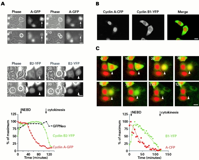
To follow the kinetics of cyclin A destruction in real time, cyclin A was tagged with GFP at its COOH terminus, and the expression plasmid was injected into G2-synchronized HeLa cells (see Materials and Methods). Fig. 2 shows that Cyclin A-GFP concentrated in the nucleus (and formed an active protein kinase with Cdk2 in vitro [data not shown]) and is thus likely to behave similarly to wild-type cyclin A. The injected cells started to accumulate nuclear fluorescence ∼2 h after microinjection of the expression plasmid and entered mitosis at about the same time as neighboring uninjected cells. The fluorescence signal of cyclin A-GFP started to decline uniformly throughout the cell after nuclear envelope breakdown (NEBD). Cyclin A-GFP signals were very much reduced in metaphase cells (Fig. 2 A, top).
Figure 2.
Degradation of cyclin-GFP in living cells. (A) Expression vectors for cyclin A-GFP (top) and cyclin B2-YFP (bottom) were injected into G2 phase HeLa cells and followed by time-lapse videomicroscopy. The graph shows the quantified data for cyclin A-GFP, cyclin B2-YFP, and GFPNeo-expressing cells from prophase until cytokinesis. (B) Dual color GFP tagging of cyclins. Cyclin A-CFP was coexpressed with cyclin B1-YFP and analyzed by sequential image acquisition. (C) Cyclin A (red) and cyclin B1 (green) destruction. Cyclin A-CFP and cyclin B1-YFP were injected into G2 phase HeLa cells and analyzed by time-lapse fluorescence microscopy. Images were pseudocolored and merged. The time course and quantified data of one of the cells going through mitosis (arrow) are shown. Bars, 20 μm.
For comparison, a similar study was made using mouse cyclin B2-YFP. Cyclin B2-YFP was located in the cytoplasm in interphase cells and translocated into the nucleus ∼10 min before NEBD. Cyclin B2-YFP levels continued to rise slightly in prometaphase localized to the mitotic spindle and were stable until metaphase and declined rapidly in anaphase (Fig. 2 A, bottom).
The expression of cyclin A-GFP often delayed the onset of anaphase (we return to this point below; see Fig. 9) which, in the cell shown in Fig. 2 A, occurred ≥20 min later than in the cell expressing cyclin B2-YFP. A comparison of cyclin A and B degradation rates was therefore only possible between cells in which the duration of mitosis was not grossly altered. Fig. 2 A compares the kinetics of loss of GFP-tagged cyclins A and B in two different cells that had a similar length of mitosis from NEBD to cytokinesis (the graph shows fluorescence values for one out of four quantified cells). Cyclin A destruction preceded that of cyclin B by 30–40 min. To enable simultaneous measurements to be made in the same cell, cyclin A was fused to CFP and mouse cyclin B1 to YFP, and the plasmids were coinjected into G2-synchronized HeLa cells (see Materials and Methods). Fig. 2 B shows that the CFP and YFP signals were well separated, and Fig. 2 C shows that cyclin A started to be degraded shortly after NEBD and reached low levels in metaphase when cyclin B1 started to become unstable. The graph in Fig. 2 C shows the YFP and CFP levels of one out of three quantified cells and indicates that cyclin A degradation precedes that of cyclin B1 by ∼30 min. These observations agree with those of den Elzen and Pines in this issue (2001).
Figure 9.
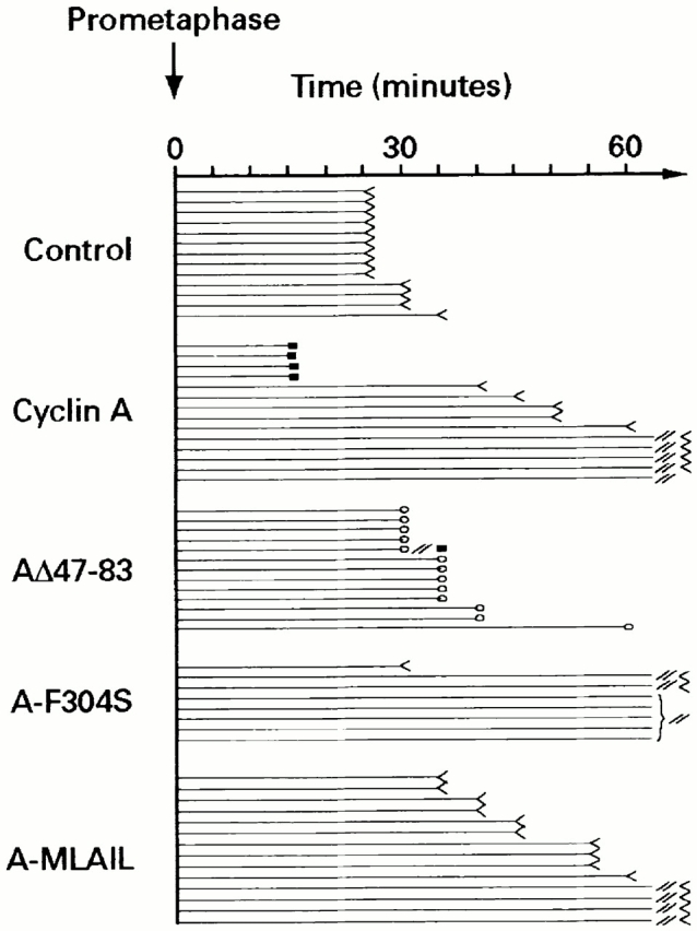
Timing of anaphase onset. Cyclin A degradation is required for the onset of anaphase. H2B-GFP–expressing cells were injected with expression plasmids for cyclin A, AΔ47-83, A-F304S, or A-MLAIL together with TRITC-labeled dextran and monitored by time-lapse videomicroscopy. The horizontal lines represent the time that individual cells spent in mitosis from prometaphase until the onset of anaphase. Each line represents a single cell: <, onset of anaphase; 0, anaphase arrest; //<, delay of ≤90 min until anaphase onset; //, delay of ≤200 min or the end of the recording time. Filled squares denote cell death.
Cyclin A Destruction Is Dependent on the APC/C but Is Insensitive to the Spindle Assembly Checkpoint
To test if the APC/C was required for the degradation of cyclin A, we injected G2-synchronized HeLa cells with affinity-purified anti-Cdc27 antibodies which arrested a high fraction of them at metaphase (Fig. 3 A). Of 68 cells injected with anti-Cdc27 antibodies, 55 arrested in mitosis. None of the control IgG-injected cells had arrested when analyzed 14 h after injection. To determine the effect of anti-Cdc27 antibodies on cyclin levels, cells were fixed with methanol and stained for cyclin A and B1. Cyclin A was present at high levels in the majority of these cells (69 out of 88 injected and arrested cells), suggesting that it was a target of the APC/C (Fig. 3 B). Next, antibodies against human Cdc20 or Cdh1 were injected into the nucleus of synchronized HeLa cells and the cells were stained for cyclin A and B1 14 h later. Injection of anti-Cdc20 antibodies also caused cells to accumulate in metaphase but with lower efficiency than anti-Cdc27 antibodies, probably indicating that the arrest was transient. Nevertheless, both cyclin A (19 out of 27 injected cells) and B1 (16 out of 16 injected cells) were readily detectable (Fig. 3 C) in the anti-Cdc20 metaphase-arrested cells. Anti-Cdh1 antiserum had no detectable effect on mitosis or cyclin degradation (data not shown). When cyclin A-GFP expression plasmid (25 ng/μl) was coinjected with either anti-Cdc27 or anti-Cdc20 antiserum, the GFP signal remained high in metaphase-arrested cells (data not shown).
Figure 3.
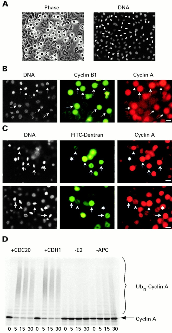
Cyclin A is an APC/C substrate. (A and B) HeLa cells were injected with antibodies against Cdc27 and stained with Hoechst 33342 after 14 h. A compares phase–contrast and Hoechst fluorescence images. Panel B shows staining for cyclin A, B1, and Hoechst 33342. Injected cells (arrow) were arrested at metaphase with high levels of cyclin A. (C) Anti-Cdc20 antibodies were injected with FITC-labeled dextran. Cells were analyzed for cyclin A 14 h later. The asterisk indicates a noninjected metaphase cell that lacks cyclin A. The arrowheads indicate cells that were injected with lower amounts of antibodies and contain low amounts of cyclin A; the full arrows indicate arrested cells with high levels of cyclin A. (D) Ubiquitylation assay. 35S-labeled cyclin A was added to an ubiquitylation reaction containing immunopurified APC/C activated by Cdc20 or Cdh1 as indicated. Samples were analyzed at the indicated times by SDS-PAGE and autoradiography. Bars, 20 μm.
These results suggested that cyclin A is a target of the Cdc20-associated APC/C in human cells. To test more directly whether the APC/C is able to recognize cyclin A as a substrate, we set up an in vitro ubiquitylation reaction for cyclin A in which immunopurified APC/C from activated Xenopus laevis egg extracts was added to radiolabeled cyclin A (see Materials and Methods). Addition of recombinant Cdc20 or Cdh1 promoted rapid and efficient conversion of cyclin A into high molecular weight ubiquitin conjugates (Fig. 3 D), confirming that cyclin A is a target of the Cdc20-activated APC/C.
The activity of the APC/C is controlled by the spindle assembly checkpoint and disruption of the mitotic spindle by microtubule poisons leads to Mad2-dependent inactivation of the APC/C's ability to ubiquitylate securin and cyclin B. Fig. 4 A shows that in cells exposed to nocodazole or taxol cyclin B1 levels were maintained, but the levels of cyclin A fell. The low levels of cyclin A in taxol- or nocodazole-treated cells were due to proteolytic degradation, since addition of the proteasome inhibitor MG132 led to accumulation of cyclin A in the arrested cells (25 of 27 cells were positive, whereas only 2 of 34 cells were positive before addition of MG132) (Fig. 4 A bottom; data not shown). Even when cyclin A-GFP was overexpressed from a strong heterologous promoter, it did not accumulate in nocodazole-arrested cells, whereas cyclin B1-YFP did (data not shown).
Figure 4.
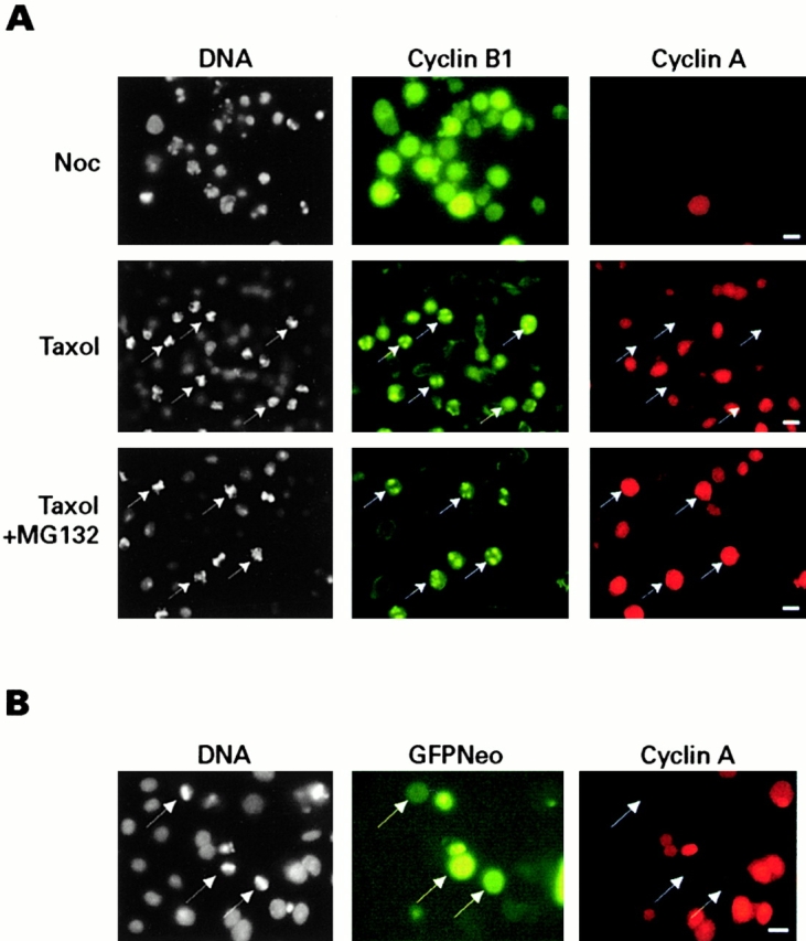
Cyclin A is unstable in nocodazole- and taxol-treated cells. (A) HeLa cells exposed to inhibitors. G2 phase HeLa cells were treated with nocodazole (Noc) for 14 h or taxol for 4 h until cells accumulated in mitosis. MG132 (100 μM) was added to the taxol-treated cells and incubated for an additional 2 h, after which cells were stained for DNA and cyclin B1 and A. (B) HeLa cells transfected with Mad1 and 2. G2 HeLa cells were injected with an expression vector for human Mad1 and a bicistronic expression vector that expressed Mad2 and GFPNeo (pTSIGN-Mad2) and were stained for DNA or cyclin A 14 h later. Metaphase-arrested GFP positive cells are indicated by arrows. Bars, 20 μm.
We next asked whether overexpression of spindle assembly checkpoint effector proteins, which are thought to act by inhibiting Cdc20-APC/C, would stabilize cyclin A. Expression plasmids for hsMad1 and hsMad2 were microinjected into synchronized HeLa cells during G2 phase and filmed by time-lapse videomicroscopy. A metaphase arrest of ≥6 h was observed, but cells were apt to die after this. Cyclin B1 was detected in all of the arrested cells (data not shown), whereas significant levels of cyclin A were only seen in 3 of 21 arrested cells, and most of the metaphase-arrested cells had very low levels (Fig. 4 B). This indicates that the mitotic block imposed by overexpression of Mad1 and Mad2 or the activation of the spindle assembly checkpoint with microtubule poisons inhibits the degradation of cyclin B1 but does not delay the proteolysis of cyclin A.
The Delay in the Degradation of Cyclin B1 Relative to Cyclin A Depends on the Mitotic Checkpoint
In animal cells, the spindle assembly checkpoint is activated during prometaphase (Chen et al. 1996) and remains active until all the chromosomes have established a bipolar orientation on the mitotic spindle (Rieder et al. 1994, Rieder et al. 1997). The observation that cyclin A is destabilized in prometaphase and is a target of the APC/C suggests that the APC/C is active in prometaphase. To test if the spindle assembly checkpoint is responsible for delaying cyclin B1 degradation, we expressed a dominant negative version of the mouse checkpoint kinase Bub1 together with cyclin B1-YFP. Fig. 5 shows that dnBub1 caused precocious disappearance of cyclin B1-YFP which was now degraded shortly after NEBD. The difference in cyclin B1 degradation relative to that of cyclin A thus seems to depend on the spindle assembly checkpoint which selectively delays the degradation of cyclin B, and presumably that of securin, until chromosomes are correctly aligned on the mitotic spindle.
Figure 5.
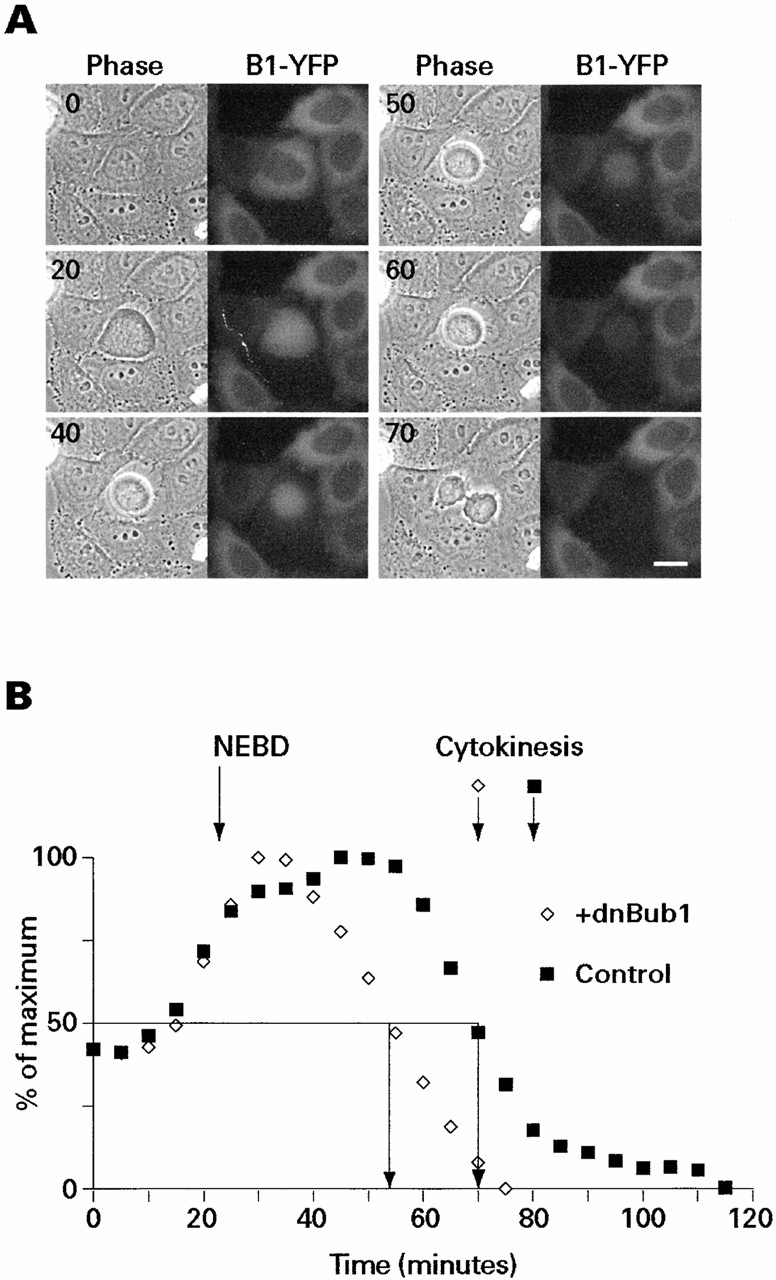
Dominant negative Bub1 accelerates the degradation of cyclin B1-YFP. Expression plasmids for the NH2-terminal domain of mouse Bub1 and cyclin B1-YFP were coinjected into G2 phase HeLa cells and followed through mitosis. (A) A sequence of images of a cell going through mitosis. (B) Quantitation of B1-YFP signal during mitosis with one out of four cells monitored. Filled squares indicate cyclin B1-YFP fluorescence in control cells; open diamonds indicate the presence of dnBub1. Bar, 20 μm.
The D-Box of Cyclin A
To understand how the APC/C is able to distinguish between cyclin A and B, we analyzed the D-box of cyclin A. Like the B-type cyclins, cyclin A possesses a sequence in its NH2 terminus that was identified as a D-box because mutation or deletion of the conserved RXXL sequence in clam cyclin A or Xenopus cyclin A1 strongly stabilized the protein (Luca et al. 1991; Lorca et al. 1992; Stewart et al. 1994). We used Xenopus egg extracts to analyze the D-box of human cyclin A2. These extracts are arrested at metaphase due to the activity of cytostatic factor (CSF) and can be released into anaphase by addition of CaCl2 (see Murray 1991). Surprisingly, human cyclin A was unstable in the majority of egg extracts, although addition of CaCl2 accelerated its proteolysis (Fig. 6 B). This suggests that cytostatic factor–arrested extracts resemble nocodazole-arrested cells, in that APC/C activity is present at a level sufficient to degrade cyclin A, although cyclin B is stable. Point mutations in the conventional D-box motif (Adb3, R47A/L50A/N57A) or even deletion of residues 45–58, which comprise the entire canonical D-box (Fig. 6 B), failed to stabilize human cyclin A2 in the egg extracts. Deletion of the NH2-terminal 60 residues did not stabilize human cyclin A, but deletions of the first 80 residues gave a stable protein (data not shown). In contrast, a double point mutation in the D-box of mouse cyclin B1 (RTALG to GTAVG, labeled B1dm in Fig. 6) completely stabilized it under the same conditions (Fig. 6 B).
Figure 6.
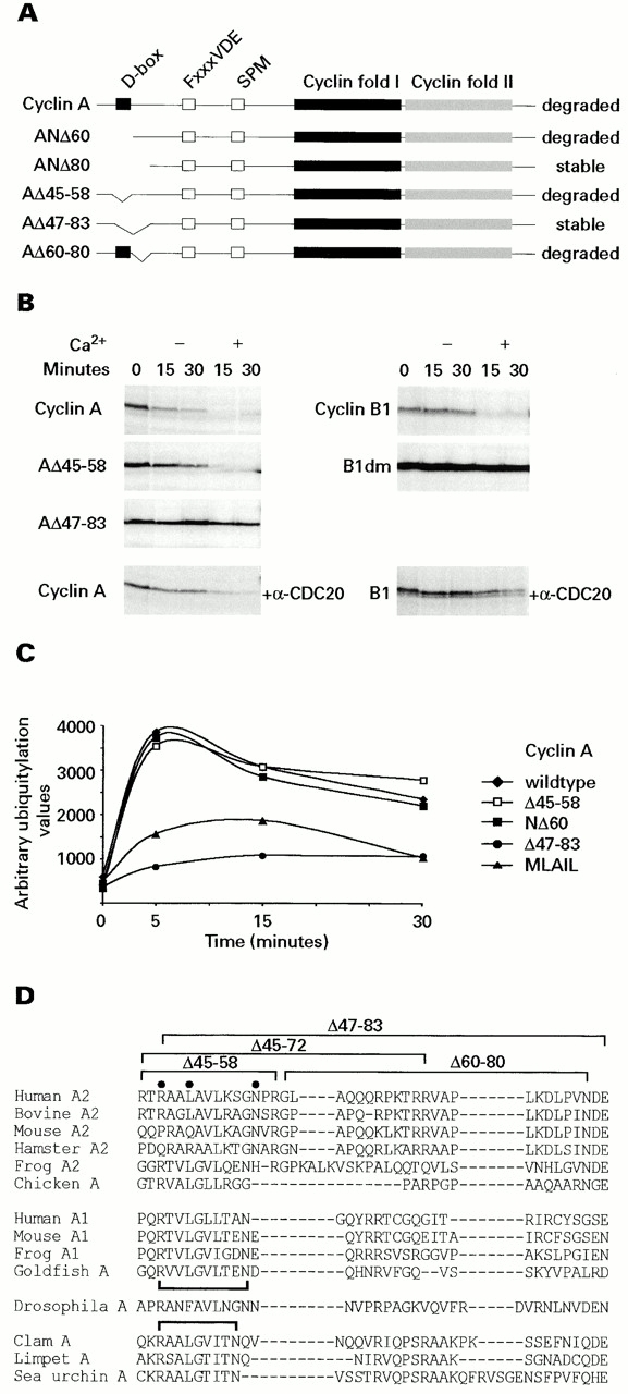
Cyclin A2 has a novel D-box. (A) Cyclin A deletion mutants. Schematic representation of cyclin A and mutants analyzed in this study. (B) Destruction assays. In vitro–translated and –labeled cyclins were added to frog egg extracts and incubated at 23°C in the presence or absence of 0.4 mM CaCl2. Samples were analyzed by SDS-PAGE and autoradiography. (C) Ubiquitylation assays. Cdc20-dependent ubiquitylation in vitro. High molecular weight forms of cyclin A were quantified by fluorography of the dried SDS gels. (D) D-box alignments. Alignment of the NH2-terminal regions of A-type cyclins surrounding the D-box, whose conserved residues (R47, L50, and N57) are marked by filled circles. The deletion mutants referred to in the text are indicated.
Deletion of residues 47–83 or 45–72 stabilized human cyclin A (Fig. 6 B), whereas Δ60-80 was still unstable (data not shown). Thus, the cis element of cyclin A required for proteolysis starts with the canonical D-box and includes an additional motif present between residues 60 and 72. As we found previously for Xenopus cyclin A1, degradation of human cyclin A2 in vitro required binding to a Cdk, since mutants defective in binding a Cdk partner, for example, a cyclin box mutant (R211L) or a small COOH-terminal deletion (ΔC28), were stable in the frog extract assay (data not shown).
The degradation of both cyclin A and B1 was delayed but not abolished when affinity-purified anti-Cdc20 antibodies were added to the extract 30 min before adding the substrates, indicating that both are dependent on Cdc20-associated APC/C (Fig. 6 B). We also tested the cyclin A mutants in the in vitro ubiquitylation assay (see Fig. 3 D). Fig. 6 C shows that wild-type cyclin A, mutant Δ45-58, and the NH2-terminal deletion mutant ANΔ60 were efficiently (>50%) converted into high molecular weight ubiquitylated forms after 5 min. In contrast, the R211L (MLAIL) and AΔ47-83 deletion mutant, which were stable in frog anaphase extracts, were very poorly ubiquitylated.
Fig. 6 D shows an alignment of A-type cyclins from a variety of species and reveals that the extended D-box motif in human cyclin A is conserved among the somatic A-type cyclins of human, cattle, mouse, and hamster but not in Xenopus cyclin A2 or chicken cyclin A (Fig. 6 D). It is also absent in all embryonic or A1-type cyclins, suggesting that the degradation of cyclins A1 and A2 might be regulated differently.
The stability of some of these mutants was checked in living cells using GFP fusion proteins. The mutant of cyclin A lacking just the classical D-box (AΔ45-58–GFP), like wild-type cyclin A-GFP was rapidly degraded in mitosis, and did not accumulate in nocodazole-arrested cells (data not shown), indicating that deletion of the classical D-box does not stabilize cyclin A in vivo_._ In contrast, cyclin AΔ47-83–GFP was stable in mitosis (see below) and accumulated in nocodazole-arrested cells (data not shown).
Nondegradable Cyclin A Arrests Cells in Anaphase
We next tested the effects of nondegradable cyclin A on cell cycle progression. HeLa cells were transfected with an expression vector for various cyclin constructs together with a GFPNeo marker. Neither wild-type cyclin A nor an inactive (Cdk nonbinding mutant R211L [Stewart et al. 1994]) arrested cells in mitosis when GFP-positive cells were analyzed 48–72 h after transfection (Fig. 7 A). By contrast, the nondegradable forms of cyclin A, ANΔ170 or AΔ47-83, arrested cells in mitosis, similarly to the cyclin B1 D-box mutant (Fig. 7 A). We generated cell lines with tetracycline-regulated expression of cyclin ANΔ170. After induction for 24 h, cyclin ANΔ170 accumulated to three to five times the levels of endogenous cyclin A (Fig. 7 B), and cells rounded up and arrested in mitosis (Fig. 7 B, bottom).
Figure 7.
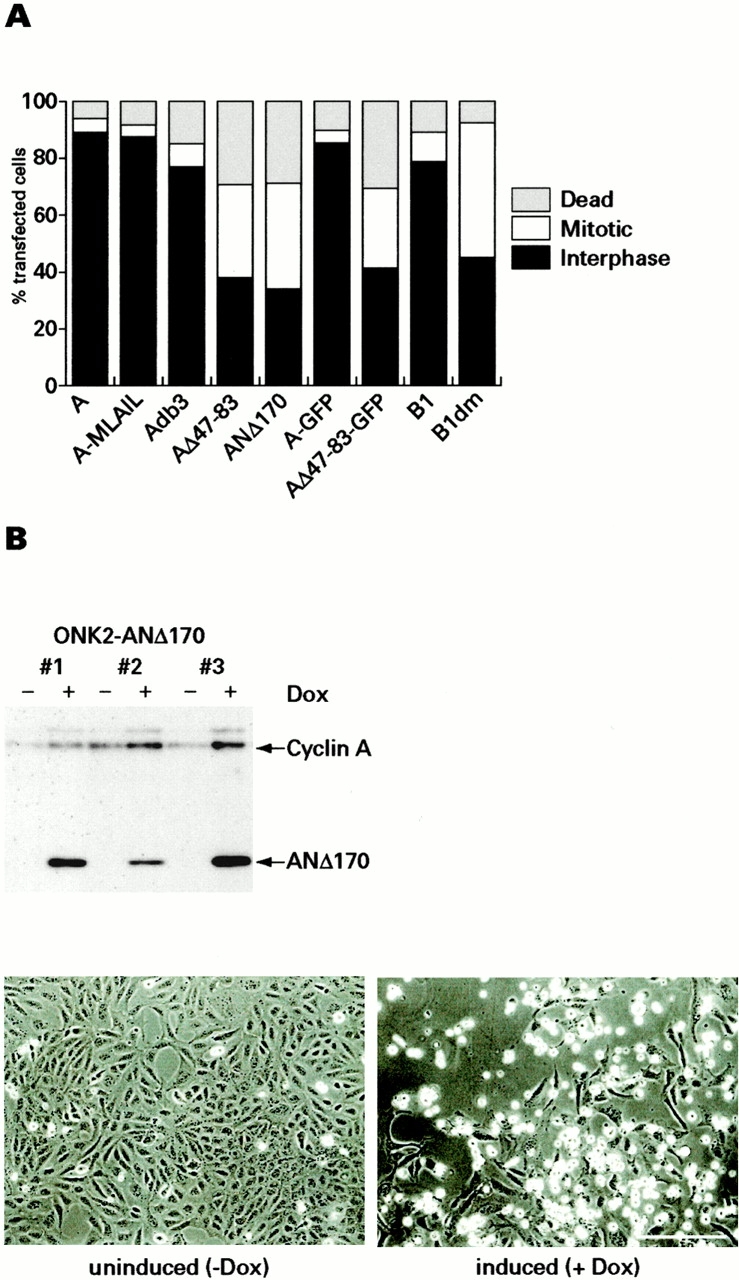
Stable cyclin A arrests cells in mitosis. (A) HeLa cells were transfected with a mixture of GFPNeo and cyclin constructs as indicated and cultured for 48 h. Cyclin-GFP plasmids were transfected without a marker. Cells were fixed, stained for DNA, and counted as being in interphase (black bars), mitosis (white bars), or dead (gray bars) as judged by Hoechst 33342 staining. (B) Tetracycline-inducible cyclin ANΔ170 expression. Cyclin A-NΔ170–inducible clones derived from HTB96 cells were induced with 200 ng/ml doxycycline (Dox) for 24 h and analyzed by immunoblotting for cyclin A. Phase–contrast images of clone 3 cells grown in the absence (left) or presence (right) of 200 ng/ml doxycycline for 24 h. Cells arrested in mitosis appear round and refractile. Bar, 200 μm.
To follow chromosome behavior in real time in cells expressing indestructible cyclin A, we generated a cell line that expressed a histone H2B-GFP fusion protein (Kanda et al. 1998). Fig. 8 A shows that these cells normally take 30 min from the first sign of chromosome condensation to the onset of anaphase. Synchronized HTB96H2B-GFP cells were microinjected with expression plasmids for cyclin AΔ47-83 and rhodamine-labeled dextran as an injection marker. Fig. 8 B shows cells expressing AΔ47-83 (arrowhead) which showed a slight delay in the onset of anaphase and final arrest in anaphase. After initiation of anaphase (Fig. 8 B, arrowhead A) cells apparently separated their sister chromatids, but cytokinesis did not occur. Instead, arrested cells exhibited rapid movements of chromosomes that appeared to be clustered in two main groups, indicating that they were still attached to their spindle poles. This is best seen in the videos available at http://www.jcb.org/cgi/content/full/153/1/137/DC1.
Fig. 9 summarizes the kinetics of mitosis in several HTB96H2B-GFP cells expressing wild-type cyclin A or various cyclin A mutants. Surprisingly, the onset of anaphase in cells expressing wild-type cyclin A was often delayed for up to several hours before mitosis was completed. A similar transient inhibitory effect on the onset of anaphase was also observed in cells expressing cyclin A-MLAIL and A-F304S, a mutant that activates Cdk2 very poorly (data not shown). This indicates that kinase activity associated with cyclin A is not required for this delay, whereas its D-box is necessary since cells expressing cyclin AΔ47-83 entered anaphase with almost the same kinetics as control cells (Fig. 9).
Discussion
In this paper, we analyze the timing and mechanism of cyclin A proteolysis in cultured somatic cells. We show that cyclin A degradation starts as cyclin B enters the nucleus, is insensitive to the spindle assembly checkpoint, and normally precedes the proteolysis of cyclin B by ∼30 min. The metaphase to anaphase transition is not initiated until cyclin A levels are extremely low and overexpression of wild-type cyclin A blocked cells in metaphase. Surprisingly, however, this arrest did not depend on the kinase activity normally associated with cyclin A and expression of active forms of cyclin A containing deletions of the D-box blocked cells in anaphase.
Despite the striking differences between the timing of cyclin A and B proteolysis, they both appear to be degraded as a result of polyubiquitylation by the Cdc20-dependent form of the APC/C. The ubiquitylation of cyclin A in cell-free assays was rapid and efficient with immunopurified APC/C and Cdc20 (or Cdh1), and there is presently no good evidence for alternative pathways for cyclin A proteolysis. Thus, even though a fraction of cyclin A is associated with the Skp1/Cullin/F-box protein complex (SCF) ubiquitin ligase (Zhang et al. 1995; Bai et al. 1996), cyclin A is a relatively stable protein with a half-life of >12 h during S and G2 phase. Cells of Skp2-deficient mice accumulate cyclin E but not cyclin A (Nakayama et al. 2000). We are tempted to speculate that the cyclin A/Cdk2 in the SCF complex is the protein kinase that tags substrates (for example, Cdc6) for degradation (Petersen et al. 1999; Willems et al. 1999; Coverley et al. 2000), although it is also possible that cyclin A activity is regulated by the SCF (Yam et al. 1999).
In Drosophila, cyclin A degradation requires the fzy gene (Dawson et al. 1995; Sigrist et al. 1995) and in both Saccharomyces cerevisiae and Xenopus (Fang et al. 1998b), Cdc20 (fzy) is required for the activation of the APC/C, and it was recently reported that Cdc20 bound directly to the NH2 terminus of cyclin A, although the D-box was not required for this interaction (Ohtoshi et al. 2000). Substrates of the APC/C are typically characterized by their possession of a nine-residue motif known as the D-box (RxxLxxxxN) (Glotzer et al. 1991). The NH2 terminus of cyclin A contains a D-box–like sequence, R47 AALAVLKSGN57. Unlike the embryonic form of cyclin A1 in Xenopus (Lorca et al. 1992; Stewart et al. 1994), human cyclin A2 was not stabilized even by deletion of the entire “classical” D-box (Δ45-58). Further mutational analysis revealed that an adjacent domain 10–20 residues downstream was required for the degradation of cyclin A. Thus, the D-box of human cyclin A appears to be significantly larger than those of frog cyclin A1 or the B-type cyclins. This enlarged D-box seems to require a correctly folded cyclin box for efficient recognition by the APC/C, which may or may not reflect a need for binding to a Cdk partner.
Cyclin A Degradation Is Independent of the Spindle Assembly Checkpoint
Even when cyclin A was expressed from a strong heterologous promoter, it could not be detected in nocodazole-arrested cells. Thus, cyclin A is degraded when the spindle assembly checkpoint is strongly activated. Even overexpression of Mad2, which inhibited the metaphase–anaphase transition and stabilized cyclin B1, did not prevent cyclin A proteolysis. Conversely, dominant negative Bub1, which inhibits the mitotic checkpoint, advanced the degradation of cyclin B1-YFP so that it showed kinetics of dissappearance similar to those of cyclin A under normal conditions (although for reasons of technical difficulty, we have not examined the kinetics of cyclin A proteolysis in cells expressing dnBub1). We suspect that, even under these conditions, cyclin A would still disappear earlier than cyclin B, as it does in Xenopus egg extracts in which the mitotic checkpoint is not operative (Minshull et al. 1990, Minshull et al. 1994). We conclude from these experiments that the APC/C is activated as cells enter mitosis, and the mitotic checkpoint selectively delays the degradation of cyclin B1 (and by implication, that of securin) until all of the chromosomes are correctly aligned on a well-formed mitotic spindle.
Currently, the simplest way to explain the differences between the proteolysis of cyclin A and other APC/C substrates is that cyclin A is simply a better substrate for the APC/C than securin and cyclin B, so that cyclin A proteolysis can occur even when the activity of the APC/C is held in check by the spindle assembly checkpoint. We imagine that a long polyubiquitin chain is required for efficient delivery to the proteasome and that long chains result from long dwell times at the APC/C. Thus, substrates with a lower dissociation constant are preferentially degraded and may even be degraded under conditions when the activity of the APC/C is reduced by the operation of the checkpoint. This view is consistent with the long delays in metaphase produced by overexpression of wild-type but not NH2-terminally truncated cyclin A. Apparently, securin and cyclin B are unable to compete with cyclin A for the APC/C. Testing this hypothesis will require further work, but it is consistent with the differences in the D-boxes. Larger D-boxes potentially make more contacts, and the additional requirement for functional cyclin A–Cdk complexes as substrates suggests the existence of additional contacts between the APC/C and cyclin A that would promote tighter binding and longer dwell times, allowing more highly processive addition of ubiquitin moieties.
Cyclin A Degradation Is Required for the Onset of Anaphase and Exit from Mitosis
The ability of overexpressed cyclin A to delay the onset of anaphase was not dependent on functional kinase activity, since cyclin A mutants unable to bind to or activate Cdks also efficiently blocked cells at metaphase. In addition, nondegradable cyclin A lacking its D-box (Δ47-83) arrested cells in anaphase without grossly affecting sister chromatid separation. This differs from what has been reported for Drosophila embryos, where expression of stable cyclin A caused a significant delay at metaphase (Sigrist et al. 1995). As the D-box of Drosophila cyclin A is not defined and coexpression of stable cyclin B caused Drosophila cells to arrest in anaphase, it is possible that the NH2-terminal deletion mutant of cyclin A used in this study was still somewhat unstable, causing a similar phenotype to the one we observe using wild-type cyclin A.
In summary, wild-type cyclin A seems to compete for the degradation of substrates required for the onset of anaphase, whereas stable cyclin A arrests cells in anaphase. We speculate that the cyclin A–dependent delay in the onset of metaphase is due to direct competition with other substrates of the APC/C such as securin. It is also possible that overexpression of cyclin A interferes with a yet unknown proteolytic pathway that is required to establish a stable chromosome alignment at the metaphase plate, as suggested by den Elzen and Pines 2001 in this issue.
Further experiments are required to define how cyclin A interferes with the onset of anaphase and how the differences between cyclin A and B destruction and the apparent substrate specificity of the mitotic checkpoint revealed by this analysis can be explained.
Supplemental Material
[Supplemental Material Index]
Acknowledgments
We thank M. Brandeis, C. Norbury, J. Pines, S. Taylor, G. Wahl, and T. Jeang for plasmids and T. Lorca for anti-Xenopus Cdc20 antibodies. R. Pepperkok, D. Zicha, C. Gray, and D. Aubyn gave wonderful support for live cell imaging, and members of the Hunt laboratory gave excellent discussions and help with the paper. We are grateful to Jonathan Pines and Nicole den Elzen for keeping in touch throughout the work.
This work was supported by an Erwin Schrödinger fellowship of the Austrian Science Promotion Fund to S. Geley, a Training and Mobility of Researchers grant FMRX-CT98-0179 to T. Hunt, and by Boehringer Ingelheim and grants from the Austrian Industrial Research Promotion Fund and the Austrian Science Promotion Fund to J.-M. Peters.
Footnotes
The online version of this article contains supplemental material.
Abbreviations used in this paper: APC/C, anaphase-promoting complex/cyclosome; CFP, cyan fluorescent protein; CHX, cycloheximide; D-box, destruction box; GFP, green fluorescent protein; NEBD, nuclear envelope breakdown; SCF, Skp1/Cullin/F-box protein complex; YFP, yellow fluorescent protein.
References
- Ausubel F.M., Brent R., Kingston R.E., Moore D.D., Seidman J.G., Smith J.A., Struhl K. Current Protocols in Molecular Biology. John Wiley & Sons, Inc; New York: 1999. [Google Scholar]
- Bai C., Sen P., Hofmann K., Ma L., Goebl M., Harper J.W., Elledge S.J. SKP1 connects cell cycle regulators to the ubiquitin proteolysis machinery through a novel motif, the F-box. Cell. 1996;86:263–274. doi: 10.1016/s0092-8674(00)80098-7. [DOI] [PubMed] [Google Scholar]
- Cardoso M.C., Leonhardt H., Nadal-Ginard B. Reversal of terminal differentiation and control of DNA replicationcyclin A and Cdk2 specifically localize at subnuclear sites of DNA replication. Cell. 1993;74:979–992. doi: 10.1016/0092-8674(93)90721-2. [DOI] [PubMed] [Google Scholar]
- Chen R.H., Waters J.C., Salmon E.D., Murray A.W. Association of spindle assembly checkpoint component XMAD2 with unattached kinetochores. Science. 1996;274:242–246. doi: 10.1126/science.274.5285.242. [DOI] [PubMed] [Google Scholar]
- Cohen-Fix O., Peters J.M., Kirschner M.W., Koshland D. Anaphase initiation in Saccharomyces cerevisiae is controlled by the APC-dependent degradation of the anaphase inhibitor Pds1p. Genes Dev. 1996;10:3081–3093. doi: 10.1101/gad.10.24.3081. [DOI] [PubMed] [Google Scholar]
- Coverley D., Pelizon C., Trewick S., Laskey R.A. Chromatin-bound Cdc6 persists in S and G2 phases in human cells, while soluble Cdc6 is destroyed in a cyclin A-cdk2 dependent process. J. Cell Sci. 2000;113:1929–1938. doi: 10.1242/jcs.113.11.1929. [DOI] [PubMed] [Google Scholar]
- Dawson I.A., Roth S., Artavanis-Tsakonas S. The Drosophila cell cycle gene fizzy is required for normal degradation of cyclins A and B during mitosis and has homology to the CDC20 gene of Saccharomyces cerevisiae . J. Cell Biol. 1995;129:725–737. doi: 10.1083/jcb.129.3.725. [DOI] [PMC free article] [PubMed] [Google Scholar]
- den Elzen N., Pines J. The destruction of cyclin A is required for progression through mitosis. J. Cell Biol. 2001;153:121–135. [Google Scholar]
- Deuschle U., Meyer W.K., Thiesen H.J. Tetracycline-reversible silencing of eukaryotic promoters. Mol. Cell. Biol. 1995;15:1907–1914. doi: 10.1128/mcb.15.4.1907. [DOI] [PMC free article] [PubMed] [Google Scholar]
- Erlandsson F., Linnman C., Ekholm S., Bengtsson E., Zetterberg A. A detailed analysis of cyclin A accumulation at the G(1)/S border in normal and transformed cells. Exp. Cell Res. 2000;259:86–95. doi: 10.1006/excr.2000.4889. [DOI] [PubMed] [Google Scholar]
- Fang G., Yu H., Kirschner M.W. The checkpoint protein MAD2 and the mitotic regulator CDC20 form a ternary complex with the anaphase-promoting complex to control anaphase initiation Genes Dev. 12 1998. 1871 1883a [DOI] [PMC free article] [PubMed] [Google Scholar]
- Fang G., Yu H., Kirschner M.W. Direct binding of CDC20 protein family members activates the anaphase-promoting complex in mitosis and G1 Mol. Cell 2 1998. 163 171b [DOI] [PubMed] [Google Scholar]
- Funabiki H., Yamano H., Kumada K., Nagao K., Hunt T., Yanagida M. Cut2 proteolysis required for sister-chromatid seperation in fission yeast. Nature. 1996;381:438–441. doi: 10.1038/381438a0. [DOI] [PubMed] [Google Scholar]
- Furuno N., den Elzen N., Pines J. Human cyclin A is required for mitosis until mid prophase. J. Cell Biol. 1999;147:295–306. doi: 10.1083/jcb.147.2.295. [DOI] [PMC free article] [PubMed] [Google Scholar]
- Gieffers C., Peters B.H., Kramer E.R., Dotti C.G., Peters J.M. Expression of the CDH1-associated form of the anaphase-promoting complex in postmitotic neurons. Proc. Natl. Acad. Sci. USA. 1999;96:11317–11322. doi: 10.1073/pnas.96.20.11317. [DOI] [PMC free article] [PubMed] [Google Scholar]
- Girard F., Strausfeld U., Fernandez A., Lamb N.J. Cyclin A is required for the onset of DNA replication in mammalian fibroblasts. Cell. 1991;67:1169–1179. doi: 10.1016/0092-8674(91)90293-8. [DOI] [PubMed] [Google Scholar]
- Glotzer M., Murray A.W., Kirschner M.W. Cyclin is degraded by the ubiquitin pathway. Nature. 1991;349:132–138. doi: 10.1038/349132a0. [DOI] [PubMed] [Google Scholar]
- Gossen M., Freundlieb S., Bender G., Muller G., Hillen W., Bujard H. Transcriptional activation by tetracyclines in mammalian cells. Science. 1995;268:1766–1769. doi: 10.1126/science.7792603. [DOI] [PubMed] [Google Scholar]
- Holloway S.L., Glotzer M., King R.W., Murray A.W. Anaphase is initiated by proteolysis rather than by the inactivation of maturation-promoting factor. Cell. 1993;73:1393–1402. doi: 10.1016/0092-8674(93)90364-v. [DOI] [PubMed] [Google Scholar]
- Howe J.A., Howell M., Hunt T., Newport J.W. Identification of a developmental timer regulating the stability of embryonic cyclin A and a new somatic A-type cyclin at gastrulation. Genes Dev. 1995;9:1164–1176. doi: 10.1101/gad.9.10.1164. [DOI] [PubMed] [Google Scholar]
- Hunt T., Luca F.C., Ruderman J.V. The requirements for protein synthesis and degradation, and the control of destruction of cyclins A and B in the meiotic and mitotic cell cycles of the clam embryo. J. Cell Biol. 1992;116:707–724. doi: 10.1083/jcb.116.3.707. [DOI] [PMC free article] [PubMed] [Google Scholar]
- Irniger S., Piatti S., Michaelis C., Nasmyth K. Genes involved in sister chromatid separation are needed for B-type cyclin proteolysis in budding yeast. Cell. 1995;81:269–278. doi: 10.1016/0092-8674(95)90337-2. [DOI] [PubMed] [Google Scholar]
- Jiang W., Wells N.J., Hunter T. Multistep regulation of DNA replication by Cdk phosphorylation of HsCdc6. Proc. Natl. Acad. Sci. USA. 1999;96:6193–6198. doi: 10.1073/pnas.96.11.6193. [DOI] [PMC free article] [PubMed] [Google Scholar]
- Jin D.Y., Spencer F., Jeang K.T. Human T cell leukemia virus type 1 oncoprotein Tax targets the human mitotic checkpoint protein MAD1. Cell. 1998;93:81–91. doi: 10.1016/s0092-8674(00)81148-4. [DOI] [PubMed] [Google Scholar]
- Kanda T., Sullivan K.F., Wahl G.M. Histone-GFP fusion protein enables sensitive analysis of chromosome dynamics in living mammalian cells. Curr. Biol. 1998;8:377–385. doi: 10.1016/s0960-9822(98)70156-3. [DOI] [PubMed] [Google Scholar]
- King R.W., Peters J.M., Tugendreich S., Rolfe M., Hieter P., Kirschner M.W. A 20S complex containing CDC27 and CDC16 catalyzes the mitosis-specific conjugation of ubiquitin to cyclin B. Cell. 1995;81:279–288. doi: 10.1016/0092-8674(95)90338-0. [DOI] [PubMed] [Google Scholar]
- King R.W., Glotzer M., Kirschner M.W. Mutagenic analysis of the destruction signal of mitotic cyclins and structural characterization of ubiquitinated intermediates. Mol. Biol. Cell. 1996;7:1343–1357. doi: 10.1091/mbc.7.9.1343. [DOI] [PMC free article] [PubMed] [Google Scholar]
- Klotzbücher A., Stewart E., Harrison D., Hunt T. The ‘destruction box’ of cyclin A allows B-type cyclins to be ubiquitinated, but not efficiently destroyed. EMBO (Eur. Mol. Biol. Organ.) J. 1996;15:3053–3064. [PMC free article] [PubMed] [Google Scholar]
- Knoblich J.A., Lehner C.F. Synergistic action of Drosophila cyclins A and B during the G2-M transition. EMBO (Eur. Mol. Biol. Organ.) J. 1993;12:65–74. doi: 10.1002/j.1460-2075.1993.tb05632.x. [DOI] [PMC free article] [PubMed] [Google Scholar]
- Kramer E.R., Gieffers C., Holzl G., Hengstschlager M., Peters J.M. Activation of the human anaphase-promoting complex by proteins of the CDC20/Fizzy family. Curr. Biol. 1998;8:1207–1210. doi: 10.1016/s0960-9822(07)00510-6. [DOI] [PubMed] [Google Scholar]
- Kramer E.R., Scheuringer N., Podtelejnikov A.V., Mann M., Peters J.M. Mitotic regulation of the APC activator proteins CDC20 and CDH1. Mol. Biol. Cell. 2000;11:1555–1569. doi: 10.1091/mbc.11.5.1555. [DOI] [PMC free article] [PubMed] [Google Scholar]
- Krek W., DeCaprio J.A. Cell synchronization. In: Vogt P.K., Verma I.M., editors. Oncogene Techniques. Vol. 254. Academic Press, Inc; San Diego: 1995. pp. 114–124. [DOI] [PubMed] [Google Scholar]
- Krude T., Jackman M., Pines J., Laskey R.A. Cyclin/Cdk-dependent initiation of DNA replication in a human cell-free system. Cell. 1997;88:109–119. doi: 10.1016/s0092-8674(00)81863-2. [DOI] [PubMed] [Google Scholar]
- Lehner C.F., O'Farrell P.H. Expression and function of Drosophila cyclin A during embryonic cell cycle progression. Cell. 1989;56:957–968. doi: 10.1016/0092-8674(89)90629-6. [DOI] [PMC free article] [PubMed] [Google Scholar]
- Li Y., Gorbea C., Mahaffey D., Rechsteiner M., Benezra R. MAD2 associates with the cyclosome/anaphase-promoting complex and inhibits its activity. Proc. Natl. Acad. Sci. USA. 1997;94:12431–12436. doi: 10.1073/pnas.94.23.12431. [DOI] [PMC free article] [PubMed] [Google Scholar]
- Liu D., Matzuk M.M., Sung W.K., Guo Q., Wang P., Wolgemuth D.J. Cyclin A1 is required for meiosis in the male mouse. Nat. Genet. 1998;20:377–380. doi: 10.1038/3855. [DOI] [PubMed] [Google Scholar]
- Lorca T., Devault A., Colas P., Van Loon A., Fesquet D., Lazaro J.B., Dorée M. Cyclin A-Cys41 does not undergo cell cycle-dependent degradation in Xenopus extracts. FEBS Lett. 1992;306:90–93. doi: 10.1016/0014-5793(92)80844-7. [DOI] [PubMed] [Google Scholar]
- Luca F.C., Shibuya E.K., Dohrmann C.E., Ruderman J.V. Both cyclin A delta 60 and B delta 97 are stable and arrest cells in M-phase, but only cyclin B delta 97 turns on cyclin destruction. EMBO (Eur. Mol. Biol. Organ.) J. 1991;10:4311–4320. doi: 10.1002/j.1460-2075.1991.tb05009.x. [DOI] [PMC free article] [PubMed] [Google Scholar]
- Minshull J., Golsteyn R., Hill C.S., Hunt T. The A- and B-type cyclin associated cdc2 kinases in Xenopus turn on and off at different times in the cell cycle. EMBO (Eur. Mol. Biol. Organ.) J. 1990;9:2865–2875. doi: 10.1002/j.1460-2075.1990.tb07476.x. [DOI] [PMC free article] [PubMed] [Google Scholar]
- Minshull J., Sun H., Tonks N.K., Murray A.W. A MAP kinase-dependent spindle assembly checkpoint in Xenopus egg extracts. Cell. 1994;79:475–486. doi: 10.1016/0092-8674(94)90256-9. [DOI] [PubMed] [Google Scholar]
- Murphy M., Stinnakre M.G., Senamaud-Beaufort C., Winston N.J., Sweeney C., Kubelka M., Carrington M., Brechot C., Sobczak-Thepot J. Delayed early embryonic lethality following disruption of the murine cyclin A2 gene. Nat. Genet. 1997;15:83–86. doi: 10.1038/ng0197-83. [DOI] [PubMed] [Google Scholar]
- Murray A.W. Cell cycle extracts. In: Kay B.K., Peng H.B., editors. Xenopus laevis: Practical Uses in Cell and Molecular Biology. Vol. 36. Academic Press, Inc; San Diego: 1991. pp. 573–597. [Google Scholar]
- Nakayama K., Nagahama H., Minamishima Y.A., Matsumoto M., Nakamichi I., Kitagawa K., Shirane M., Tsunematsu R., Tsukiyama T., Ishida N., Kitagawa M., Hatakeyama S. Targeted disruption of Skp2 results in accumulation of cyclin E and p27(Kip1), polyploidy and centrosome overduplication. EMBO (Eur. Mol. Biol. Organ.) J. 2000;19:2069–2081. doi: 10.1093/emboj/19.9.2069. [DOI] [PMC free article] [PubMed] [Google Scholar]
- Ohtoshi A., Maeda T., Higashi H., Ashizawa S., Hatakeyama M. Human p55(CDC)/Cdc20 associates with cyclin A and is phosphorylated by the cyclin A-Cdk2 complex. Biochem. Biophys. Res. Commun. 2000;268:530–534. doi: 10.1006/bbrc.2000.2167. [DOI] [PubMed] [Google Scholar]
- Pagano M., Pepperkok R., Verde F., Ansorge W., Draetta G. Cyclin A is required at two points in the human cell cycle. EMBO (Eur. Mol. Biol. Organ.) J. 1992;11:961–971. doi: 10.1002/j.1460-2075.1992.tb05135.x. [DOI] [PMC free article] [PubMed] [Google Scholar]
- Pear W.S., Nolan G.P., Scott M.L., Baltimore D. Production of high-titer helper-free retroviruses by transient transfection. Proc. Natl. Acad. Sci. USA. 1993;90:8392–8396. doi: 10.1073/pnas.90.18.8392. [DOI] [PMC free article] [PubMed] [Google Scholar]
- Pepperkok R., Squire A., Geley S., Bastiaens P.I. Simultaneous detection of multiple green fluorescent proteins in live cells by fluorescence lifetime imaging microscopy. Curr. Biol. 1999;9:269–272. doi: 10.1016/s0960-9822(99)80117-1. [DOI] [PubMed] [Google Scholar]
- Peters J.M. Subunits and substrates of the anaphase-promoting complex. Exp. Cell Res. 1999;248:339–349. doi: 10.1006/excr.1999.4443. [DOI] [PubMed] [Google Scholar]
- Petersen B.O., Lukas J., Sorensen C.S., Bartek J., Helin K. Phosphorylation of mammalian CDC6 by cyclin A/Cdk2 regulates its subcellular localization. EMBO (Eur. Mol. Biol. Organ.) J. 1999;18:396–410. doi: 10.1093/emboj/18.2.396. [DOI] [PMC free article] [PubMed] [Google Scholar]
- Pines J., Hunter T. Human cyclin A is adenovirus E1A-associated protein p60 and behaves differently from cyclin B. Nature. 1990;346:760–763. doi: 10.1038/346760a0. [DOI] [PubMed] [Google Scholar]
- Pines J., Hunter T. Human cyclins A and B1 are differentially located in the cell and undergo cell cycle–dependent nuclear transport. J. Cell Biol. 1991;115:1–17. doi: 10.1083/jcb.115.1.1. [DOI] [PMC free article] [PubMed] [Google Scholar]
- Poon R.Y.C., Yamashita K., Howell M., Erschler M.A., Belyavsky A., Hunt T. Cell cycle regulation of the p34cdc2/p33cdk2-activating kinase p40MO15 . J. Cell Sci. 1994;107:2789–2799. doi: 10.1242/jcs.107.10.2789. [DOI] [PubMed] [Google Scholar]
- Rieder C.L., Schultz A., Cole R., Sluder G. Anaphase onset in vertebrate somatic cells is controlled by a checkpoint that monitors sister kinetochore attachment to the spindle. J. Cell Biol. 1994;127:1301–1310. doi: 10.1083/jcb.127.5.1301. [DOI] [PMC free article] [PubMed] [Google Scholar]
- Rieder C.L., Khodjakov A., Paliulis L.V., Fortier T.M., Cole R.W., Sluder G. Mitosis in vertebrate somatic cells with two spindlesimplications for the metaphase/anaphase transition checkpoint and cleavage. Proc. Natl. Acad. Sci. USA. 1997;94:5107–5112. doi: 10.1073/pnas.94.10.5107. [DOI] [PMC free article] [PubMed] [Google Scholar]
- Rimmington G., Dalby B., Glover D.M. Expression of N-terminally truncated cyclin B in the Drosophila larval brain leads to mitotic delay at late anaphase. J. Cell Sci. 1994;107:2729–2738. doi: 10.1242/jcs.107.10.2729. [DOI] [PubMed] [Google Scholar]
- Rudner A.D., Murray A.W. The spindle assembly checkpoint. Curr. Opin. Cell Biol. 1996;8:773–780. doi: 10.1016/s0955-0674(96)80077-9. [DOI] [PubMed] [Google Scholar]
- Rudner A.D., Murray A.W. Phosphorylation by Cdc28 activates the Cdc20-dependent activity of the anaphase-promoting complex. J. Cell Biol. 2000;149:1377–1390. doi: 10.1083/jcb.149.7.1377. [DOI] [PMC free article] [PubMed] [Google Scholar]
- Sigrist S., Jacobs H., Stratmann R., Lehner C.F. Exit from mitosis is regulated by Drosophila fizzy and the sequential destruction of cyclins A, B and B3. EMBO (Eur. Mol. Biol. Organ.) J. 1995;14:4827–4838. doi: 10.1002/j.1460-2075.1995.tb00164.x. [DOI] [PMC free article] [PubMed] [Google Scholar]
- Stewart E., Kobayashi H., Harrison D., Hunt T. Destruction of Xenopus cyclins A and B2, but not B1, requires binding to p34cdc2. EMBO (Eur. Mol. Biol. Organ.) J. 1994;13:584–594. doi: 10.1002/j.1460-2075.1994.tb06296.x. [DOI] [PMC free article] [PubMed] [Google Scholar]
- Surana U., Amon A., Dowzer C., McGrew J., Byers B., Nasmyth K. Destruction of the CDC28/CLB mitotic kinase is not required for the metaphase to anaphase transition in budding yeast. EMBO (Eur. Mol. Biol. Organ.) J. 1993;12:1969–1978. doi: 10.1002/j.1460-2075.1993.tb05846.x. [DOI] [PMC free article] [PubMed] [Google Scholar]
- Sweeney C., Murphy M., Kubelka M., Ravnik S.E., Hawkins C.F., Wolgemuth D.J., Carrington M. A distinct cyclin A is expressed in germ cells in the mouse. Development. 1996;122:53–64. doi: 10.1242/dev.122.1.53. [DOI] [PubMed] [Google Scholar]
- Tugendreich S., Tomkiel J., Earnshaw W., Hieter P. CDC27Hs colocalizes with CDC16Hs to the centrosome and mitotic spindle and is essential for the metaphase to anaphase transition. Cell. 1995;81:261–268. doi: 10.1016/0092-8674(95)90336-4. [DOI] [PubMed] [Google Scholar]
- Whitfield W.G., Gonzalez C., Maldonado-Codina G., Glover D.M. The A- and B-type cyclins of Drosophila are accumulated and destroyed in temporally distinct events that define separable phases of the G2-M transition. EMBO (Eur. Mol. Biol. Organ.) J. 1990;9:2563–2572. doi: 10.1002/j.1460-2075.1990.tb07437.x. [DOI] [PMC free article] [PubMed] [Google Scholar]
- Willems A.R., Goh T., Taylor L., Chernushevich I., Shevchenko A., Tyers M. SCF ubiquitin protein ligases and phosphorylation-dependent proteolysis. Philos. Trans. R. Soc. Lond. B Biol. Sci. 1999;354:1533–1550. doi: 10.1098/rstb.1999.0497. [DOI] [PMC free article] [PubMed] [Google Scholar]
- Yam C.H., Ng R.W., Siu W.Y., Lau A.W., Poon R.Y. Regulation of cyclin A-Cdk2 by SCF component Skp1 and F-box protein Skp2. Mol. Cell. Biol. 1999;19:635–645. doi: 10.1128/mcb.19.1.635. [DOI] [PMC free article] [PubMed] [Google Scholar]
- Yamano H., Gannon J., Hunt T. The role of proteolysis in cell cycle progression in Schizosaccharomyces pombe . EMBO (Eur. Mol. Biol. Organ.) J. 1996;15:5268–5279. [PMC free article] [PubMed] [Google Scholar]
- Yang R., Morosetti R., Koeffler H.P. Characterization of a second human cyclin A that is highly expressed in testis and in several leukemic cell lines. Cancer Res. 1997;57:913–920. [PubMed] [Google Scholar]
- Zachariae W., Nasmyth K. Whose end is destructioncell division and the anaphase-promoting complex. Genes Dev. 1999;13:2039–2058. doi: 10.1101/gad.13.16.2039. [DOI] [PubMed] [Google Scholar]
- Zhang H., Kobayashi R., Galaktionov K., Beach D. p19Skp1 and p45Skp2 are essential elements of the cyclin A-Cdk2 S phase kinase. Cell. 1995;82:915–925. doi: 10.1016/0092-8674(95)90271-6. [DOI] [PubMed] [Google Scholar]
- Zou H., McGarry T.J., Bernal T., Kirschner M.W. Identification of a vertebrate sister-chromatid separation inhibitor involved in transformation and tumorigenesis. Science. 1999;285:418–422. doi: 10.1126/science.285.5426.418. [DOI] [PubMed] [Google Scholar]
Associated Data
This section collects any data citations, data availability statements, or supplementary materials included in this article.
Supplementary Materials
[Supplemental Material Index]
