Where CD4+CD25+ T reg cells impinge on autoimmune diabetes (original) (raw)
Abstract
Foxp3 is required for the generation and activity of CD4**+CD25+** regulatory T (T reg) cells, which are important controllers of autoimmunity, including type-1 diabetes. To determine where T reg cells affect the diabetogenic cascade, we crossed the Foxp3 scurfy mutation, which eliminates T reg cells, with the BDC2.5 T cell receptor (TCR) transgenic mouse line. In this model, the absence of T reg cells did not augment the initial activation or phenotypic characteristics of effector T cells in the draining lymph nodes, nor accelerate the onset of T cell infiltration of the pancreatic islets. However, this insulitis was immediately destructive, causing a dramatic progression to overt diabetes. Microarray analysis revealed that T reg cells in the insulitic lesion adopted a gene expression program different from that in lymph nodes, whereas T reg cells in draining or irrelevant lymph nodes appeared very similar. Thus, T reg cells primarily impinge on autoimmune diabetes by reining in destructive T cells inside the islets, more than during the initial activation in the draining lymph nodes.
Type-1 diabetes (T1D) is a prototypical organ-specific autoimmune disease, caused by T cell induced autoimmune destruction of the insulin-producing β-cells of the islets of Langerhans of the pancreas. Many of the critical features of T1D in humans, especially the immunological aspects, are closely mimicked by the diabetes that spontaneously arises in nonobese diabetic (NOD) mice. Autoimmune diabetes in both humans and mice exhibits two clearly distinguishable stages: (a) a clinically occult phase termed insulitis, featuring infiltration of autoreactive T lymphocytes and other inflammatory cells into the islets; and (b) an overt diabetes phase, when extensive destruction of β-cells results in a deficiency in insulin production and ultimately hyperglycemia. The silent insulitic state, reflected by circulating autoantibodies to pancreas constituents, can persist for long periods of time, suggesting that immunoregulatory controls are able to keep autoreactive T cells in check.
The asynchrony of the disease in NOD mice can make it difficult to dissect elements controlling the initiation and progression of autoimmune diabetes. To simplify the NOD model, several groups have developed lines of TCR transgenic mice with exaggerated reactivity to pancreas autoantigens. One of these is the BDC2.5 line, which expresses the TCR of a diabetogenic CD4+, Th1-like, T cell clone (1, 2). The BDC2.5 TCR recognizes an unknown antigen presented by the NOD MHC class II molecule, Ag7. In BDC2.5/NOD mice, the vast majority of T lymphocytes are reactive against islets. The disease initiation phase is very synchronous in these animals: massive insulitis begins sharply between 2 and 3 wk of age in all mice, after an initial activation of clonotypic T cells in the draining pancreatic lymph nodes (PLNs) (2–4). On the other hand, islet destruction and full-blown diabetes appear highly regulated in BDC2.5/NOD mice, most animals never progressing to full-blown diabetes. Furthermore, when the BDC2.5 transgene was introduced into NOD.Rag o/o mice, fully penetrant and rapid diabetes was observed (5), suggesting the presence of protective lymphocytes. Indeed, cells with protective capacity have been demonstrated in the BDC2.5/NOD mouse in multiple studies (5–8).
The nature and diversity of T lymphocytes with regulatory properties remain poorly understood (9,10), but the best characterized of these cells are undoubtedly those conditioned by the Foxp3 transcription factor. The importance of Foxp3 in regulatory processes was realized when it became clear that loss-of-function mutations in its gene were responsible for a severe autoimmune syndrome in humans that commonly manifests as lymphadenopathy and multi-organ autoimmune damage Similarly, in mice, null mutations of Foxp3 cause massive lymphoproliferation and widespread inflammatory infiltration of organs (for review see reference 11). Foxp3 is expressed at the highest level in CD4+CD25+ T reg cells, a population recognized as having potent regulatory capacity (for review see reference 12). Indeed, recent results indicate that Foxp3 behaves as a master regulator of the T reg phenotype (for review see reference 13).
A question currently under intensive investigation is precisely how Foxp3-dependent T reg cells control the development of autoimmune diseases in general and type-1 diabetes in particular. In vitro studies have demonstrated that T reg cells, which do not themselves actively divide, suppress the proliferation of effector T (T eff) cells and their production of cytokines (9, 12). However, it is now accepted that their proliferative incapacity is particular to in vitro conditions, as T reg cells proliferated readily in vivo in several experimental contexts (14, 15). In vivo, the protective role of T reg cells has been established in a variety of autoimmune diseases (12), including T1D (6–8, 16–21), but just how this protection occurs remains unclear. Several mechanisms have been proposed, e.g., preventing proper localization of T eff cells in the draining LNs; suppressing their activation, proliferation or differentiation into T eff cell subsets; inducing anergy in pathogenic T cells; modulating the function of APCs; inhibiting memory T cell accumulation; and generally restraining inflammatory pathology (10, 12, 22, 23). However, most in vivo studies of T reg cells to date have used experimental systems where they were transferred, together with effector cells, into lymphopenic hosts. This situation has greatly complicated the interpretation of the results: pathology that arose likely entailed enhancement of self-reactivity in the effector population through the acquisition of heightened responsiveness associated with homeostatic expansion (24, 25). Because T reg cells are known to interfere with homeostatic expansion (26, 27), it was possible that the regulatory activity was simply a side effect of preventing homeostatic expansion (27).
Knowing the location and mechanism of T reg cell action in vivo is essential to understanding its role in immune regulation, and to potentially harnessing its capacities for therapeutic purposes. In this study, we bred a null mutation of the Foxp3 gene onto the NOD genetic background, and used the BDC2.5 model to analyze the impact of Foxp3 and T reg cells on the development of autoimmune diabetes, in particular as it evolves between disease initiation in the secondary lymphoid organs and execution in the target tissues.
Results
Fulminant diabetes in Foxp3-deficient BDC2.5/NOD mice
NOD.Foxp3 sf congenic mice that carry a null mutation of the Foxp3 gene, scurfy (28), were generated by backcrossing the mutation (originally from the C57BL/6 genetic background) onto the NOD/Lt background. Experimental animals came from the 7–11th generation of backcrossing. All idd loci were verified to be homozygous for the NOD alleles by the sixth generation. As do Foxp3-deficient mice on other genetic backgrounds (11), NOD.Foxp3 sf animals developed severe lymphoproliferation and inflammatory infiltration of multiple organs, particularly the liver, skin, and lungs. At 2 wk old, the NOD mutants developed exocrine pancreatitis and occasional peri-insulitis, but invasive insulitis or diabetes was not detected by 3 wk of age, when they began to die.
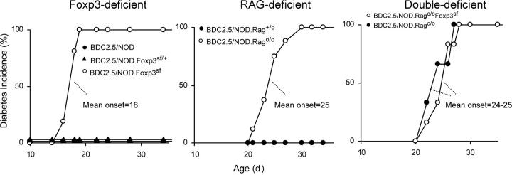
To generate BDC2.5 TCR transgenic mice lacking T reg cells, we crossed NOD.Foxp3 sf with BDC2.5/NOD animals. The presence of the BDC2.5 TCR transgene dramatically ameliorated the lymphoproliferation and multi-organ infiltration (unpublished data). On the other hand, diabetes occurred in a dramatic fashion in BDC2.5/NOD.Foxp_3_sf mice, with onset as early as 16 d of age, and 100% incidence by 21 d (Fig. 1, left). No diabetes was detected in BDC2.5/NOD male or BDC2.5/NOD.Foxp_3_sf/ + female littermates.
Figure 1.
Very early diabetes onset in Foxp3-deficient BDC2.5/NOD mice. (left) Diabetes development in BDC2.5/NOD.Foxp3 sf mice (n = 20) compared with BDC2.5/NOD (n = 21) or BDC2.5/NOD.Foxp3 sf/+ (n = 25) littermates. (middle) Incidence of diabetes in BDC2.5/NOD.Rag o/o mice (n = 8) compared with that in BDC2.5/NOD.Rag + /o controls (n = 7). (right) Diabetes development in BDC2.5/NOD.Rag o/o Foxp3sf and BDC2.5/NOD.Rag o/o littermates (n = 6 in each group).
We then compared diabetes development in BDC2.5/NOD.Rag o/o mice, which is another model of aggressive diabetes that is attributed to a lack of regulatory T cells. Indeed, these mutants had few cells with a regulatory CD25+CD69− phenotype (unpublished data). Diabetes developed rapidly in BDC2.5/NOD.Rag o/o mice, but was slower than in their Foxp3-deficient counterparts (Fig. 1, middle; 18 ± 1 vs. 25 ± 3 mean age of onset, P = 6 × 10⁻¹¹).
To test whether Foxp3 has a function in pathogenic effector T cells, we crossed the Foxp3-null mutation into the BDC2.5/NOD.Rag o/o line. Diabetes developed with identical kinetics in the BDC2.5/NOD.Rag o/o and BDC2.5/NOD.Rag o/o Foxp3sf groups (Fig. 1, right), indicating that Foxp3 affects only the T reg compartment in this system, and plays no required role in pathogenic effector cells. The subtle delayed diabetes in BDC2.5/NOD.Rag o/o and BDC2.5/NOD.Rag o/o Foxp3sf mice is most likely caused by the slightly later onset of insulitis in the RAG-deficient context (reference 5; unpublished data).
A deficit in CD25**+** T reg cells in Foxp3-deficient BDC2.5 mice
At this stage, it was important to validate the experimental model by confirming that, as anticipated (29), the Foxp3 deficiency led to a dearth of T reg cells in BDC2.5/NOD.Foxp3 sf animals. Flow cytometric analyses demonstrated that the differentiation, lineage commitment, and accumulation of clonotype-positive thymocytes were normal, as was their export to the secondary lymphoid organs (unpublished data). On the other hand, BDC2.5/NOD.Foxp3 sf mice exhibited a deficit in CD25hi cells of the T reg phenotype (Fig. 2 a); those CD25+ T cells that remained, displayed the lower level of CD25 typical of activated cells. The deficit was manifest in both the spleen and the PLN.
Figure 2.
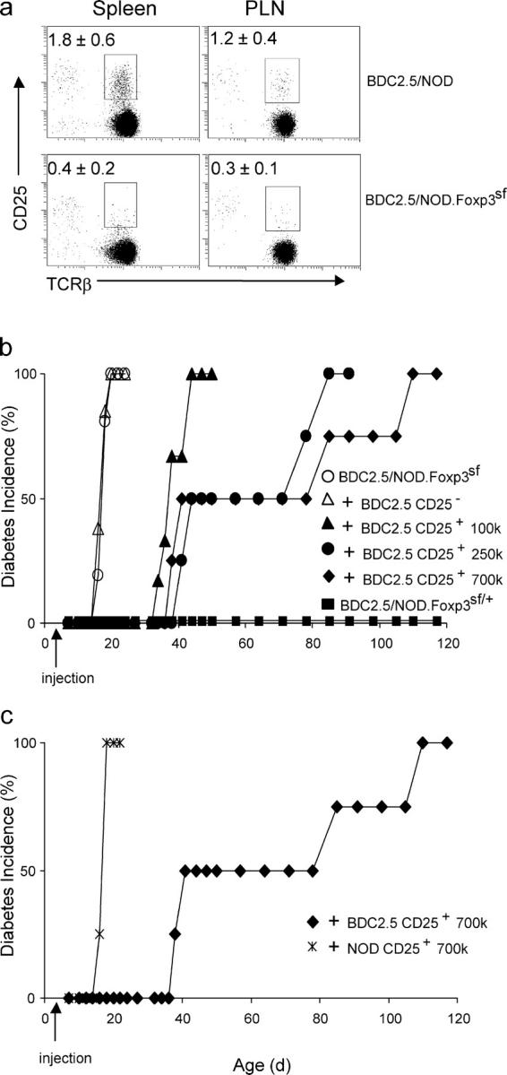
A deficit in T reg cells in Foxp3-deficient BDC2.5/NOD mice caused accelerated diabetes. (a) The T reg deficiency in BDC2.5/NOD.Foxp3 sf mice. CD4+CD25+ T cells from 12-d-old BDC2.5/NOD.Foxp3 sf and BDC2.5/NOD littermates were enumerated by flow cytometry. Plots were gated on lymphoid, CD4+CD8−CD69−B220−CD11b−7AAD− cells. Each group consisted of four to five mice from two to three independent experiments. The number within the plot refers to the mean percentage (±SD) of cells in the counting gate. (b) Treatment with antigen-specific T reg cells from BDC2.5/NOD mice rescued BDC/NOD.Foxp3 sf mice from early diabetes. (c) Nonspecific T reg cells from NOD mice were not effective in suppressing diabetes in BDC2.5/NOD.Foxp3 sf mice. The CD25− group represents a composite of five BDC2.5/NOD.Foxp3 sf mice injected with 7 × 10⁵ BDC2.5 cells and three mice with 105 cells. Each of the CD25+ groups consists of four to six animals. The BDC2.5/NOD.Foxp3 sf/+ group consists of 15 mice.
If the accelerated diabetes of BDC2.5/NOD.Foxp3 sf mice results from the absence of CD25+ T reg cells, it should be inhibitable by complementation with this population. Thus, we reconstituted neonatal BDC2.5/NOD.Foxp3 sf mice with freshly isolated T reg cells from BDC2.5/NOD mice. Injection of 7 × 10⁵ CD4+CD25+ cells from BDC2.5/NOD mice protected all BDC2.5/NOD.Foxp3 sf recipients from early onset diabetes (Fig. 2 b); however, this protection waned with time, the mice becoming progressively diabetic between 40 and 120 d. The duration of the protection was dose dependent, as the transfer of lower numbers of CD4+CD25+ cells resulting in earlier escape from protection (Fig. 2 b). Previous studies have reported that T reg cells derived from BDC2.5 mice, with their enriched antiislet repertoire, are far more effective in protecting from diabetes than those from standard NOD mice (6, 7). This enhanced effectiveness was also observed in this neonatal transfer system, as no protection was conferred by equivalent numbers of CD4+CD25+ cells from nontransgenic NOD donors (Fig. 2 c).
No impact of T reg cells on T cell activation in the PLN
The different kinetics of diabetes development displayed by BDC2.5/NOD versus BDC2.5/NOD.Foxp3 sf mice reflect solely the absence of T reg cells in the latter, providing a clean model to test where the regulatory population impinges on pathogenesis. In particular, perturbation induced by transfer of cells into lymphopenic hosts is not at play. Here, we analyzed the unfolding of diabetes in BDC2.5/NOD.Foxp3 sf mice, focusing on the checkpoints known to demarcate the initiation and progression of disease in the BDC2.5 model.
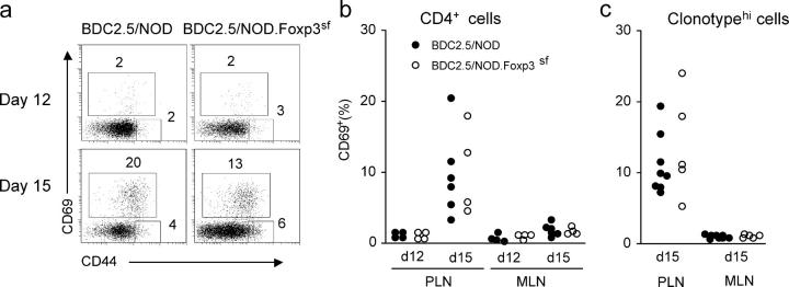
A possible explanation for the more aggressive diabetes of BDC2.5/NOD.Foxp3 sf mice is that T reg cells blocked the initial encounter of antigen by pathogenic T cells in the PLN, a very possible scenario since it has been proposed that T reg cells perturb DC maturation and antigen presentation (18), or block localized interaction between APCs and T eff cells in the PLN (22). We tested this possibility by evaluating the expression of the CD69 and CD44 activation markers in the PLN of BDC2.5/NOD.Foxp3 sf mice or Foxp3-positive control littermates. At day 12, only a minimal number of CD4+ T cells expressed CD69 in either type of mice (Fig. 3, a and b). By day 15, 5–30% of CD4+ T cells in the PLN became CD69+, but there was no notable difference between BDC2.5/NOD.Foxp3 sf mice and BDC2.5/NOD controls (Fig. 3 b). Also, no difference was found when gating the analysis on those CD4+ cells staining at high levels with a monoclonal antibody specific for the BDC2.5 clonotype (Fig. 3 c), a population that was demonstrated to be diabetogenic (30).
Figure 3.
Naturally occurring T reg cells did not suppress initial T cell activation. Similar kinetics of T cell activation in the PLN of BDC2.5/NOD and BDC2.5/NOD.Foxp3 sf littermates, as indicated by the percentage of cells expressing CD44 and CD69 activation markers. Gated on either CD4+ (a and b) or BDC2.5 clonotypehi T cells (c). Cells from the nondraining MLNs were used as negative controls; b and c derive from independent experiments. Each dot represents one animal.
One of the hallmarks of T reg cells is their ability to nonspecifically suppress the proliferation of T cells stimulated through their TCRs, at least in vitro (9, 12). Thus, it was conceivable that T reg cells limit the activation of BDC2.5/NOD T cells, and that the release of this damper by the elimination of T reg cells in BDC2.5/NOD.Foxp3 sf mice led to runaway proliferation. In vivo proliferation of BDC2.5 effectors in the PLN of BDC2.5/NOD and BDC2.5/NOD.Foxp3 sf mice was compared by quantitating the incorporation of 5-bromo-2-deoxyuridine (BrdU). Mice were injected with BrdU at 15 d old, and BDC2.5-clonotypehi T cells from the PLN were analyzed for incorporation of BrdU after a 6-h labeling period. As shown in Fig. 4 (a and b), the clonotypehi effectors proliferated to an equivalent extent in BDC2.5/NOD and BDC2.5/NOD.Foxp3 sf littermates. This observation was consistent with the absolute number of BDC2.5 clonotypehi cells, which was equivalent in the PLN of the two types of mice (for BDC2.5/NOD, 4.1 ± 0.9 × 10⁴, n = 8; for BDC2.5/NOD.Foxp3 sf, 3.6 ± 1.8 × 10⁴, n = 5). In contrast, although we could recover 2,000–5,000 T cells from the pancreas of a 15-d-old BDC2.5/NOD.Foxp3 sf mouse, we could isolate only 50–100 T cells from a BDC2.5/NOD control.
Figure 4.

T reg cells did not suppress T cell proliferation, Th1–cytokine production, costimulatory molecule, and chemokine receptor expression in the PLN. (a) Representative plots for BrdU incorporation analysis. Each group consists of five to eight mice. The PLN from mice that did not receive BrdU (right) served as a negative control. The numbers in the plots are percentages of the gated population. Plots are derived from BDC2.5 clonotypehiCD8−B220−CD11b− population. (b) Percentages (mean ± SD) of BrdU-labeled cells in the CD69+ and CD69− subsets shown in panel a. (c and d) PCR Quantification of IL2 (c) and IFNγ (d) mRNA in BDC2.5 clonotypehiCD25− from the PLN of 15-d-old BDC2.5/NOD versus BDC2.5/NOD.Foxp3 sf mice. Data are from two experiments with five mice in each group. (e and f) Frequencies of BDC2.5 clonotypehi cells expressing the costimulatory molecule CD134 (OX40) and chemokine receptor CD195 (CCR5), respectively, in the PLN of 15-d-old BDC2.5/NOD versus BDC2.5/NOD.Foxp3 sf mice. The dashed line indicates background staining. Each dot represents one animal.
Although the activation and proliferation of autoreactive T cells did not seem to be affected by the presence of T reg cells in the PLN, it is possible to hypothesize that their differentiation status and cytokine profile might be altered—for example, inhibiting the differentiation along a pathogenic Th1 pathway. Flow cytometric and quantitative RT-PCR were performed on clonotypehiCD25−/lo cells from the PLN of BDC2.5/NOD.Foxp3 sf mice and control littermates. No difference was seen in the levels of mRNAs encoding IL-2 or IFNγ (Fig. 4, c and d). No IL-4 mRNA was detected in either group (unpublished data). There was no difference in the expression of costimulatory molecules: CD134 was found in low and variable numbers of clonotypehi cells; there was some variability between mice, which is understandable because the very first stages of insulitis were examined, but with no notable difference between mutant and control mice (Fig. 4 e); no expression of ICOS was detected in either group. A previous study suggested that T reg cells inhibit chemokine receptor CXCR3 by T eff cells in the PLN and thus control the trafficking of these cells to the pancreas (19). We could not detect substantial CXCR3 expression on T cells from the PLN of the 15-d-old mice with or without T reg cells (unpublished data). The expression of another chemokine receptor, CCR5, also implicated in diabetes progression (31), was not substantially different between BDC2.5/NOD and BDC2.5/NOD.Foxp3 sf mice (Fig. 4 f).
Also, no impact of T reg cells on the PLN activation and proliferation of T eff cells in a transfer system
Our analysis of BDC2.5/NOD.Foxp3 sf mice indicated that T reg cells did not have a substantial negative influence on the activation of autoreactive T cells in the draining PLN, rather seeming to exert their effects within the pancreatic lesion, in apparent contradiction with some previous papers (18, 22, 32). This discrepancy might be linked to the use of transfer systems in previous analyses, whereas we relied on a genetic mutation to generate otherwise identical mice with or without T reg cells. Therefore, we examined the impact of T reg cells in settings of adoptive transfer. First, we analyzed activation of the effector T cells in 15-d-old BDC2.5/NOD.Foxp3 sf mice that had received CD25+ T reg cells as neonates (i.e., under conditions in which they are protected from diabetes), as shown in Fig. 2. Donor cells came from Thy1.1 congenic BDC2.5/NOD mice in order to distinguish them from the host, and amounted to roughly 30% of the T reg frequency in BDC2.5/NOD mice. Accumulation of transferred T reg cells in the PLN was no more than that in the nondraining mesenteric LNs (MLNs; Fig. 5 a). The transferred T reg cells maintained CD25 expression (not depicted). As illustrated in Fig. 5 a, the BDC2.5 T reg cells, although they were sufficient to inhibit diabetes, did not suppress the PLN-specific induction of CD69 on host T cells.
Figure 5.
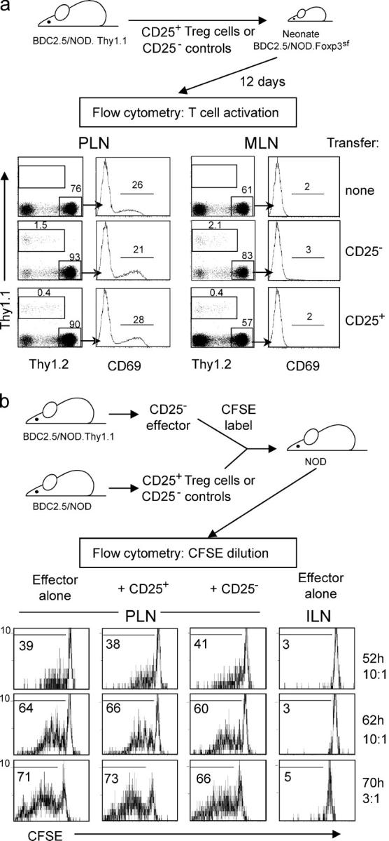
Transferred T reg cells did not inhibit the activation and proliferation of effector T cells in the PLN. (a) CD4+CD25+ or CD4+CD25− cells isolated from Thy1.1 congenic BDC2.5/NOD mice were transferred to 3-d-old BDC2.5/NOD.Foxp3 sf mice, and the activation of endogenous Thy1.2+ T cells in the PLN was analyzed at day 15. The numbers indicate the percentage of cells in the respective counting gate. Plots are gated on lymphoid, B220−CD8− cells. Data represent three experiments with four to five mice in each group receiving 105 or 2.5 × 10⁵ cells. (b) CD25− T cells from Thy1.1-congenic BDC2.5/NOD mice were labeled with CFSE, and were injected alone, or were mixed with CD4+CD25+ or CD4+CD25− cells from BDC2.5/NOD mice at a 10:1 or 3:1 ratio, into standard NOD mice. Dilution of CFSE in the transferred Thy1.1+ effectors in the PLN or inguinal LN was analyzed from 52 to 70 h after injection. The numbers within the plots indicate the percentages of cells that exhibit CFSE dilution. Data are from three experiments.
Second, as one might argue that the large number of clonotype-positive cells in the host might outnumber the transferred regulatory cells, we tested the effect of T reg cells on the activation of cotransferred BDC2.5/NOD effector T cells. A low number of naive BDC2.5/NOD effector T cells, labeled with carboxyfluorescein succinimide ester (CFSE), were transferred alone or mixed with CD4+CD25+ T reg or CD4+CD25− control cells, into 5-wk-old NOD mice; the proliferation of transferred effector cells was assessed by measuring CFSE dilution. As shown in Fig. 5 b, autoreactive T cells proliferated vigorously in the PLN but not in irrelevant inguinal LNs. In the three experiments presented in Fig. 5 b, no impact of T reg cells on cotransferred T eff cells could be distinguished, regardless of the ratio or readout time. This remained true even at 200 h after transfer (unpublished data).
T reg cells' effect on the progression, but not the timing, of insulitis
A potential explanation for the accelerated diabetes in BDC2.5/NOD.Foxp3 sf mice was that the T reg cell deficiency accelerated disease initiation, allowing T cells to infiltrate the islets at an earlier time. Insulitis begins very sharply in BDC2.5/NOD mice, ∼2 wk of age, probably in response to perturbation in islet physiology (2–4). As shown in Fig. 6, the time course of insulitis was similar in BDC2.5/NOD and BDC2.5/NOD.Foxp3 sf animals: no islet infiltration was detected in 12-d-old mice; but, at day 15, islets showed clear infiltration. The insulitis of BDC2.5/NOD.Foxp3 sf mice differed in that it was immediately aggressive (Fig. 6 a, bottom right), compared with the “respectful” disposition of invading cells in animals with T reg cells (Fig. 6 a, bottom left).
Figure 6.
Naturally occurring T reg cells regulated the aggressiveness of insulitis. (a) Precipitous progression of Insulitis in Foxp3-deficient BDC2.5/NOD mice. Pancreatic sections stained with hematoxylin and eosin (original magnification, 200), representative of four to five mice in each group. (b) Scores of insulitis at 12 and 15 d old. Each bar represents one animal.
Microarray analysis of T reg cell activity in the PLN and pancreas
Hence, all lines of evidence indicate that Foxp3-dependent T reg cells dampen the progression of autoimmune diabetes by controlling the aggressiveness of autoreactive T eff cells in the target organ, but have no detectable impact on initial priming and activation in the draining LNs. To test the robustness of this conclusion, we sought an independent approach. We have previously shown that a substantial number of T reg cells are found in the insulitic lesion of BDC2.5/NOD mice, identified by their CD25+CD69− phenotype and by their strong expression of Foxp3 (8). If the target organ is the location where T reg cells show their main action, it is reasonable to expect that an alteration in their gene expression profiles might subtend this functional role. Therefore, we performed a comparative microarray analysis of three different tissues from 3- to 4-wk-old BDC2.5/NOD mice: the MLN, an irrelevant LN devoid of antigenic stimulus for BDC2.5 T cells; the draining PLN, where priming and activation of BDC2.5 cells occurs; and the pancreas, where infiltrate cells are in direct contact with the antigen source. For all three locations, we isolated both the CD4+CD25+CD69− T reg cell population (∼80–90% stain positive for intracellular Foxp3 protein and there is no difference among the cells isolated from the three locations) and the remainder of the CD4+ cells (only 2–5% are Foxp3+; Fig. 7 a). The six populations should define profiles that distinguish, for all three organs, T reg cells from their conventional counterparts. Total RNA was prepared from purified cells, amplified, and used to generate probes for Affymetrix Mu74Av2 chips. Data were preprocessed with the RMA algorithm, and averaged between at least three independent replicates for each condition. False discovery rates were estimated by generating randomized datasets based on the intra-replicate distribution of variance.
Figure 7.
Gene expression profiles of T reg cells in the respectful insulitic lesion and in lymph nodes. (a) Sorting gates for the CD4+CD25+ or CD4+CD25− cells isolated from MLNs or PLNs, or from the pancreatic infiltrate. (b) Definition of the T reg signature. Genes were identified by comparing microarray datasets from the pairwise comparison of populations shown in panel a (left boxes) or from published datasets: lymph node GFP+CD25+ T reg cells versus naive GFP-CD25− cells from the Foxp3 gfp knockin mice (33), or CD103+CD25+ versus CD103+CD25− cells from B6 mice (reference 34). Genes detected as differentially expressed in two of the three datasets were included in the signature gene set. (c) Biparameter plots comparing expression values of genes from the T reg signature in T reg cells of the MLNs, PLNs, or pancreatic infiltrate. The number of genes over- or underexpressed in either condition is shown in the plot.
We initially sought to define a general “T reg signature,” a set of genes whose over- or underexpression distinguishes the CD4+CD25+ population from conventional CD4+ T cells (hereafter Tconv) in different contexts. For a robust definition of this signature, we used the present datasets together with T reg expression profiles from two other independent sources. Fontenot et al. have performed a microarray analysis of T reg cells identified by a fluorescent protein reporter “knocked-in” the Foxp3 locus (33); these data were obtained with lymph node T reg cells from unchallenged mice (hereafter referred to as the Seattle data). Huehn et al. (34) have analyzed different phenotypic variants of T reg cells, in particular those marked by the expression of CD103 (αEβ7 integrin), which reside preferentially in inflamed tissues (hereafter referred to as the Berlin data). These additional datasets allowed us to perform a combined selection procedure, diagramed in Fig. 7 b, where validation is provided by discovery in independent datasets. The Seattle datasets provide a perspective on “central” T reg cells residing in lymphoid organs, whereas the Berlin datasets highlight genes particularly expressed in “peripheral” T reg cells in the tissues. In practice, we first identified all genes in the BDC2.5/NOD datasets that were overexpressed in T reg relative to Tconv cells by 1.4-fold in any one of the three comparisons: MLN, PLN, or pancreatic infiltrate (this initial fold-change cutoff, which corresponds to a false-discovery rate of 0.18 for single datasets, or 0.25 in any one of the three, was voluntarily chosen as being nonstringent). This primary set of 556 genes was then cross-matched with the other two T reg/Tconv comparisons. 212 genes were found to be overexpressed also by T reg cells in either the Berlin or Seattle data groups. The same operation was performed for genes underexpressed in T reg cells: 831 primary calls, of which 96 were shared with either the Berlin or Seattle data. These cross-comparisons resulted in highly notable assignments, because the same process run on randomized datasets resulted in only 22 and 4 genes apparently over- or underexpressed in the T reg population. In addition, 15 genes were internally reproduced, as different features on the chip yielded identical results. (Table S1, available at http://www.jem.org/cgi/content/full/jem.20051409/DC1).
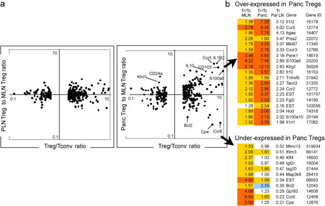
We then asked how the genes in the T reg signature compared with BDC2.5/NOD T reg cells from the MLN, PLN, and pancreatic infiltrate. As illustrated by the expression values plotted in Fig. 7 c, the profiles of the two LN T reg populations were much more similar to each other than they were to the profile of infiltrating T reg cells (altogether 30 vs. 5 genes varying by twofold or more, the latter being equivalent to normalized background). This finding was confirmed in the displays of Fig. 8, which plots “T regness” on the x axis (the max ratio of expression between T reg and T conv, calculated within each tissue pair) versus the expression ratio between T reg populations on the y axis. Several points can be made from this means of displaying the data. First, many of the T reg signature genes were distinctly over- or underexpressed in islet-infiltrating T reg cells relative to T reg cells in the control MLN (Fig. 8 a, right). In contrast, there was much less difference between the PLN and MLN T reg cells (Fig. 8 a, left). These differentially expressed genes included IL-10, an important mediator of T reg cell activity, CD103, and S100a6, which have previously been described as characteristic of effector/memory T reg cells (34); and chemokine receptors that would account for their different location (up-regulated, Ccr5, Cxcr3, and Ccr2; down-regulated, Ccr6). Second, this differential expression primarily concerned the overexpressed genes of the T reg signature (Fig. 8 b, top); genes under-expressed in T reg cells were more stable overall (Fig. 8 b, bottom). Third, the genes whose expression diverged most in the infiltrating T reg cells tended to be those with the highest T reg/T conv differential. Thus, the T reg cells present in the pancreatic infiltrate, at the location where they are effectively suppressing tissue damage, have a phenotype quite distinct from that of their counterparts residing in the lymph nodes, and one that seems to amplify T reg characteristics.
Figure 8.
Nature of the genes that distinguish T reg cells in the pancreatic infiltrate. (a) The biparameter plots show, for all genes of the T reg signature, the ratio of expression values in T reg or T conv cells (T reg/T conv, x-axis) versus the ratio of expression in pancreatic infiltrate T reg cells versus MLN T reg cells (right); as a control, the ratio between T reg cells in PLN and MLN is shown on the left. (b) Nature of the genes whose expression is altered in T reg cells of the pancreatic infiltrate relative to LNs T reg cells. The first three columns give the ratio of expression in T reg cells versus conventional CD4+ cells of the MLN or pancreas (Tr/Tc MLN and Tr/Tc Panc, respectively), and the ratio of pancreatic versus MLN T reg cells (Tr Pa/LN). The NCBI gene symbol is used, along with the NCBI GeneID identifier. Yellow and orange highlights denote genes overexpressed by >1.35- or 2.5-fold, respectively; blue highlight denotes underexpression by ⩽0.6 fold.
Discussion
Among the shared characteristics of T1D in humans and mice is the slow progression of the disease in most cases. In humans, serum autoantibodies against islet antigens, indicators of islet inflammation, can appear years before overt diabetes. In NOD mice, insulitis begins at ∼3–4 wk after birth, but diabetes does not manifest until 12 wk of age. Studies with the BDC2.5 model have highlighted two checkpoints in disease progression, as well as a critical role for Foxp3-dependent CD25+ T reg cells (6–8). Here, we asked where and how T reg cells exert their control on the unfolding of diabetes, under experimental conditions not influenced by homeostatic expansion of the effector population. Several lines of evidence converged to demonstrate that T reg cells do not delay or hinder the activation and expansion of antiislet T cells in the PLN, nor the timing of their infiltration of the islets. Although we detected no change in initial activation, proliferation, cytokine production, chemokine receptor, and costimulatory molecule expression of T eff cells in the PLN, we cannot formally exclude a subtle effect on T eff cells in the LN that would precede and predict their mode of response once they home to the islet. Yet the most obvious impact is in the target tissue, arguing that this is the location where T reg cells primarily act, to control an otherwise destructive infiltration. In keeping with this tissue-localized activity, we found a clear shift in the gene expression profiles of islet-resident T reg cells from the T reg signature characteristic of lymphoid-tissue cells.
The scurfy model offers complete but isolated ablation of Foxp3-dependent T reg cells, which is different from the situation with other genetically engineered or transfer systems that induce variations in their number or activity, or where the mutation may have other consequences. For example, the numbers of CD25+ T reg cells in CD28−/− or _B7−/−_mice are greatly reduced, but substantial populations still exist (16) and antigen presentation is also affected. Adoptive transfer models often take the absence of CD25 to denote effector cells, but CD25+ T regs can emerge from CD25− populations after transfer (35).
As suggested by Tarbell et al. (7), the physiological function of CD25+ T reg cells needs to be addressed under conditions independent of lymphopenia-induced homeostasis, and BDC2.5/NOD.Foxp3 sf animals meet this condition. We compared the initial antiislet T cell activation in the PLN with or without T reg cells, under conditions where both T eff and T reg cells have matured in vivo without perturbation. In BDC2.5/NOD mice, and most likely in standard NOD mice as well (36), activation of BDC2.5 T cells in the PLN is under tight temporal control. Before 12 d old, there is no notable T cell activation (3), and we have suggested that overcoming this “checkpoint 1” is linked to a ripple of developmentally regulated β-cell apoptosis (4). Although it is reasonable to propose the counterinterpretation that T reg cells are involved in this delay, in keeping with suggestions that they can keep DCs from maturing until released by a CD40-dependent T cell help signal (18), our data refute this scenario because the absence of T reg cells did not accelerate the timing of the initial antiislet T cell activation and insulitis.
The absence of an impact of T reg cells on the initial antiislet T cell activation phases in the draining PLN contrasts with previous findings in other models of autoimmune diabetes, where it has been suggested that T reg cells suppressed the homing of effector T cells to the PLN or activation or proliferation therein. On the basis of results from experiments performed with in vitro expanded islet-specific T reg cells, it was suggested that BDC2.5 T reg cells intervene in the PLN by excluding localized interaction between BDC2.5 effector T cells and APCs (22). However, this result may have reflected the pharmacological effect of in vitro activation and expansion of the T reg cells and/or numeric effects beyond their physiological role. In the same vein, another paper proposed that T reg cells controlled the pathogenic effect of an islet-specific, CD8+ TCR transgene–encoded T cell specificity by inhibiting DC maturation in the PLN (18), but this study did not directly demonstrate the impact of transferred T reg cells on the activation of the CD8+ effector cells in the PLN. In another study (32), thymectomized NOD.B7-2o/o recipients were treated with anti-CD25 antibodies to deplete T reg cells, and the division of CFSE-labeled naive BDC2.5 T cells transferred into these recipients was measured 2 wk later in the PLN. Compared with control animals, the PLN of anti-CD25-treated mice harbored about twofold more BDC2.5 T cells that had undergone division, suggesting that the T reg cells suppress the activation of autoreactive T cells in the draining lymph nodes (32). However, this impact was modest and anti-CD25 treatment is not without other effects.
On the other hand, results from several systems are compatible with a role for T reg cells in preventing the locally damaging consequences of autoimmune infiltration. In a transgenic model where diabetes was provoked by islet-specific expression of TNFα and CD80, the transfer of T reg cells protected the recipients from diabetes and invasive but nondestructive insulitis was detected in the pancreata of the protected mice (17). In one of our previous studies, sorted CD4+CD25−/lo effector cells from pancreata of BDC2.5/NOD mice transferred diabetes into NOD.scid recipients. The diabetes was inhibited by cotransfer of CD25+ T reg cells that were also isolated from pancreata of BDC2.5/NOD mice; insulitis was clearly present in the protected mice, and had acquired the respectful aspect characteristic of the stably balanced lesions of BDC2.5/NOD mice (8). In another study, diabetes occurred a few weeks after transfer of splenocytes from diabetic NOD mice into NOD.scid recipients, but disease was suppressed for months by cotransfer of BDC2.5/NOD CD25+ T reg cells expanded in vitro with DCs plus a mimotope peptide (7). Histological examination revealed that insulitis was clearly present, but appeared controlled, in the protected mice (7), again pointing to local control. The delayed onset of insulitis may have been caused by an effect of T reg cells in suppressing lymphopenia-induced homeostatic expansion. Recently, Sarween et al. (19) have reported the action of T reg cells in a model where diabetes was induced in RAG-deficient mice carrying the RIP-mOVA transgene and transferred with ovalbumin-specific transgenic T cells. As here, T reg cells had little or no impact on the activation of effector cells. The results differed, however, in that T reg cells appeared to have an effect on IFNγ production by T eff cells in the PLN, which was not the case here. In addition, T reg cells seemed to slow the migration of OVA-specific cells into the islets, perhaps through a reduction in CXCR3 in the PLN. Again, this was not the case here, as insulitis appeared with the same kinetics in the presence and absence of T reg cells. The difference might be explained by the different antigens involved (natural pancreatic autoantigen vs. transgenic foreign antigen), and the latter also requiring an additional immunization in adjuvant to break a state of “clonal ignorance.”
That T reg cells predominantly affect effector cells in a delayed manner and in the target tissues has also been suggested in other contexts. Huehn et al. showed that regulatory T cells with a memory phenotype akin to those of tissular lymphocytes were more effective suppressors of arthritis in vivo than were their naive counterparts (34). With hemagglutinin as a foreign antigen, T reg cells had no impact on the early phase of the response, but prevented the later accumulation of effector cells (15). T reg cells have also been detected, and shown to be active, in local infiltrates marking ear infection by Leishmania major (37) or in tolerated cardiac allografts (38). They were also present in lymphoid populations infiltrating tumors, where they were thought to hamper the efficacy of tumor-specific effectors (39). Consistent with the present findings, T reg cells did not inhibit proliferation and IFNγ production by T eff cells in the draining LNs, in a mouse model of tumor rejection by transferred antigen-specific CD8+ T cells (40). Finally, CD25+ T reg cells could inhibit local inflammation even when there were no T cells involved (and per force no activation in the draining lymph node), such as in the _Helicobacter_-dependent colitis model in Rag2−/− mice (41).
Consistent with the predominant role of T reg cells in the insulitic lesion, we found that they acquired a distinct gene expression profile, relative to T reg cells residing in the LNs. Some of the changes were consistent with their tissular localization, and overlapped to a large extent the transcriptional differences between circulating and tissue-homing T reg cells observed by Huenh et al. (34). Others were more evocative of physiological changes linked to their antiinflammatory function, most notably a strong increase in IL-10 transcripts. Although these observations are correlative, they are quite consistent with a view where the tissular action of T regs is accompanied by an amplification of their specific phenotype.
In conclusion, the dramatic diabetes of BDC2.5/NOD. Foxp3 sf mice illustrates the requirement for Foxp3-dependent CD25+ T reg cells in maintaining a balanced insulitis. This effect illustrates the great therapeutic potential of T reg cells, and also argues that it will be highly desirable to target their action to the tissue where they may be most effective.
Materials and Methods
Mice.
B6.Cg-Foxp3 sf mice on the C57BL/6 background, provided by F. Ramsdell (Celltech R&D, Inc., Bothell, WA) were backcrossed onto the NOD/Lt background. The Foxp3 sf mutation was identified by PCR (primers: 5′-TCAGGCCTCAATGGACAAGA-3′ and 5′-GCCCAAGGCTATGCATTTAT-3′ for WT Foxp3; 5′-TCAGGCCTCAATGGACAAAA and GCCCAAGGCTATGCATTTAT-3′ for Foxp3 sf mutant). Breeders in the backcross were also genotyped for microsatellite markers linked to diabetes susceptibility/resistance loci Idd1 through Idd20. The loci that have a major impact on diabetes incidence were fixed to NOD alleles early in the backcrossing. By the sixth generation of backcrossing (N6), all Idd loci were homozygous for the NOD alleles. The Foxp3 gene is located on the X chromosome at the 2.1 cM position. Although there is no Idd locus on the X chromosome, we minimized the contamination of non-NOD genes from the X chromosome. A large number of offspring from the N6 generation were screened for recombination on the X chromosome by typing microsatellite markers near the Foxp3 gene. One female carrier of Foxp3 sf mutation was found to be homozygous for the NOD alleles at both DXMit136 (located at 2.8cM) and DXMit166 (located at 15.5cM). This founder was selected for further backcrossing. Experimental mice are from the 7–11th generations. NOD.Foxp3 sf mice were crossed with BDC2.5/NOD to generate BDC2.5 mice deficient in Foxp3. This line was also crossed with NOD.Rag1 o/o (The Jackson Laboratory) and F1 offspring were intercrossed to generate NOD.Rag o/o Foxp3sf/+ mice. NOD.Rag o/o Foxp3sf/+ mice were further crossed with BDC2.5/NOD.Rag o/o mice protected from early diabetes by normal NOD splenocytes (5), to generate BDC2.5/NOD.Rag o/o Foxp3sf mice on the NOD background. NOD.Thy1.1 mice (The Jackson Laboratory) were crossed with BDC2.5/NOD; backcrossing F1 offspring with NOD.Thy1.1 generated BDC2.5/NOD.Thy1.1 mice. Mice were maintained in the Joslin Diabetes Center barrier facility (protocol 99–19, 99–20 approved by the Joslin Diabetes Center's International Animal Care and Use Committee).
Diabetes monitoring and insulitis scoring.
Urine glucose levels were tested every 2– 3 d with Uristix (Bayer Diagnostics). Positive reads were confirmed by blood glucose measuring (>250 mg/dl) with a Glucometer 3 reader (Bayer Diagnostics). To score insulitis, pancreata were fixed in formalin solution (Sigma-Aldrich). Paraffin-embedded sections were stained with hematoxylin and eosin and examined by microscopy. Multiple hematoxylin and eosin sections of pancreata and at least 40 islets per animal were assessed. Peri-insulitis refers to the presence of inflammatory cells in the immediate vicinity of the islets and insulitis includes all invasive infiltration in the islets.
Flow cytometry.
Single cell suspensions of lymphoid organs or blood leukocytes were prepared and blocked with anti-CD16/32 (2.4G2) and normal mouse sera (Jackson ImmunoResearch) against Fc-mediated nonspecific binding of antibodies. The samples were then stained with antibody conjugates with a standard procedure. The following antibody conjugates were used: biotinylated anti-BDC2.5 TCR; FITC conjugated anti-CD44, anti-TCRβ, anti-Thy1.1, anti-CD25 (BD Biosciences); PE conjugated anti-CD62L, anti-CD69, anti-CD25, anti-Thy1.2, anti-ICOS (BD Biosciences), anti-CD134 (OX40) (eBioscience), and streptavidin (Biosource International); PE-Cy5 conjugated anti-CD8, anti-CD69, anti-CD11b (Caltag), anti-B220 (BD Biosciences) and anti-Thy1.2 (eBioscience); PE-Texas Red conjugated anti-CD4, anti-CD8 and anti-B220 (Caltag). Cell-surface expression of CXCR3 and CCR5 and intracellular expression of Foxp3 were examined with PE-conjugated monoclonal antibodies (R&D Systems, BD Biosciences, and eBioscience, respectively) according to the manufacturers' protocols. Staining with 7-aminoactinomycin D (7-AAD; Sigma-Aldrich) was used to exclude dead cells. Stained samples were analyzed with a flow cytometer (EPICS XL; Beckman Coulter).
For assessment of T cell proliferation by incorporation of BrdU, 0.5 mg BrdU was injected intraperitoneally into 15-d-old mice. 6 h later, animals were killed and cells from the PLN were stained for BDC2.5 clonotype and CD69, and then for incorporated BrdU with anti-BrdU antibody (BD Biosciences) following the manufacturer's protocols.
Cell sorting and adoptive transfer.
Splenocytes of NOD or BDC2.5/NOD mice were blocked against Fc-binding as stated above, stained with anti-CD4-APC, CD-25-PE, CD8-FITC, B220-FITC, CD11b-FITC, CD69-FITC (BD Biosciences) and sorted with a MoFlo cytometer and Summit software (DakoCytomation) for CD8−B220−CD11b−CD69−CD4+ cells that are either CD25+ or CD25− (purity >95%). Sorted cells were resuspended in PBS and injected intraperitoneally into 3-d-old BDC2.5/NOD.Foxp3 sf recipients. To examine cytokine expression, subsets of BDC2.5 clonotypehi were sorted from PLN of 15-d-old mice. RNA was extracted from these cells with TRIzol (Invitrogen). Ex vivo analysis of IL2 and IFNγ production were done using quantitative RT-PCR with sorted PLN BDC2.5-clonotypehiCD25−/lo effector cells, which included both CD69+ CD25lo and CD69− cells, as described previously (8).
For the experiment of cotransferring T eff and T reg cells, splenocytes from BDC2.5/NOD.Thy1.1 mice were labeled with CFSE and then depleted of CD25+ and CD69+ cells with MACS-beads (Miltenyi Biotec). CD4+CD25− or CD4+CD25+ were sorted as above from the spleen of BDC2.5/NOD mice. CFSE-labeled effectors (0.6- to 1.2 × 10⁶ cells) alone, or mixture of CFSE-labeled effectors with CD25+ T reg cells or CD25− control cells at 10:1 or 3:1 ratio, were injected into normal 5-wk-old NOD mice. Dilution of CFSE in Thy1.1+ cells was analyzed by flow cytometry.
Gene expression profiling.
Pancreata, PLNs, or MLNs were isolated from 3–4 wk-old BDC2.5/NOD mice (n = 10-20 mice per RNA sample). Cells were stained with CD4-PECy7 (GK1.5), B220-PE–Texas red (RA3-6B2), 7AAD live cell dye, CD25-APC (PC61), and CD69-FITC (H1.2F3; BD Biosciences). Live B220−CD4+ cells were sorted to separate the CD4+CD25+CD69− population (T reg) and the combined population of all other CD4+ cells (CD4+CD25−CD69− and CD4+CD25lo/−CD69+ cells), using a MoFlo flow cytometer and Summit® software. Sorted cells were collected into media with 5% fetal bovine serum, and assessed for purity. If necessary, cells were resorted to achieve >98% purity. Cells (typically 104–105 cells per experiment) were lysed in TRIzol, and total RNA prepared according to manufacturer's instructions (Invitrogen). RNA was amplified twice using the MessageAmp RNA kit (Ambion), except the biotinylation step was performed using the BioArray HighYield RNA Transcript Labeling Kit (T7) (Enzo Life Sciences). 5–15 μg of an RNA probe was hybridized to murine genome U74Av2 array GeneChips as per manufacturer's instructions (Affymetrix) by the Joslin Genomics Core. The raw data were processed with the RMA algorithm for probe-level normalization (8) (S+ ArrayAnalyzer, Insightful, modified by R. Park, D.M. and C.B.) and composite expression values were calculated for each of the genes on the chip (averaging after outlier elimination, “Distill” software). Gene-wise p-values were calculated with Welch's modified t test. A randomized dataset was generated using inter-replicate CVs to estimate false-discovery rates. Prior data from earlier pancreas infiltrate samples (8) (GEO database accession no. GSE1419), was pooled and distilled together with newly generated pancreas samples prepared in parallel with the PLN and MLN samples generated for this study (GSE1419).
Online supplemental material.
Specific genes identified by microarray analysis of T reg and T eff cells are presented in Table S1. Online supplemental material is available at http://www.jem.org/cgi/content/full/jem.20051409/DC1.
Acknowledgments
We thank R. Melamed, E. Hyatt, V. Bruklich, K. Hattori, G. Losyev, J. Lavecchia, and A. Pinkhasov for assistance with data analysis, mice, flow cytometry, and histology.
This work was supported by funds from the National Institutes of Health (NIH) (PO1 AI39671-09) and the William T. Young Chair to D. Mathis and C. Benoist, and by core services of Joslin's National Institute of Diabetes and Digestive and Kidney Diseases–funded DERC. Z. Chen has been supported by postdoctoral fellowships from Juvenile Diabetes Research Foundation and the Program in AIDS Research (Dana-Farber Cancer Institute/NIH). A. Herman was supported by training grants from the Dana-Farber Cancer Institute and the Joslin Diabetes Center.
The authors have no conflicting financial interests.
Abbreviations used in this paper: 7AAD, 7-aminoactinomycin; BrdU, 5-bromo-2-deoxyuridine; CFSE, carboxyfluorescein succinimide ester; MLN, mesenteric LN; NOD, nonobese diabetic; PLN, pancreatic LN; T1D, type-1 diabetes; T eff, effector T cell; T reg, regulatory T cell.
Z. Chen and A.E. Herman contributed equally to this work.
A.E. Herman's present address is Genomics Institute of the Novartis Research Foundation, San Diego, CA 92121.
References
- 1.Haskins, K., M. Portas, B. Bergman, K. Lafferty, and B. Bradley. 1989. Pancreatic islet-specific T-cell clones from nonobese diabetic mice. Proc. Natl. Acad. Sci. USA. 86:8000–8004. [DOI] [PMC free article] [PubMed] [Google Scholar]
- 2.Katz, J.D., B. Wang, K. Haskins, C. Benoist, and D. Mathis. 1993. Following a diabetogenic T cell from genesis through pathogenesis. Cell. 74:1089–1100. [DOI] [PubMed] [Google Scholar]
- 3.Hoglund, P., J. Mintern, C. Waltzinger, W. Heath, C. Benoist, and D. Mathis. 1999. Initiation of autoimmune diabetes by developmentally regulated presentation of islet cell antigens in the pancreatic lymph nodes. J. Exp. Med. 189:331–339. [DOI] [PMC free article] [PubMed] [Google Scholar]
- 4.Turley, S., L. Poirot, M. Hattori, C. Benoist, and D. Mathis. 2003. Physiological beta cell death triggers priming of self-reactive T cells by dendritic cells in a type-1 diabetes model. J. Exp. Med. 198:1527–1537. [DOI] [PMC free article] [PubMed] [Google Scholar]
- 5.Gonzalez, A., I. Andre-Schmutz, C. Carnaud, D. Mathis, and C. Benoist. 2001. Damage control, rather than unresponsiveness, effected by protective DX5+ T cells in autoimmune diabetes. Nat. Immunol. 2:1117–1125. [DOI] [PubMed] [Google Scholar]
- 6.Tang, Q., K.J. Henriksen, M. Bi, E.B. Finger, G. Szot, J. Ye, E.L. Masteller, H. McDevitt, M. Bonyhadi, and J.A. Bluestone. 2004. In vitro–expanded antigen-specific regulatory T cells suppress autoimmune diabetes. J. Exp. Med. 199:1455–1465. [DOI] [PMC free article] [PubMed] [Google Scholar]
- 7.Tarbell, K.V., S. Yamazaki, K. Olson, P. Toy, and R.M. Steinman. 2004. CD25+ CD4+ T cells, expanded with dendritic cells presenting a single autoantigenic peptide, suppress autoimmune diabetes. J. Exp. Med. 199:1467–1477. [DOI] [PMC free article] [PubMed] [Google Scholar]
- 8.Herman, A.E., G.J. Freeman, D. Mathis, and C. Benoist. 2004. CD4+ CD25+ T regulatory cells dependent on ICOS promote regulation of effector cells in the prediabetic lesion. J. Exp. Med. 199:1479–1489. [DOI] [PMC free article] [PubMed] [Google Scholar]
- 9.Shevach, E.M. 2002. CD4+ CD25+ suppressor T cells: more questions than answers. Nat. Rev. Immunol. 2:389–400. [DOI] [PubMed] [Google Scholar]
- 10.von Boehmer, H. 2005. Mechanisms of suppression by suppressor T cells. Nat. Immunol. 6:338–344. [DOI] [PubMed] [Google Scholar]
- 11.Ramsdell, F., and S.F. Ziegler. 2003. Transcription factors in autoimmunity. Curr. Opin. Immunol. 15:718–724. [DOI] [PubMed] [Google Scholar]
- 12.Sakaguchi, S. 2004. Naturally arising CD4+ regulatory t cells for immunologic self-tolerance and negative control of immune responses. Annu. Rev. Immunol. 22:531–562. [DOI] [PubMed] [Google Scholar]
- 13.Fontenot, J.D., and A.Y. Rudensky. 2005. A well adapted regulatory contrivance: regulatory T cell development and the forkhead family transcription factor Foxp3. Nat. Immunol. 6:331–337. [DOI] [PubMed] [Google Scholar]
- 14.Gavin, M.A., S.R. Clarke, E. Negrou, A. Gallegos, and A. Rudensky. 2002. Homeostasis and anergy of CD4(+)CD25(+) suppressor T cells in vivo. Nat. Immunol. 3:33–41. [DOI] [PubMed] [Google Scholar]
- 15.Klein, L., K. Khazaie, and H. von Boehmer. 2003. In vivo dynamics of antigen-specific regulatory T cells not predicted from behavior in vitro. Proc. Natl. Acad. Sci. USA. 100:8886–8891. [DOI] [PMC free article] [PubMed] [Google Scholar]
- 16.Salomon, B., D.J. Lenschow, L. Rhee, N. Ashourian, B. Singh, A. Sharpe, and J.A. Bluestone. 2000. B7/CD28 costimulation is essential for the homeostasis of the CD4+CD25+ immunoregulatory T cells that control autoimmune diabetes. Immunity. 12:431–440. [DOI] [PubMed] [Google Scholar]
- 17.Green, E.A., Y. Choi, and R.A. Flavell. 2002. Pancreatic lymph node-derived CD4(+)CD25(+) Treg cells: highly potent regulators of diabetes that require TRANCE-RANK signals. Immunity. 16:183–191. [DOI] [PubMed] [Google Scholar]
- 18.Serra, P., A. Amrani, J. Yamanouchi, B. Han, S. Thiessen, T. Utsugi, J. Verdaguer, and P. Santamaria. 2003. CD40 ligation releases immature dendritic cells from the control of regulatory CD4+CD25+ T cells. Immunity. 19:877–889. [DOI] [PubMed] [Google Scholar]
- 19.Sarween, N., A. Chodos, C. Raykundalia, M. Khan, A.K. Abbas, and L.S. Walker. 2004. CD4+CD25+ cells controlling a pathogenic CD4 response inhibit cytokine differentiation, CXCR-3 expression, and tissue invasion. J. Immunol. 173:2942–2951. [DOI] [PubMed] [Google Scholar]
- 20.Peng, Y., Y. Laouar, M.O. Li, E.A. Green, and R.A. Flavell. 2004. TGF-beta regulates in vivo expansion of Foxp3-expressing CD4+ CD25+ regulatory T cells responsible for protection against diabetes. Proc. Natl. Acad. Sci. USA. 101:4572–4577. [DOI] [PMC free article] [PubMed] [Google Scholar]
- 21.Jaeckel, E., H. von Boehmer, and M.P. Manns. 2005. Antigen-specific FoxP3-transduced T-cells can control established type 1 diabetes. Diabetes. 54:306–310. [DOI] [PubMed] [Google Scholar]
- 22.Bluestone, J.A. 2005. Regulatory T-cell therapy: is it ready for the clinic? Nat. Rev. Immunol. 5:343–349. [DOI] [PubMed] [Google Scholar]
- 23.Maloy, K.J., and F. Powrie. 2001. Regulatory T cells in the control of immune pathology. Nat. Immunol. 2:816–822. [DOI] [PubMed] [Google Scholar]
- 24.Goldrath, A.W., L.Y. Bogatzki, and M.J. Bevan. 2000. Naive T cells transiently acquire a memory-like phenotype during homeostasis-driven proliferation. J. Exp. Med. 192:557–564. [DOI] [PMC free article] [PubMed] [Google Scholar]
- 25.Cho, B.K., V.P. Rao, Q. Ge, H.N. Eisen, and J. Chen. 2000. Homeostasis-stimulated proliferation drives naive T cells to differentiate directly into memory T cells. J. Exp. Med. 192:549–556. [DOI] [PMC free article] [PubMed] [Google Scholar]
- 26.Annacker, O., R. Pimenta-Araujo, O. Burlen-Defranoux, T.C. Barbosa, A. Cumano, and A. Bandeira. 2001. CD25+ CD4+ T cells regulate the expansion of peripheral CD4 T cells through the production of IL-10. J. Immunol. 166:3008–3018. [DOI] [PubMed] [Google Scholar]
- 27.Barthlott, T., G. Kassiotis, and B. Stockinger. 2003. T cell regulation as a side effect of homeostasis and competition. J. Exp. Med. 197:451–460. [DOI] [PMC free article] [PubMed] [Google Scholar]
- 28.Brunkow, M.E., E.W. Jeffery, K.A. Hjerrild, B. Paeper, L.B. Clark, S.A. Yasayko, J.E. Wilkinson, D. Galas, S.F. Ziegler, and F. Ramsdell. 2001. Disruption of a new forkhead/winged-helix protein, scurfin, results in the fatal lymphoproliferative disorder of the scurfy mouse. Nat. Genet. 27:68–73. [DOI] [PubMed] [Google Scholar]
- 29.Fontenot, J.D., M.A. Gavin, and A.Y. Rudensky. 2003. Foxp3 programs the development and function of CD4+CD25+ regulatory T cells. Nat. Immunol. 4:330–336. [DOI] [PubMed] [Google Scholar]
- 30.Kanagawa, O., A. Militech, and B.A. Vaupel. 2002. Regulation of diabetes development by regulatory T cells in pancreatic islet antigen-specific TCR transgenic nonobese diabetic mice. J. Immunol. 168:6159–6164. [DOI] [PubMed] [Google Scholar]
- 31.Carvalho-Pinto, C., M.I. Garcia, L. Gomez, A. Ballesteros, A. Zaballos, J.M. Flores, M. Mellado, J.M. Rodriguez-Frade, D. Balomenos, and A. Martinez. 2004. Leukocyte attraction through the CCR5 receptor controls progress from insulitis to diabetes in non-obese diabetic mice. Eur. J. Immunol. 34:548–557. [DOI] [PubMed] [Google Scholar]
- 32.Bour-Jordan, H., B.L. Salomon, H.L. Thompson, G.L. Szot, M.R. Bernhard, and J.A. Bluestone. 2004. Costimulation controls diabetes by altering the balance of pathogenic and regulatory T cells. J. Clin. Invest. 114:979–987. [DOI] [PMC free article] [PubMed] [Google Scholar]
- 33.Fontenot, J.D., J.P. Rasmussen, L.M. Williams, J.L. Dooley, A.G. Farr, and A.Y. Rudensky. 2005. Regulatory T cell lineage specification by the forkhead transcription factor foxp3. Immunity. 22:329–341. [DOI] [PubMed] [Google Scholar]
- 34.Huehn, J., K. Siegmund, J.C. Lehmann, C. Siewert, U. Haubold, M. Feuerer, G.F. Debes, J. Lauber, O. Frey, G.K. Przybylski, et al. 2004. Developmental stage, phenotype, and migration distinguish naive- and effector/memory-like CD4+ regulatory T cells. J. Exp. Med. 199:303-313. [DOI] [PMC free article] [PubMed] [Google Scholar]
- 35.Curotto de Lafaille, M.A., A.C. Lino, N. Kutchukhidze, and J.J. Lafaille. 2004. CD25− T cells generate CD25+Foxp3+ regulatory T cells by peripheral expansion. J. Immunol. 173:7259–7268. [DOI] [PubMed] [Google Scholar]
- 36.Stratmann, T., N. Martin-Orozco, V. Mallet-Designe, D. McGavern, G. Losyev, C. Dobbs, M.B.A. Oldstone, K. Yoshida, H. Kikutani, D. Mathis, et al. 2003. Susceptible MHC alleles, not background genes, select an autoimmune T cell reactivity. J. Clin. Invest. 112:902–914. [DOI] [PMC free article] [PubMed] [Google Scholar]
- 37.Suffia, I., S.K. Reckling, G. Salay, and Y. Belkaid. 2005. A role for CD103 in the retention of CD4+CD25+ Treg and control of Leishmania major infection. J. Immunol. 174:5444–5455. [DOI] [PubMed] [Google Scholar]
- 38.Lee, I., L. Wang, A.D. Wells, M.E. Dorf, E. Ozkaynak, and W.W. Hancock. 2005. Recruitment of Foxp3+ T regulatory cells mediating allograft tolerance depends on the CCR4 chemokine receptor. J. Exp. Med. 201:1037–1044. [DOI] [PMC free article] [PubMed] [Google Scholar]
- 39.Curiel, T.J., G. Coukos, L. Zou, X. Alvarez, P. Cheng, P. Mottram, M. Evdemon-Hogan, J.R. Conejo-Garcia, L. Zhang, M. Burow, et al. 2004. Specific recruitment of regulatory T cells in ovarian carcinoma fosters immune privilege and predicts reduced survival. Nat. Med. 10:942–949. [DOI] [PubMed] [Google Scholar]
- 40.Chen, M.L., M.J. Pittet, L. Gorelik, R.A. Flavell, R. Weissleder, H. von Boehmer, and K. Khazaie. 2005. Regulatory T cells suppress tumor-specific CD8 T cell cytotoxicity through TGF-beta signals in vivo. Proc. Natl. Acad. Sci. USA. 102:419–424. [DOI] [PMC free article] [PubMed] [Google Scholar]
- 41.Maloy, K.J., L. Salaun, R. Cahill, G. Dougan, N.J. Saunders, and F. Powrie. 2003. CD4+CD25+ T(R) cells suppress innate immune pathology through cytokine-dependent mechanisms. J. Exp. Med. 197:111–119. [DOI] [PMC free article] [PubMed] [Google Scholar]
