A maternally methylated CpG island in KvLQT1 is associated with an antisense paternal transcript and loss of imprinting in Beckwith–Wiedemann syndrome (original) (raw)
Abstract
Loss of imprinting at IGF2, generally through an _H19_-independent mechanism, is associated with a large percentage of patients with the overgrowth and cancer predisposition condition Beckwith–Wiedemann syndrome (BWS). Imprinting control elements are proposed to exist within the KvLQT1 locus, because multiple BWS-associated chromosome rearrangements disrupt this gene. We have identified an evolutionarily conserved, maternally methylated CpG island (KvDMR1) in an intron of the KvLQT1 gene. Among 12 cases of BWS with normal H19 methylation, 5 showed demethylation of KvDMR1 in fibroblast or lymphocyte DNA; whereas, in 4 cases of BWS with H19 hypermethylation, methylation at KvDMRl was normal. Thus, inactivation of H19 and hypomethylation at KvDMR1 (or an associated phenomenon) represent distinct epigenetic anomalies associated with biallelic expression of IGF2. Reverse transcription–PCR analysis of the human and syntenic mouse loci identified the presence of a _KvDMR1_-associated RNA transcribed exclusively from the paternal allele and in the opposite orientation with respect to the maternally expressed KvLQT1 gene. We propose that KvDMR1 and/or its associated antisense RNA (KvLQT1-AS) represents an additional imprinting control element or center in the human 11p15.5 and mouse distal 7 imprinted domains.
Genomic imprinting describes the process by which a subset of mammalian genes is “marked” during gametogenesis such that they are expressed differentially in somatic cells depending on their parental origin (1–3). This mark may be differential methylation, because DNA methylation is necessary for the proper regulation of imprinted genes (4). Furthermore, some differentially methylated regions (DMRs) are thought to represent gametic imprints, because they are differentially methylated in male and female germ cells and remain so throughout development (5–10). Nevertheless, the mechanism by which the primary information in these DMRs is used to regulate genomic imprinting is understood only partially. The DMRs of most imprinted genes are associated with short, G-rich, direct repeat sequences (11, 12), which have been postulated to facilitate heterochromatization and gene silencing at imprinted loci (11). A more recently identified characteristic of imprinted genes is their association, in some cases, with imprinted antisense RNA transcripts. At the paternally expressed mouse Igf2 and Zpf127 (and human homologue) loci, antisense transcripts that are also expressed paternally have been identified and overlap with the protein coding gene (13–15). For the maternally expressed Igf2r and UBE3A genes, overlapping antisense transcripts have been found and are oppositely imprinted with respect to the protein coding gene (16, 17). It has been proposed that antisense transcripts serve to regulate overlapping genes by promoter or transcript occlusion or by competing with these loci for regulatory elements such as transcription factors or enhancers (18). An allele-specific competitive advantage for each transcript on the respective alleles would lead to imprinted gene expression (19).
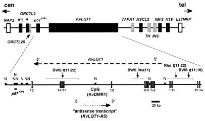
Normal mammalian development requires the correct parental contribution of imprinted genes. A lack of biparental contribution or aberrant expression of imprinted genes leads to a variety of developmental abnormalities in the mouse (20–22) and humans (22–27). Human chromosome 11p15.5 contains at least seven imprinted genes (Fig. 1), six of which are preferentially or exclusively expressed from the maternally derived chromosome and one (IGF2) of which is expressed primarily from the paternal allele (22). BWS, an overgrowth and cancer predisposition condition that maps to this region, results from the aberrant expression of one or more of these imprinted loci. BWS has a complex genetic etiology and can arise from paternal uniparental disomy (UPD), paternal duplication of 11p15.5, maternally inherited coding mutations in the _p57_KIP2 gene, or maternal chromosome rearrangements (26, 28). However, the most common mechanism resulting in BWS is the loss of imprinting (LOI) of IGF2 without apparent chromosomal abnormalities (26, 29–32). In contrast to Wilms’ tumors where LOI at IGF2 is usually accompanied by hypermethylation and silencing of the H19 gene (33, 34), biallelic expression of IGF2 in BWS usually occurs independently of changes in methylation or expression at H19 (32).
Figure 1.
Map of the 1,000-kb 11p15.5 imprinted domain. The relative location and sizes of genes are drawn approximately to scale, with genes shown to be regulated by genomic imprinting as solid black boxes. Imprinted expression of ORCTL2S, TAPA1(CD81), TH, and INS (gray boxes) has not yet been established, although Tapa1 and Ins have restricted allele-specific expression in the mouse (22). Presented below the map is an enlargement of the KvLQT1 locus showing its exon–intron structure (nomenclature from Lee et al.; ref. 38) as determined by comparison of the mRNA (GenBank accession no. U89364) and genomic sequences (GenBank accession nos. AC001228, AC003675, U90095, AC002403, AC000377, and AC003693), and the position of the intronic, differentially methylated CpG island KvDMR1 and _Not_I (N) sites. The direction of transcription of the maternally expressed KvLQT1 gene and the paternally expressed antisense transcript (KvLQT1-AS) are indicated. The approximate locations of the Beckwith–Wiedemann syndrome (BWS) and rhabdoid tumor (Rhd) 11p15.5 rearrangement breakpoints are shown. cen, centromere; tel, telomere.
The identification of an _H19_-independent pathway for LOI at IGF2 in patients with BWS suggests that at least one additional imprinting control region exists in chromosome band 11p15.5 (31, 32, 35). The existence of additional imprinting control elements is supported by the finding that, although targeted deletion of H19 affects the imprinting of Igf2 and Ins2 (36), the more distant Mash2, Kvlqt1, and _p57_Kip2 genes are unaffected (37). Although effects at more removed loci cannot be excluded, the disruption of the KvLQT1 gene in multiple cases of BWS patients with chromosome rearrangements suggests that the KvLQT1 locus may harbor imprinting control elements (38, 39). This report describes the identification and characterization of a region within the human and mouse KvLQT1 genes that has characteristics of an imprinting control element. We find that the imprinted methylation at this locus is disrupted in a majority of patients with BWS lacking other known genetic defects, and we suggest that this loss of methylation may be causally related to the disease phenotype.
MATERIALS AND METHODS
Cell Lines, Patient Samples, and Mice.
Cell lines with names containing the prefix “GM-” were obtained from the NIGMS Human Genetic Mutant Cell Repository. Peripheral blood lymphocytes were obtained from laboratory staff. Wilms’ tumor samples were provided by the National Wilms’ Tumor Study Group Tissue Bank. Cell lines from patients with BWS carrying the inv(11), t(11;16), and t(11;22) rearrangements as well as a cell line from the rhabdoid tumor with the t(11;22) rearrangement have been described by Sait et al. (40). Fibroblast or lymphocyte DNA samples from nonrearrangement BWS cases were from patients who have been described (31, 32). Human testis, sperm, and ovaries were obtained as described by Driscoll and Migeon (41). Other fetal tissues were acquired from the Brain and Tissues Banks for Developmental Disorders (University of Maryland). C57BL/6J and PWK inbred mouse strains were provided by Rosemary Elliott (Roswell Park Cancer Institute).
DNA Preparation and Southern Hybridization.
Genomic DNA was prepared from cell lines and most tissues by standard proteinase K digestion and phenol extraction. Sperm DNA was isolated from semen as described (41). Probes BX2 and DMRP were generated by PCR from PAC clone pdJ-74K15 (39) by using primers BX2f (5′-TCCATGCAGGGGATCGG-3′) plus BX2r (5′-GCAGTCCACATGGAAGGGCCAAACG-3′) and DMRP2f (5′-TCCTGGGGAGGTAGAAATG-3′) plus DMRP2r (5′-TGTCTGCCTGCTTCCTCTG-3′), respectively. PCR products were cloned into pCRII-TOPO (Invitrogen). Southern hybridizations were generally carried out as described (40) with two final washes of 0.2× SSC/0.5% SDS at 65°C. Hybridization signal was detected by using a Storm PhosphorImager (Molecular Dynamics). Quantitation of band intensity was done by using National Institutes of Health image software (rsb.info.nih.gov/nih-image/).
Isolation and Characterization of the Mouse Locus.
A portion (subclone 34; sc34) of the corresponding mouse locus was isolated by low-stringency (final washes: 2× SSC/0.5% SDS at 55°C) hybridization of the human locus to a _Pst_I subclone library of a murine PAC clone known to span the syntenic region on mouse distal chromosome 7 (42). The locus was expanded by rescreening the subclone library with a probe generated from the mouse PAC by using a linker-mediated PCR method (39) and a unique primer (sc34.3: 5′-GGTCCTGAATATAACTAGAAACCC-3′) designed from the sequence of sc34.
Reverse Transcription–PCR (RT-PCR).
Total RNA was isolated from cell lines and tissues by using RNeasy Mini Kits (Qiagen, Chatsworth, CA), and ≈6 μg of RNA was treated with RNase-free DNase (GIBCO/BRL). Half of the treated RNA was used for first-strand cDNA synthesis with Superscript II (GIBCO/BRL) reverse transcriptase (+RT) by using either oligo(dT) or an antisense strand-specific (sense with respect to KvLQT1 mRNA) primer. The remainder of the treated RNA was incubated in a similar manner but without reverse transcriptase (−RT). The sequence of primers used for expressed sequence tag (EST) “connection,” “transcript sampling,” and expression analysis in single chromosome 11 hybrids (see Fig. 2 for primer positions) as well as those used for strand-specific reverse transcription are available on request from the authors.
Figure 2.
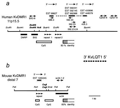
The human and mouse KvDMR1 loci. (a) Physical map of the human locus showing restriction sites with respect to hybridization probes DMRP and BX2 (solid black boxes), the KvDMR1 CpG island, the position of two direct repeat structures, and several ESTs transcribed in the opposite (antisense) direction compared with KvLQT1. The dashed line connecting these ESTs indicates that amplification products joining these cDNA fragments could be obtained by RT-PCR by using EST-specific primers. The bidirectional arrows (primer names indicated above) correspond to positive RT-PCR assays used to “sample” the genomic DNA sequence for expressed sequences. (b) Physical map of the syntenic mouse locus (same symbols as in a). A region of homology (83% identity over 437 bp) between the mouse and human loci is indicated by the striped box (the open end indicates that the downstream extent of homology is unknown, as the mouse sequence is not complete).
Analysis of Allelic Expression Patterns in the Mouse.
An expressed polymorphism in the mouse antisense transcript was detected between C57BL/6J and PWK mice by single-strand conformation polymorphism analysis by using genomic DNA and primers sc34.1 (5′-TTGCCTGAGGATGGCTGTG-3′) and sc34.2 (5′-CTTTCCGCTGTAACCTTTCTG-3′) with 0.5× MDE gels (FMC; 32 W; 3.5 h; 4°C). Cloning into pCRII-TOPO and sequencing identified a single base pair (A–G) polymorphism (position 4,231 in GenBank accession no. AF119385) between C57BL/6J and PWK mice, respectively. Allelic expression analysis was performed by using the same primers but with cDNA made from F1 fetal tissue RNA. Polymorphism characterization and assessment of imprinting at Kvlqt1 were carried out as described above but with primers Kvlqt1.5 (5′-GGGTAGAGCCTGACTCCTTCATTC-3′) and Kvlqt1.6 (5′-TAGGGTGGACAGTGGACAATCC-3′). Two polymorphisms between C57BL/6J and PWK mice were found: a dinucleotide substitution, GC/CA in C57BL/6J and PWK, respectively, at position 2,460–2,461 in the 3′ untranslated region of the Kvlqt1 mRNA (GenBank accession no. U70068) and an insertion–deletion polymorphism (C) at position 2,481.
Bioinformatics.
CpG islands were located by using grail (avalon.epn.ornl.gov/Grail-bin/EmptyGrailForm). Database searches were performed by using blast at the National Center for Biotechnology Information web site (www.nlm.nih.gov/cgi-bin/BLAST/nph-blast?Jform=0). Direct repeat structures (see Fig. 2 for locations) were identified by dot matrix analysis by using the pustell dna matrix module (window = 40 min; percentage score = 57) in macvector (Oxford Molecular Group, Campbell, CA).
RESULTS
Maternal-Specific Methylation at KvDMR1.
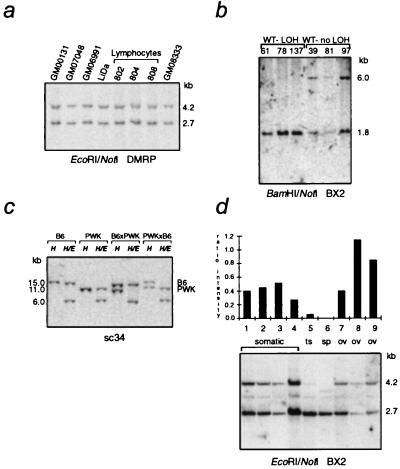
One-third (>325 kb) of the 1-Mb imprinted domain in 11p15.5 is occupied by the KvLQT1 gene, in which protein-coding mutations result in Romano–Ward and Jervell and Lange–Nielsen syndromes (43). The human and mouse (Kvlqt1) genes are regulated by genomic imprinting in a developmental and tissue-specific manner (37, 38, 44–46); however, features characteristic of imprinted genes, such as differentially methylated CpG-rich regions (DMRs) and short direct tandem repeat structures (11, 12), have not been reported. Large-scale sequencing of PAC clone pdJ-74K15 (GenBank accession no. U90058; ref. 39) and computer analysis identified a _Not_I-site-containing CpG island (designated KvDMR1) in intron 10 of KvLQT1, which also contained two direct repeat sequence motifs (Figs. 1 and 2a). Two probes, DMRP and BX2 (Fig. 2a), were developed by PCR and hybridized to Southern blots of normal human DNA digested with the methylation-sensitive enzymes _Not_I or _Bss_HII and either _Eco_RI or _Bam_HI. The 4.2-kb _Eco_RI and 6.0-kb _Bam_HI fragments detected by these probes were digested only partially with _Not_I or _Bss_HII, presumably because of differential methylation (Fig. 3a and data not shown). A similar experiment with DNA from Wilms’ tumors that had LOH in 11p15 showed that the uncut 6.0-kb _Bam_HI fragments were absent or greatly reduced (Fig. 3b). Because virtually all 11p15 LOH observed in Wilms’ tumors involves the maternal chromosome, these results suggest that the observed differential methylation was likely due to complete methylation on the maternally derived chromosome.
Figure 3.
Differential methylation of KvDMR1. (a) DNA isolated from normal lymphoblastoid (GM00131, GM07048, and GM06991), peripheral blood lymphocyte (802, 804, and 808), or fibroblast (GM08333) cells was digested with _Eco_RI and _Not_I and hybridized with DMRP (see Fig. 2a). (b) Southern blot of _Bam_HI/_Not_I-digested DNA from Wilms’ tumors (WT) with loss of heterozygosity (LOH) or without LOH (no LOH) in 11p15 and hybridized with the BX2 probe (Fig. 2a). (c) Hybridization of sc34 to _Hin_dIII (H) and _Hin_dIII/_Eag_I (H/E) double digests of adult kidney DNA from reciprocal [C57BL/6J (B6) × PWK]F1 animals (for F1 hybrids, the maternal parent is specified first). (d) The BX2 probe was hybridized to _Eco_RI/_Not_I digests of human DNA from somatic tissues (peripheral blood lymphocytes or brain), testes (ts), sperm (sp), and fetal ovaries (ov). The ratio of the intensity of the upper and lower bands is shown in the accompanying histogram. The faint band at 3–3.5 kb present in all lanes is a cross-hybridizing locus observed when experiments are done at reduced stringency. The additional band seen in the sperm lane is likely due to a heterogeneous methylation at this cross-reacting locus.
To confirm the parental origin of the methylated KvDMR1 allele and to show that this methylation was regulated by genomic imprinting, the mouse locus was identified and used to analyze DNA isolated from adult kidney. sc34 (Fig. 2b) detected a _Hin_dIII restriction fragment length polymorphism between C57BL/6J and PWK mice. In (C57BL/6J × PWK)F1 interspecific animals, only the 11.0-kb PWK (paternal) allele was cleaved by the methylation-sensitive restriction enzyme _Eag_I, whereas, in F1 DNA from the reciprocal cross, the paternally derived 15.0-kb C57BL/6J allele was cut by _Eag_I (Fig. 3c). The observed allele-specific methylation illustrates that KvLQT1 has a DNA methylation imprint typically associated with imprinted genes (11, 12), providing evidence for maternal-specific methylation in the human 11p15.5/mouse distal 7 imprinted domain.
To determine whether the methylation at KvDMR1 represents an imprinting mark established in the germ line, BX2 was hybridized to _Eco_RI/_Not_I digests of human somatic and germ-line DNA samples (Fig. 3d). In this experiment, the average ratio of the intensity of the uncut (methylated) to cut (unmethylated) band for somatic cell DNA was 0.4. Methylation at this locus was virtually absent in sperm DNA but enriched in two (ratio = 0.8 and 1.1) of three fetal ovary DNA samples. Because ovary samples typically contain 70% somatic cells (41), these results are consistent with the _Not_I site being completely methylated in human oocytes. The lack of enrichment in one ovarian specimen may reflect a larger than average contribution of somatic tissue in this dissection. Although definitive proof of maternal methylation during gametogenesis awaits analysis of purified oocytes, these results are consistent with maternal-specific methylation at KvDMR1 and, together with the finding of differential methylation at this site in murine embryonic stem cells (data not shown), suggest that this epigenetic difference represents a true gametic imprinting mark.
Identification of Human (KvLQT1) and Mouse (Kvlqt1) Antisense Transcripts.
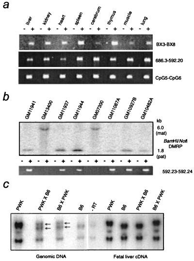
Further characterization of the mouse locus (GenBank accession no. AF119385) located the differentially methylated _Eag_I site(s) within a CpG island and identified a direct repeat sequence (Fig. 2b) in a position analogous to the human CpG island. Sequence alignment uncovered a region of 83% identity over >400 bp between human and mouse loci (see Fig. 2); however, no consensus splice sites could be found. blast analysis of KvDMR1 and flanking sequences identified several ESTs in human and one in mouse representing sequences transcribed in the opposite orientation with respect to KvLQT1 (Fig. 2). RT-PCR analysis with oligo(dT)-primed cDNA from human fetal liver RNA suggested that these cDNAs represented fragments of the same transcript (Fig. 4a). All EST and RT-PCR sequences were continuous with genomic DNA and showed no evidence of exon–intron boundaries. For each RT-PCR experiment, identical results were obtained when cDNA synthesis was carried out with primers specific for transcripts from the antisense strand (with respect to the direction of transcription of KvLQT1). Although RT-PCR detected transcripts in all human fetal tissues tested (Fig. 4a), corresponding transcripts were not detectable on Northern blots (CLONTECH) made from fetal or adult RNAs. Mouse EST 1265245 and human EST 68627 contained potential ORFs (150–400 bp) but are unlikely to reflect protein-coding potential, considering that no homology (at the nucleotide or amino acid level) exists between them. Although its length and potential overlap with KvLQT1 exons remain to be determined, we have designated this transcript KvLQT1-AS (KvLQT1 antisense).
Figure 4.
Expression of an antisense transcript associated with KvDMR1. (a) RNA was isolated from human fetal tissues and analyzed by RT-PCR with the indicated primer pairs (see Fig. 2a for location of primers). Primers 686.3 and 592.20 were designed from EST 68627 and EST 592241, respectively, and were used to show the connection of these two ESTs. The + and − indicate that the PCR templates were from cDNA (+RT) or mock cDNA (−RT), respectively. (b, Upper) Southern blot of _Bam_HI/_Not_I-digested DNA from a panel of eight single human chromosome 11 somatic cell hybrids (47) hybridized with DMRP. (b, Lower) RT-PCR analysis of same hybrids with primers specific for EST 592241. (c) Paternal-specific expression at the mouse KvDMR1 locus. Arrows indicate the presence of both alleles in DNA from F1 animals. In F1 fetal liver RNA from a C57BL/6J × PWK mating, as well as in the reciprocal cross, only the paternal allele was detected. The weak bands in the −RT lane result from contaminating DNA in the RNA samples, because the PCR primers do not amplify across an intron.
Imprinted Expression of KvLQT1/Kvlqt1-AS.
Because of the proximity to KvDMR1, we wished to determined whether KvLQT1-AS was imprinted. However, no polymorphisms were detected in 12 individuals after single-strand conformation polymorphism scanning of 450 bp and restriction-endonuclease-fingerprinting analysis of 2,600 bp (data not shown). We therefore took advantage of a recently developed panel of single human chromosome 11 somatic cell hybrids that have been characterized with respect to their expression of the imprinted H19 and IGF2 genes and methylation at KvDMR1 (47). Primers designed for ESTs 68627, 592241, and 435896 (Fig. 2a) were used in oligo(dT)-primed RT-PCR analysis of these hybrids, and, as illustrated for the EST 592241 primer pair (Fig. 4b), expression was observed only in the six hybrids shown to contain an unmethylated chromosome 11 (i.e., paternal). Identical results were obtained when this experiment was repeated with a primer designed for reverse transcription of the antisense RNA. By using an expressed sequence polymorphism, RT-PCR analysis with either strand-specific or oligo(dT)-primed cDNA of embryonic day-14.5 fetal liver from (C57BL/6J × PWK)F1 offspring identified exclusive paternal expression of the mouse antisense transcript (Fig. 4c), confirming the pattern of expression for the human transcript. Furthermore, we found that Kvlqt1-AS is also paternally expressed in embryonic day-14.5 mouse kidney, lung, gut, and heart (data not shown), the same fetal tissues shown in earlier studies (44–46) to have exclusive maternal expression of Kvlqt1.
Loss of Imprinting at KvDMR1 in Patients with BWS.
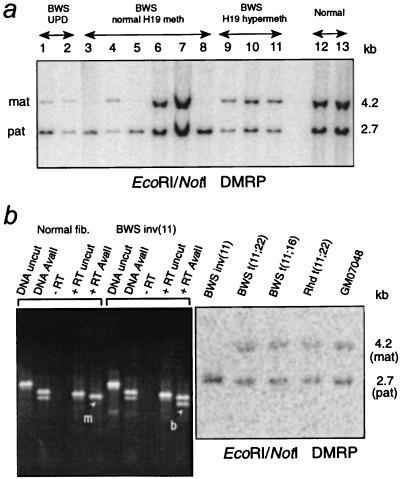
Because all BWS chromosome rearrangements in BWS breakpoint cluster 1 (BWSCR1) are located within the KvLQT1 gene (38, 39), it has been postulated that the disruption of the KvLQT1 genomic region affects the imprinting of IGF2 and perhaps other genes in the 11p15 domain (38). Because epigenetic changes at KvDMR1 might be related to this deregulation, the methylation status of this locus was tested in several classes of patients with BWS. Reduced methylation at KvDMR1 was observed in three patients with paternal UPD (Fig. 5a and not shown), reflecting the mosaic nature of UPD in BWS (28). Of 12 patients without UPD and with normal methylation at H19 (31, 32), 5 showed complete loss of the methylated band, whereas all 4 patients with BWS and hypermethylation at H19 (31, 32) showed normal methylation at KvDMR1 (Fig. 5a and data not shown). Of the five patients with loss of methylation at KvDMR1, two were informative for the _Apa_I/_Ava_II polymorphism described in IGF2 (48) and both showed LOI at IGF2 (32). All seven samples with normal methylation at KvDMR1 and H19 were uninformative at IGF2, precluding assessment of imprinting in these patients. However, three of these have been shown to have mutations in the _p57_KIP gene (A.C.S. and E.R.M., unpublished work). DNA from an aborted fetus with BWS, a maternally inherited inv(11)(p13;p15.5) (40), and LOI at IGF2 also showed loss of methylation at KvDMR1 (Fig. 5b), indicating that the inv(11) affected imprinted loci separated by 500 kb and on either side of the breakpoint. Two additional BWS translocations and one rhabdoid tumor translocation (40) showed normal methylation at KvDMR1 (Fig. 5b); the allelic expression pattern of IGF2 could not be determined in these samples, because none were informative at IGF2. The BWS t(11;22) has, however, been shown to disrupt the asynchronous replication pattern at IGF2 (28), suggesting that rearrangements in this domain might affect imprinting by additional mechanisms not connected to aberrant methylation at KvDMR1.
Figure 5.
(a) Southern blot of _Eco_RI/_Not_I digests of DNA from patients with nonrearrangement BWS and normal controls hybridized with DMRP. Densitometric analysis showed an increase in the cleaved unmethylated (paternal) band with respect to the uncleaved methylated (maternal) band in patients with BWS known to have paternal UPD (lanes 1 and 2) compared with normal individuals (lanes 12 and 13). Methylation at KvDMR1 was absent in the patients with BWS shown in lanes 3, 5, and 8. (b) Loss of imprinting at IGF2 and KvDMR1 in a BWS-associated inv(11). (Left) PCR and RT-PCR analysis at the _Ava_II/_Apa_I polymorphism in IGF2 (48). The lane labeled DNA _Ava_II illustrates that the BWS inv(11) cells and normal skin fibroblast control cells are heterozygous for the _Ava_II site. The presence of both alleles in the RNA +RT _Ava_II lane indicates that IGF2 is biallelically expressed. +RT and −RT indicate that the PCR templates were from cDNA (+RT) or mock cDNA (−RT). m, monoallelic; b, biallelic. (Right) Southern hybridization of the DMRP probe to _Eco_RI/_Not_I digests of DNA from individuals with BWS with the inv(11), a t(11;22), and t(11;16), as well as a rhabdoid tumor with a t(11;22) (40), all of which disrupt KvLQT1 (39). DNA from the inv(11) fetus with BWS showed an absence of the methylated allele.
DISCUSSION
This work describes an imprinted CpG island in an intron of KvLQT1 that is methylated on the active (maternal) allele of this gene and associated with an oppositely oriented RNA transcript expressed from the repressed (paternal) KvLQT1 locus. This situation is reminiscent of the “imprinting box” in region 2 of the mouse Igf2r locus (8), which has recently been shown to be necessary for the correct imprinted expression of Igf2r transgenes (16). Moreover, transgenes showing repression of Igf2r after paternal transmission expressed an antisense transcript dependent on this CpG island (16). Thus, KvLQT1 can be added to the increasing number of endogenous imprinted genes shown to overlap with imprinted antisense transcripts (13–17). Similar to the situation at the Igf2r and UBE3A loci, and unlike that at Igf2 and Zpf127/ZPF127 (13–15), KvLQT1-AS/Kvlqt1-AS is imprinted in the opposite direction compared with the associated protein-coding gene and therefore conforms to the expression competition model of genomic imprinting (19, 49). On the other hand, if the overlapping antisense transcripts associated with the similarly imprinted Igf2 and ZPF127/Zpf127 loci carry out regulatory functions, these functions may not be related to genomic imprinting and/or are likely to act through a different mechanism. It is interesting to note that the three oppositely imprinted antisense RNAs described to date are associated with maternally expressed imprinted genes (16, 17, and this study).
Based on expression analysis in _Dnmt_1−/− mice, Caspary et al. (37) showed Kvlqt1 to be an indirect target of methylation and predicted the existence of a maternally methylated locus and associated paternally expressed RNA within Kvlqt1, with the paternal transcript competing with Kvlqt1 for expression. One possibility is that Kvlqt1-AS is an imprintor gene that competes with the target-imprinted gene (19) Kvlqt1 for expression and is silenced directly by DNA methylation. Down-regulation of Kvlqt1-AS expression during developmental relaxation of Kvlqt1 imprinting (37, 44–46) would lend support to the notion of a functional role for the antisense RNA transcription in Kvlqt1 imprinting. A second possibility is that KvDMR1 acts as an insulator or boundary element as recently suggested for the core element upstream of the H19 gene (50, 51). In this model, KvDMR1 would block the promoter of Kvlqt1 and/or other genes in the vicinity from interacting with enhancers, and antisense RNA transcript levels would not necessarily change during development. Although Kvlqt1 has maternal-specific expression during early embryonic growth in all mice tested, the developmental regulation of Kvlqt1 imprinting varies considerably between strains (37, 44–46). Whether this variability is related to differences in methylation at KvDMR1 or elsewhere within the gene is unknown; however, consistent with KvDMR1 being a imprinting control element, differential methylation is maintained (at least at the _Eag_I sites tested; Fig. 3c) in adult kidney DNA where biallelic expression of Kvlqt1 is evident in the same tissue from (C57BL/6J × PWK)F1 offspring (G.V.F. and M.J.H., unpublished work). In this respect, it will also be important to compare the expression of KvLQT1 and KvLQT1-AS in BWS patients with and without methylation at KvDMR1.
The H19 locus is not a domain-wide imprinting control element, because, unlike Ins2 and Igf2, the imprinting of _p57_Kip2, Kvlqt1, and Mash2 is unaffected in H19 deletion mice (37). This finding supports earlier conclusions resulting from the observation that the majority of informative patients with BWS have LOI at IGF2 (32) but retain normal methylation and monoallelic expression at H19 (30–32). This lack of reciprocity was also noted for the BWS inv(11) reported by Brown et al. (35) as well as the inv(11) case described here. These observations suggest the existence of an _H19_-independent mechanism for the regulation of IGF2 imprinting and, together with the results of the study of the H19 knockout mouse (36, 37), predict the existence of a separately regulated imprinted domain and at least one additional cis-acting imprinting control element or center in 11p15.5 (and mouse distal 7). A subset of DMRs have been termed gametic imprints because of their establishment in the germ line and maintenance throughout development (2). Transgenic and targeted mutation analyses in the mouse have shown the importance of at least two of these sites in the regulation of genomic imprinting, namely the upstream DMR of H19 and region 2 in Igf2r (16, 50). Furthermore, the maternally methylated DMR at exon 1 of the SNRPN gene is included in the smallest microdeletions found in patients with Praeder–Willi syndrome showing imprinting-center defects (52). Although direct studies of purified oocytes are needed for confirmation, the lack of methylation at KvDMR1 in sperm and its enrichment in ovaries suggest that this locus is also a gametic imprinting mark and could therefore represent a critical control element or imprinting center in 11p15.5 (and distal chromosome 7 in the mouse). The loss of methylation at KvDMR1 in patients with BWS with normal H19 methylation and biallelic expression of IGF2 and the existence of the associated paternally expressed antisense RNA suggest the hypothesis that this locus regulates the imprinted expression of KvLQT1, IGF2, and perhaps other imprinted genes in the domain. Functional disruption of KvDMR1, as evidenced by loss of methylation, may account for a majority (five of nine patients studied here) of non-UPD, nonrearrangement BWS cases without mutations at _p57_KIP2. A limitation of this study is the general unavailability of patient tissues affected by overgrowth. It is formally possible that changes in methylation at KvDMR1 in fibroblasts and lymphocytes from patients with BWS may reflect tissue-specific differences or occasional loss of methylation in these cell types, and may not accurately reflect the methylation status in affected tissues. However, in an analysis of 17 normal individuals (Fig. 3a and data not shown) including DNA from four lymphoblast and two fibroblast cell lines, three lymphocyte samples, and eight fetal hearts, no departure from the differentially methylated pattern shown in Fig. 3a has been observed. On the other hand, the proportion of patients with BWS and epigenetic changes at KvDMR1 might even be greater if analysis of the tissues actually affected by overgrowth were available for molecular examination. Analysis of genomic sequence indicates the presence of multiple CpG islands within the 11p15.5 imprinted domain (C.D.D., G.V.F., and M.J.H., unpublished work). The assessment of methylation at these sites in both normal and patient DNA will help to determine whether their methylation patterns show allelic specificity and are as stringently controlled as the methylation pattern for KvDMR1. Definitive answers to questions regarding the function of KvDMR1 and its associated paternal transcript await the generation of mouse models with specific mutations in this region and further detailed study of the molecular pathology of patients with BWS.
Acknowledgments
We would like to thank Paul Grundy and the National Wilm’s Tumor Study Group Tissue Bank for supplying Wilm’s tumor samples; Pamela Karnes for the rhabdoid tumor cell lines; Rosemary Elliott and the Roswell Park Cancer Institute animal facility for C57BL/6J and PWK mice; Paul Soloway, Ramsi Haddad, and Bill Held for critically reading the manuscript; and the Brain and Tissues Banks for Developmental Disorders for human fetal tissues. W.R., E.R.M., and P.N.S. acknowledge the support of the Wellcome Trust, and J.A.J. acknowledges the support of the Biotechnology and Biological Sciences Research Council (United Kingdom) and the Newton Trust. This work was funded by a grant to W.R. from the Cancer Research Campaign (United Kingdom) and National Institutes of Health Grant CA63333 to M.J.H. and T.B.S.
ABBREVIATIONS
BWS
Beckwith–Wiedemann syndrome
DMR
differentially methylated region
EST
expressed sequence tag
LOI
loss of imprinting
LOH
loss of heterozygosity
RT-PCR
reverse transcription–PCR
RT
reverse transcriptase
UPD
uniparental disomy
Footnotes
This paper was submitted directly (Track II) to the Proceedings Office.
Data deposition: The sequence reported in this paper has been deposited in the GenBank database (accession no. AF119385).
References
- 1.Barlow D P. Trends Genet. 1994;10:194–199. doi: 10.1016/0168-9525(94)90255-0. [DOI] [PubMed] [Google Scholar]
- 2.Constancia M, Pickard B, Kelsey G, Reik W. Genome Res. 1998;8:881–900. doi: 10.1101/gr.8.9.881. [DOI] [PubMed] [Google Scholar]
- 3.Surani M A. Cell. 1998;93:309–312. doi: 10.1016/s0092-8674(00)81156-3. [DOI] [PubMed] [Google Scholar]
- 4.Li E, Beard C, Jaenisch R. Nature (London) 1993;366:362–365. doi: 10.1038/366362a0. [DOI] [PubMed] [Google Scholar]
- 5.Olek A, Walter J. Nat Genet. 1997;17:275–276. doi: 10.1038/ng1197-275. [DOI] [PubMed] [Google Scholar]
- 6.Tremblay K D, Duran K L, Bartolomei M S. Mol Cell Biol. 1997;17:4322–4329. doi: 10.1128/mcb.17.8.4322. [DOI] [PMC free article] [PubMed] [Google Scholar]
- 7.Shibata H, Ueda T, Kamiya M, Yoshiki A, Kusakabe M, Plass C, Held W A, Sunahara S, Katsuki M, Muramatsu M, et al. Genomics. 1997;44:171–178. doi: 10.1006/geno.1997.4877. [DOI] [PubMed] [Google Scholar]
- 8.Stoger R, Kubicka P, Liu C G, Kafri T, Razin A, Cedar H, Barlow D P. Cell. 1993;73:61–71. doi: 10.1016/0092-8674(93)90160-r. [DOI] [PubMed] [Google Scholar]
- 9.Shemer R, Birger Y, Riggs A D, Razin A. Proc Natl Acad Sci USA. 1997;94:10267–10272. doi: 10.1073/pnas.94.19.10267. [DOI] [PMC free article] [PubMed] [Google Scholar]
- 10.Shibata H, Yoda Y, Kato R, Ueda T, Kamiya M, Hiraiwa N, Yoshiki A, Plass C, Pearsall R S, Held W A, et al. Genomics. 1998;49:30–37. doi: 10.1006/geno.1998.5218. [DOI] [PubMed] [Google Scholar]
- 11.Neumann B, Kubicka P, Barlow D. Nat Genet. 1995;9:12–13. doi: 10.1038/ng0195-12. [DOI] [PubMed] [Google Scholar]
- 12.Gabriel J M, Gray T A, Stubbs L, Saitoh S, Ohta T, Nicholls R D. Mamm Genome. 1998;9:788–793. doi: 10.1007/s003359900868. [DOI] [PubMed] [Google Scholar]
- 13.Moore T, Constancia M, Zubair M, Bailleul B, Feil R, Sasaki H, Reik W. Proc Natl Acad Sci USA. 1997;94:12509–12514. doi: 10.1073/pnas.94.23.12509. [DOI] [PMC free article] [PubMed] [Google Scholar]
- 14.Jong, M. T. C., Carey, A. H., Caldwell, K. A., Lau, M. H., Handel, M. A., Driscoll, D. J., Stewart, C. L., Rinchik, E. M. & Nicholls, R. D. (1999) Hum. Mol. Genet., in press. [DOI] [PubMed]
- 15.Jong, M. T. C., Gray, T. A., Glenn, C. C., Ji, Y., Saitoh, S., Driscoll, D. J. & Nicholls, R. D. (1999) Hum. Mol. Genet., in press. [DOI] [PubMed]
- 16.Wutz A, Smrzka O, Schweifer N, Schellanders K, Wagner E, Barlow D. Nature (London) 1997;389:745–749. doi: 10.1038/39631. [DOI] [PubMed] [Google Scholar]
- 17.Rougeulle C, Cardoso C, Fontes M, Colleaux L, Lalande M. Nat Genet. 1998;19:15–16. doi: 10.1038/ng0598-15. [DOI] [PubMed] [Google Scholar]
- 18.Reik W, Constancia M. Nature (London) 1997;389:669–671. doi: 10.1038/39461. [DOI] [PubMed] [Google Scholar]
- 19.Barlow D P. EMBO J. 1997;16:6899–6905. doi: 10.1093/emboj/16.23.6899. [DOI] [PMC free article] [PubMed] [Google Scholar]
- 20.Cattanach B, Jones J. J Inherited Metab Dis. 1994;17:403–420. doi: 10.1007/BF00711356. [DOI] [PubMed] [Google Scholar]
- 21.Surani M. Curr Opin Cell Biol. 1994;6:390–395. doi: 10.1016/0955-0674(94)90031-0. [DOI] [PubMed] [Google Scholar]
- 22.Morison I M, Reeve A E. Hum Mol Genet. 1998;7:1599–1609. doi: 10.1093/hmg/7.10.1599. [DOI] [PubMed] [Google Scholar]
- 23.Ledbetter D H, Engel E. Hum Mol Genet. 1995;4:1757–1764. doi: 10.1093/hmg/4.suppl_1.1757. [DOI] [PubMed] [Google Scholar]
- 24.Lalande M. Annu Rev Genet. 1996;30:173–195. doi: 10.1146/annurev.genet.30.1.173. [DOI] [PubMed] [Google Scholar]
- 25.Issa J P, Baylin S B. Nat Med. 1996;2:281–282. doi: 10.1038/nm0396-281. [DOI] [PubMed] [Google Scholar]
- 26.Reik W, Maher E R. Trends Genet. 1997;13:330–334. doi: 10.1016/s0168-9525(97)01200-6. [DOI] [PubMed] [Google Scholar]
- 27.Nicholls R D, Saitoh S, Horsthemke B. Trends Genet. 1998;14:194–200. doi: 10.1016/s0168-9525(98)01432-2. [DOI] [PubMed] [Google Scholar]
- 28.Li M, Squire J A, Weksberg R. Am J Med Genet. 1998;79:253–259. [PubMed] [Google Scholar]
- 29.Weksberg R, Shen D R, Fei Y L, Song Q L, Squire J. Nat Genet. 1993;5:143–150. doi: 10.1038/ng1093-143. [DOI] [PubMed] [Google Scholar]
- 30.Reik W, Brown K W, Schneid H, Le Bouc Y, Bickmore W, Maher E R. Hum Mol Genet. 1995;4:2379–2385. doi: 10.1093/hmg/4.12.2379. [DOI] [PubMed] [Google Scholar]
- 31.Catchpoole D, Lam W W, Valler D, Temple I K, Joyce J A, Reik W, Schofield P N, Maher E R. J Med Genet. 1997;34:353–359. doi: 10.1136/jmg.34.5.353. [DOI] [PMC free article] [PubMed] [Google Scholar]
- 32.Joyce J A, Lam W K, Catchpoole D J, Jenks P, Reik W, Maher E R, Schofield P N. Hum Mol Genet. 1997;6:1543–1548. doi: 10.1093/hmg/6.9.1543. [DOI] [PubMed] [Google Scholar]
- 33.Rainer S, Johnson L A, Dobry C J, Ping A J, Grundy P E, Feinberg A P. Nature (London) 1993;362:747–749. doi: 10.1038/362747a0. [DOI] [PubMed] [Google Scholar]
- 34.Taniguchi T, Sullivan M J, Ogawa O, Reeve A E. Proc Natl Acad Sci USA. 1995;92:2159–2163. doi: 10.1073/pnas.92.6.2159. [DOI] [PMC free article] [PubMed] [Google Scholar]
- 35.Brown K, Villar A, Bickmore W, Clayton-Smith J, Catchpoole D, Maher E, Reik W. Hum Mol Genet. 1996;5:2027–2032. doi: 10.1093/hmg/5.12.2027. [DOI] [PubMed] [Google Scholar]
- 36.Leighton P A, Ingram R S, Eggenschwiler J, Efstratiadis A, Tilghman S M. Nature (London) 1995;375:34–39. doi: 10.1038/375034a0. [DOI] [PubMed] [Google Scholar]
- 37.Caspary T, Cleary M A, Baker C C, Guan X J, Tilghman S M. Mol Cell Biol. 1998;18:3466–3474. doi: 10.1128/mcb.18.6.3466. [DOI] [PMC free article] [PubMed] [Google Scholar]
- 38.Lee M, Hu R-J, Johnson L, Feinberg A. Nat Genet. 1997;15:181–185. doi: 10.1038/ng0297-181. [DOI] [PubMed] [Google Scholar]
- 39.Reid L H, Davies C, Cooper P R, Crider-Miller S J, Sait S N, Nowak N J, Evans G, Stanbridge E J, deJong P, Shows T B, et al. Genomics. 1997;43:366–375. doi: 10.1006/geno.1997.4826. [DOI] [PubMed] [Google Scholar]
- 40.Sait S, Nowak N, Singh-Kahlon P, Weksberg R, Squire J, Shows T, Higgins M. Genes Chromosomes Cancer. 1994;11:97–105. doi: 10.1002/gcc.2870110206. [DOI] [PubMed] [Google Scholar]
- 41.Driscoll D J, Migeon B R. Somatic Cell Mol Genet. 1990;16:267–282. doi: 10.1007/BF01233363. [DOI] [PubMed] [Google Scholar]
- 42.Day C D, Smilinich N J, Fitzpatrick G V, deJong P J, Shows T B, Higgins M J. Mamm Genome. 1999;10:182–185. doi: 10.1007/s003359900965. [DOI] [PubMed] [Google Scholar]
- 43.Wang Q, Bowles N E, Towbin J A. Mol Med Today. 1998;4:382–388. doi: 10.1016/s1357-4310(98)01320-3. [DOI] [PubMed] [Google Scholar]
- 44.Gould T D, Pfeifer K. Hum Mol Genet. 1998;7:483–487. doi: 10.1093/hmg/7.3.483. [DOI] [PubMed] [Google Scholar]
- 45.Jiang S, Hemann M A, Lee M P, Feinberg A P. Genomics. 1998;53:395–399. doi: 10.1006/geno.1998.5511. [DOI] [PubMed] [Google Scholar]
- 46.Paulsen M, Davies K R, Bowden L M, Villar A J, Franck O, Fuermann M, Dean W L, Moore T F, Rodrigues N, Davies K E, et al. Hum Mol Genet. 1998;7:1149–1159. doi: 10.1093/hmg/7.7.1149. [DOI] [PubMed] [Google Scholar]
- 47.Gabriel J M, Higgins M J, Gebuhr T C, Shows T B, Saitoh S, Nicholls R D. Proc Natl Acad Sci USA. 1998;95:14857–14862. doi: 10.1073/pnas.95.25.14857. [DOI] [PMC free article] [PubMed] [Google Scholar]
- 48.Tadokora K, Fujii H, Inoue T, Yamada M. Nucleic Acids Res. 1991;19:6967. doi: 10.1093/nar/19.24.6967. [DOI] [PMC free article] [PubMed] [Google Scholar]
- 49.Bartolomei M, Webber A L, Brunkow M E, Tilgham S M. Genes Dev. 1993;7:1663–1673. doi: 10.1101/gad.7.9.1663. [DOI] [PubMed] [Google Scholar]
- 50.Thorvaldsen J L, Duran K L, Bartolomei M S. Genes Dev. 1998;12:3693–3702. doi: 10.1101/gad.12.23.3693. [DOI] [PMC free article] [PubMed] [Google Scholar]
- 51.Webber A L, Ingram R S, Levorse J M, Tilghman S M. Nature (London) 1998;391:711–716. doi: 10.1038/35655. [DOI] [PubMed] [Google Scholar]
- 52.Ohta T, Gray T A, Rogan P K, Buiting K, Gabriel J M, Saitoh S, Muralidhar B, Bilienska B, Krajewska-Walasck M, Driscoll D J, et al. Am J Hum Genet. 1999;64:397–413. doi: 10.1086/302233. [DOI] [PMC free article] [PubMed] [Google Scholar]
