Amyloid-β Binds to the Extracellular Cysteine-rich Domain of Frizzled and Inhibits Wnt/β-Catenin Signaling (original) (raw)
Abstract
The amyloid-β peptide (Aβ) plays a major role in neuronal dysfunction and neurotoxicity in Alzheimer disease. However, the signal transduction mechanisms involved in Aβ-induced neuronal dysfunction remain to be fully elucidated. A major current unknown is the identity of the protein receptor(s) involved in neuronal Aβ binding. Using phage display of peptide libraries, we have identified a number of peptides that bind Aβ and are homologous to neuronal receptors putatively involved in Aβ interactions. We report here on a cysteine-linked cyclic heptapeptide (denominated cSP5) that binds Aβ with high affinity and is homologous to the extracellular cysteine-rich domain of several members of the Frizzled (Fz) family of Wnt receptors. Based on this homology, we investigated the interaction between Aβ and Fz. The results show that Aβ binds to the Fz cysteine-rich domain at or in close proximity to the Wnt-binding site and inhibits the canonical Wnt signaling pathway. Interestingly, the cSP5 peptide completely blocks Aβ binding to Fz and prevents inhibition of Wnt signaling. These results indicate that the Aβ-binding site in Fz is homologous to cSP5 and that this is a relevant target for Aβ-instigated neurotoxicity. Furthermore, they suggest that blocking the interaction of Aβ with Fz might lead to novel therapeutic approaches to prevent neuronal dysfunction in Alzheimer disease.
Alzheimer disease (AD)2 is a progressive neurodegenerative disorder characterized, in its early stages, by a striking inability to form new memories. Recent work indicates that cognitive and memory impairments in early AD are caused by synaptic dysfunction instigated by pathological assemblies of the amyloid-β peptide (Aβ) (for recent reviews, see Refs.1-3). Neuropathological hallmarks of AD include increased brain levels and extracellular build-up of Aβ aggregates, intraneuronal neurofibrillary tangles composed of hyperphosphorylated Tau, and, notably, synaptic loss (1). Despite the fact that Aβ has been strongly implicated in neuronal dysfunction and neurotoxicity in AD, the signal transduction mechanisms involved in the neuronal impact of Aβ remain to be fully elucidated. A major current unknown is the identity of the neuronal receptor(s) that bind Aβ and mediate neuronal dysfunction. Identification of such receptor(s) would provide considerable insight into mechanisms of pathogenesis and might reveal novel opportunities for the development of strategies to combat AD.
The Wnt signaling pathway has been recently proposed to play a role in AD (for a review, see Ref. 4). Wnts are secreted glycoproteins that bind to and signal through Frizzled (Fz) receptors and mediate cell-cell communication (5). Wnt signaling regulates a variety of biological processes, including development, cell movement, polarity, axon guidance, and synapse formation (6). Different types of Wnt·Fz complexes may signal through the so-called canonical or noncanonical Wnt pathways. Canonical Wnt/Fz signaling results in stabilization and increased intracellular levels of β-catenin, whereas the noncanonical pathway is considered to be largely β-catenin-independent, operating instead through stimulation of intracellular Ca2+ release and activation of protein kinase C and Ca2+/calmodulin-dependent kinase II (6).
Canonical Wnt signaling is essential for neuronal development and for the maintenance of the developing nervous system (7), and it has recently been implicated in adult hippocampal neurogenesis (8). Of considerable interest regarding its possible implications in synaptic dysfunction in AD, Wnt signaling is also involved in regulation of synaptic function and plasticity (reviewed in Ref. 9).
Wnt signaling initiates upon the interaction, at the cell surface, of Wnt ligands with their cognate receptor complex, consisting of one member of the Fz family of membrane receptors and low density lipoprotein receptor-related protein 5 or 6 (10). Binding of Wnt to the extracellular cysteine-rich domain of Fz (FzCRD) results in recruitment of the post-synaptic density associated protein dishevelled, leading to inhibition of glycogen synthase kinase-3β (GSK-3β), accumulation, and nuclear translocation of β-catenin. In the nucleus, β-catenin binds to components of the high mobility group family of transcription factors lymphoid enhancer factor/T-cell factor (TCF) to regulate transcription of a variety of genes involved in the wide spectrum of functions regulated by this pathway (6). In the absence of a Wnt ligand, GSK-3β phosphorylates β-catenin, targeting it for ubiquitin-proteasome degradation (11,12). As a result, intracellular β-catenin levels are diminished, thus switching off the expression of Wnt target genes (13-15).
Several lines of evidence indicate that deregulated Wnt signaling may play a role in the pathogenesis of AD (reviewed in Refs.16-19). β-Catenin levels are markedly reduced in AD patients carrying PS1-inherited mutations (20,21). Activation of protein kinase C inhibits Aβ neurotoxicity by inhibiting GSK-3β activity and increasing nuclear translocation of β-catenin, suggesting that regulation of components of the noncanonical Wnt signaling pathway by Ca2+-dependent protein kinase C may be relevant in Aβ toxicity (22). Activation of GSK-3β is involved in the phosphorylation of Tau and formation of neurofibrillary tangles, a hallmark of AD (23). Moreover, the addition of Wnt to hippocampal neurons in culture prevents Aβ neurotoxicity (24). Together, those studies suggest that the Wnt/β-catenin signaling pathway plays an important role in neuroprotection against Aβ toxicity. However, the mechanisms by which extracellular Aβ causes such intraneuronal effects have not been fully clarified.
With the goal of identifying ligands that mediate the neuronal impact of Aβ, we have used phage display of peptide libraries to identify peptides that bind to Aβ. Using this approach, we isolated a cysteine-linked heptapeptide, denoted cSP5, that binds Aβ with high affinity. Homology comparison with human protein sequences revealed that cSP5 is highly homologous to a conserved region of the FzCRD present in several members of the Fz family of Wnt receptors.
Here we show, for the first time, that Aβ directly binds to the extracellular CRD of Fz5 at or in close proximity to the Wnt-binding site. Furthermore, we show that Aβ inhibits β-catenin accumulation, nuclear translocation, and Wnt-targeted gene transcription. Interestingly, the cSP5 peptide completely blocks Aβ binding to Fz and its effects on the Wnt/Fz signaling cascade. These results indicate that the Aβ-binding site in Fz is homologous to cSP5 and that this is a relevant target for Aβ neurotoxicity. Furthermore, they suggest that blocking the interaction of Aβ with Fz might lead to novel therapeutic approaches to prevent neuronal dysfunction in AD.
EXPERIMENTAL PROCEDURES
_Reagents_—Aβ with 40 or 42 amino acid residues was purchased from Bachem (Torrance, CA). The peptide was dissolved in 50% trifluorethanol in PBS (140 mm NaCl, 2.7 mm KCl, 10 mm phosphate buffer, pH 7.3) immediately before use. Where indicated (see “Results”), the experiments were also carried out using soluble Aβ42 oligomers (also known as “Aβ-derived diffusible ligands” (ADDLs)) prepared as previously described (37). Anti-β-catenin antibody was from Sigma, anti-Aβ mAb 6E10 was from Abcam (Cambridge, MA), anti-hemagglutinin (anti-HA) was from Santa Cruz (Santa Cruz, CA), anti-cyclophilin B was from Affinity Bioreagents (Golden, CO), and recombinant Fz4CRD, Fz5CRD, anti-Fz4CRD, and anti-Fz5CRD were from R & D Systems (Minneapolis, MN).
_Biopanning against A_β—Screening of the phage display peptide library was performed as previously described (25). Briefly, Aβ40 was diluted to 0.1 μg/ml in 50% trifluorethanol/PBS, and 50-μl aliquots were dried in the wells of a 96-well plate at 37 °C under constant shaking for 16 h. To check the state of aggregation of the samples, the material from one well was extracted and submitted to SDS-PAGE electrophoresis. Silver staining of the gel indicated that most of the Aβ40 was in monomeric form, and no high molecular weight aggregates could be visualized. The wells were blocked with 0.5% bovine serum albumin in PBS for 1 h, and 10 μl of a phage display library (PhD-C7C, New England Biolabs, Ipswich, MA) were added to each well. All further procedures were carried out exactly according to the manufacturer's instructions.
A peptide with the amino acid sequence SPPLRFF (denoted SP peptide) was expressed by one of 24 Aβ-binding phages that were individually picked and amplified and had their DNA sequenced. The sequence of the SP peptide was compared with protein sequences deposited in several data banks (NCBI) using BLAST (26).
_Peptide Synthesis_—The cyclic peptides cSP5 (CSPDLRF-FLC), cSP3 (CSRDFRPFLC), and cSP6 (CSPNIETFLC) were synthesized as previously described (27).
_Cell Culture_—Murine neuroblastoma 2A cells (N2A; ATCC CCL-131) and L-cells were cultured in Dulbecco's modified Eagle's medium (DMEM; Invitrogen) supplemented with 10% fetal bovine serum (FBS, Invitrogen), 100 units/ml penicillin, and 2 mm glutamine (Invitrogen) in a humidified atmosphere of 5% CO2 at 37 °C. For experiments, N2A cells were plated in DMEM, 10%FBS for 12 h, and neuronal differentiation was initiated by incubation in serum-free DMEM supplemented with 250 μm N6, 2′-_O_-dibutyryl cAMP for 24 h.
Binding of FzCRD to A_β—In a 96-well plate, 1 μg of Aβ40 or Aβ42 in 50 μl of 50% solution of trifluorethanol in PBS were dried overnight at 37 °C in different wells, washed with PBS, and blocked for 2 h with 1% BSA in PBS. Different concentrations of Fz4CRD or Fz5CRD in PBS containing 1% BSA were added to the wells and incubated overnight at 4 °C with agitation. The wells were washed six times with PBS, incubated with the corresponding anti-Fz4CRD or anti-Fz5CRD for 1 h, washed, incubated with horseradish peroxidase-conjugated secondary antibody for 1 h, and developed with SuperSignal Femto Substrate (Pierce). Specificity of the binding assay was verified by appropriate controls carried out using wells coated with BSA alone or using a nonspecific peptide (denoted SQI) that does not bind Aβ (25). To calculate the Kd for binding of Fz5CRD to Aβ, wells of an enzyme-linked immunosorbent assay plate were coated with Aβ40 or with different concentrations of Fz5CRD alone (i.e. in the absence of Aβ). Wells coated with Aβ were then incubated with different concentrations of soluble Fz5CRD. All of the wells were incubated with peroxidase-conjugated anti-Fz5CRD and developed with SuperSignal Femto Substrate (Pierce). The luminescence values obtained for anti-Fz5CRD adhesion to wells coated with known concentrations of Fz5CRD alone allowed the construction of a standard curve that directly related luminescence counts to Fz5CRD concentration (in nm). This standard curve was then used to determine the concentrations of Fz5CRD that bound to Aβ in each of the Aβ-coated wells. Nonlinear regression of the binding data yielded a_Kd value of 105 ± 9 nm for Fz5CRD binding to Aβ.
_Transfection Experiments_—Human embryonic kidney 293T cells (ATCC) were cultured in 100-mm plates using DMEM supplemented with 10% fetal bovine serum. At 70% confluence, the cells were transfected with 10 μg of Fz5HA, Fz6HA (kindly provided by Dr. Xi He, Children's Hospital Boston, Harvard Medical School), or PCS2 (empty vector) plasmids using a standard calcium phosphate method (28). Ten hours after transfection, the medium was changed, and the cells were allowed to recover for 24 h in growth medium. After the recovery period, the intact cells were directly used in cross-linking experiments or used for membrane extraction for cross-linking experiments with Aβ40, as described below.
_Cross-linking_—2.5 μm Aβ40 was incubated for 2 h on ice with 0.5 μm Fz5CRD in PBS buffer containing 0.5 μm BSA in the absence or in the presence of 10 μm cSP5, cross-linked with EDAC as previously described (29), and immunoprecipitated with anti-Aβ and protein A-Sepharose beads (Amersham Biosciences). After SDS-PAGE, the samples were transferred to nitrocellulose, and the membranes were probed with anti-Fz5CRD.
The procedure was basically the same when Aβ40 was cross-linked to whole cells; Aβ40 was incubated for 2 h on ice with 2 × 10⁶ 293T cells transfected with Fz5HA, Fz6HA, or empty plasmid (mock transfected) in the presence or absence of cSP3, cSP5, or cSP6. The proteins were cross-linked with EDAC, and the cells were washed three times with PBS and lysed with 100 mm β-d-_n_-octyl glucoside in 50 mm Tris-Cl, pH 7.6, for 2 h at 4 °C. The material was centrifuged at 14,000 × g for 30 min, and the supernatant was immunoprecipitated with anti-Aβ.
When cell membrane extracts were cross-linked to Aβ40, 4 × 10⁶ 293T cells transfected with Fz5HA or with PCS2 (empty vector) plasmids were rinsed briefly with ice-cold detachment buffer (0.25m sucrose, 1 mm EDTA, 0.3 mm phenylmethylsulfonyl fluoride, 10 mm Tris-Cl, pH 7.0) and scraped off in the presence of 0.5 ml of detachment buffer. The samples were centrifuged at 12,000 × g for 2 min, the supernatant was removed, and the pellet was incubated with solubilization buffer (1% Triton X-100, 125 mm NaCl, 1 mm EDTA, 10 mm Tris, pH 7.0) supplemented with protease inhibitor mixture (Roche Applied Science) for 40 min at 4 °C with end-over-end mixing. After solubilization, insoluble cell debris were removed by centrifugation at 12,000 × g. The material was incubated under gentle agitation for 2 h on ice with Aβ and cSP5 (as described above) and then with Protein-A-Sepharose for another hour. The supernatant was then immunoprecipitated with anti-Aβ. After SDS-PAGE analysis, the samples were probed by Western immunoblotting with anti-HA.
_Wnt Conditioned Medium_—Wnt 3A conditioned medium was produced by L-Wnt3A cells following the ATCC protocol. Briefly, the cells were cultured in growth medium without G418 for 4 days, approximately to confluency. After this period, conditioned medium was recovered, and fresh medium was added for 3 more days. The final conditioned medium was a mixture of the media collected after 4 and 3 days of incubation with cells.
_Affinity Chromatography_—1 mg of Aβ40 was coupled to a solid matrix (UltraLink Biosupport medium; Pierce) and used for affinity chromatography experiments. The cell membrane preparations from 4 × 10⁶ 293T cells transfected with Fz5HA) or with control empty plasmid (mock transfected) were incubated overnight with the Aβ affinity gel at 4 °C with agitation. The gel was loaded into a 1-ml column and washed with 40 ml of PBS. The gel was then sequentially washed with 10 ml of 1m NaCl, 40 ml of PBS, and 10 ml each of 4 and 8 m urea solutions. The collected fractions were dialyzed, concentrated, and, after SDS-PAGE separation, the samples were probed by Western blot with anti-HA.
β_-Catenin Quantification_—N2A cells or L-cells were incubated with 15% Wnt conditioned medium or 15% control medium, 500 nm Aβ, and/or 1.5 μm cSP5 for 9 h. The cultures were then rinsed with PBS and lysed in buffer containing 100 mm Tris-HCl, pH 7.5, 1% SDS, 150 mm NaCl, 1 mm EDTA, 20 mm sodium pyrophosphate, 20 mm sodium fluoride, 1 mm sodium orthovanadate, and a mixture of protease inhibitors. Nuclear enriched fractions were prepared as described by Golan et al. (30). Protein content in the samples was measured by the BCA method using bovine serum albumin as a standard. The samples (10 μg of total protein applied per lane) were resolved by SDS-PAGE. The gel was transferred to nitrocellulose membranes and probed with anti-β-catenin antibody. As a loading control, anti-cyclophilin B was used to allow direct comparison between the total protein mass applied to different lanes.
To evaluate the amount of β-catenin in the nucleus, N2A cells or L-cells plated on coverslips were incubated as described above with Wnt, Aβ, and/or cSP5 and were fixed with 4% _p_-formaldehyde. The cells were permeabilized with 0.05% saponin in 0.5% BSA solution in PBS for 1 h and incubated with anti-β-catenin as recommended by the manufacturer for 1 h. The coverslips were washed five times with PBS, incubated with Alexa 488-labeled goat anti-rabbit IgG, and rinsed five times. The cell nuclei were stained with DAPI (Sigma). The coverslips were mounted and imaged on a Nikon Eclipse TE300 microscope. The overlap between DAPI and anti-β-catenin labeling was used to determine nuclear localization of the protein. For each experimental condition, five images were acquired from each coverslip, enabling the counting of β-catenin in the nuclei of ∼1,200 cells/experimental condition. The data were analyzed for statistically significant differences between groups using Student's t test.
_Cell Transfection and Luciferase Activity Assay_—HEK 293T cells cultured in DMEM supplemented with 10% fetal bovine serum were transfected with TOPFlash (luciferase reporter containing TCF/lymphoid enhancer factor-binding site) and pGal (for β-galactosidase expression) plasmids using FuGENE 6 transfection reagent (Roche Applied Science). 0.1 μg of TOP-Flash and 0.025 μg of pGal/well were mixed with FuGENE reagent in DMEM, according to the manufacturer's instructions. DNA/FuGENE mixture was added to cells and maintained for 24 h at 37 °C. The cells were incubated for 18 h with 15% of Wnt conditioned medium, 500 nm Aβ, and/or 1.5 μm cSP5, cSP3, or cSP6. The cells were then lysed, and the lysates were used to determine reporter gene expression using the luciferase assay kit (Promega, San Luis Obispo, CA) in a Veritas Microplate Luminometer (Turner Biosystems). Transfection efficiencies were normalized with control plasmids by using β-galactosidase reporter activity, and the results are presented as “fold increases” in TOPFlash activity relative to cells transfected with the empty vector.
_RT-PCR_—Total RNA was isolated using the TRIzol method (Invitrogen) from N2A cells incubated as described above with Wnt, Aβ, and/or cSP5. RNA concentration was determined by absorption at 260 nm, and cDNA templates were prepared from 1 μg of RNA by using the SuperScript III First Strand Synthesis System for RT-PCR (Invitrogen). RT-PCR was performed using 0.1% of the cDNA product as starting material and primers for β-actin (131-bp fragment, 5′-GCCAAGTCCTGTTTCTGCTC-3′ and 5′-TCTACAATGAGCTGCGTGTG-3′), cyclin D1 (167-bp fragment, 5′-TCTCCTGCTACCGCACAAC-3′ and 5′-TTCCTCCACTTCCCCCTC-3′), and axin 2 (5′-CTCCTTGGAGGCAAGAGC-3′ and 5′-GGCCACGCAGCACCGCTG-3′). The reaction was followed in real time by SYBR green incorporation using SYBR GreenER qPCR SuperMix Universal according to the protocol provided by the manufacturer (Invitrogen) using an Applied Biosystems 7500 real time PCR system. After RT-PCR, a first derivative melting curve analysis was performed to confirm specificity. All of the reactions were performed in triplicate. Gene expression was normalized for the expression of the housekeeping gene β-actin.
RESULTS
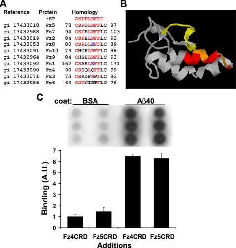
_A_β _Binds to the Cysteine-rich Domain of Frizzled_—To identify putative Aβ ligands, we initially screened a phage display peptide library and selected clones that bound specifically and with high affinities to soluble Aβ40 (see “Experimental Procedures”). The sequence of each clone was then submitted to BLAST analysis. One of the peptides thus identified, with amino acid sequence CSPPLRFFC (henceforth denoted as cSP), presented significant sequence homology to a conserved region of the extracellular CRD of several members of the human Fz family of proteins (Fig. 1_A_). Interestingly, the region of the FzCRD domain that is homologous to cSP (Fig. 1_B_, highlighted in red and orange) has been shown to be part of the Wnt-binding site in Fz (5). These initial results suggested that Aβ binds at or near the Wnt-binding site at the extracellular FzCRD.
FIGURE 1.
The cSP peptide is homologous to the conserved extracellular CRD of human Frizzled receptors. A, sequence alignments of cSP with members of the human Fz family. Identical residues are shown in red and highly conservative replacements are in blue. B, ribbon representation of the structure of the extracellular domain of Fz5. The amino acid sequence homologous to the cSP peptide is highlighted in red and_orange_. Residues involved in Wnt binding (5) are shown in_yellow_ and orange. The figure was generated using RasMol and the atomic coordinates for Fz5 (Protein Data Bank accession code 1IJY; Ref.5). C, wells in a 96-well plate were coated with either 1 μg of Aβ40 or 1 μg of BSA, blocked with 1% BSA, and incubated with 500 ng of Fz5CRD or Fz4CRD. After washing, binding was evaluated with anti-Fz4 or anti-Fz5 antibody. The plot shows results from densitometric quantification of the binding data using National Institutes of Health Image J software. Each bar represents the average of two independent experiments performed in triplicate. The blot illustrates one of the experiments performed.
To confirm the binding of Aβ to FzCRD, we performed a direct in vitro binding assay. Aβ40 and BSA were solubilized and immediately used to coat the wells of a 96-well plate. After blocking with BSA, the wells were incubated with Fz4CRD or Fz5CRD and probed with the respective anti-Fz4 or anti-Fz5 antibody. This essay revealed that Fz4CRD as well as Fz5CRD specifically bound to Aβ40 (Fig. 1_C_), suggesting that the amino acid residues that are replaced in the cSP-homologous region of Fz4CRD relative to Fz5CRD are not essential for Aβ binding.
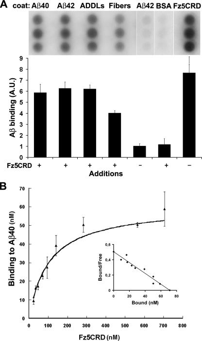
We also investigated the binding of Fz5CRD to Aβ in different aggregation states, including soluble oligomers currently accepted as the main toxins in AD and commonly known as ADDLs (37). Fz5CRD bound to wells coated with soluble Aβ40 and Aβ42, ADDLs, and fibrillar Aβ42 (Fig. 2_A_). Interestingly, Fz5CRD binding to fibrillar Aβ42 is 35% lower than binding to soluble Aβ40 and to ADDLs. It is also important to note that we cannot exclude the possibility that our Aβ fibril preparation may contain a small percentage of soluble Aβ species in equilibrium with fibrils. These results suggest that Fz5CRD interacts preferentially with soluble Aβ species.
FIGURE 2.
Fz5CRD binds Aβ40 and Aβ42. A, wells in a 96-well plate were coated with either 1 μg of Aβ40, 1 μg of Aβ42, 1 μg of soluble Aβ oligomers (ADDLs), 1 μg of Aβ fibrils, 1 μg of BSA, or 500 ng of Fz5CRD, blocked with 1% BSA and incubated with 500 ng of Fz5CRD or 1% BSA. After washing, binding was evaluated using anti-Fz5 antibody. The plot shows densitometric quantification of binding results using National Institutes of Health Image J software. Each_bar_ represents the average of two independent experiments performed in triplicate. The blot illustrates one of the experiments performed.B, wells were coated with 1 μg of Aβ40 or BSA, blocked with 1% BSA, and incubated with different concentrations of Fz5CRD as indicated. After washing, binding was evaluated with anti-Fz5 antibody. The plot represents values obtained for Fz5CRD binding to Aβ-coated wells minus background binding to BSA-coated wells. Each point represents the average of two independent experiments performed in triplicate. The inset shows a Scatchard plot of the binding data fit to a single straight line.
Aβ42 has a high propensity to aggregate and to form insoluble fibrils under physiological conditions. To minimize this problem and to avoid fibrillization and consequently loss of affinity of Fz5CRD by Aβ in the different binding assays performed, in all subsequent experiments we used Aβ40, which is much less prone to aggregation.Fig. 2_B_ shows a dose-response curve in which wells were coated with Aβ40 and incubated with different concentrations of Fz5CRD. The Kd estimated from the binding data (see “Experimental Procedures”) was 105 ± 10 nm. Because Wnt binds to Fz with a_Kd_ in the 1-10 nm range (38), these results suggest that Fz has a lower affinity for Aβ than for Wnt.
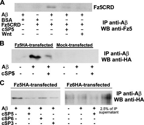
_cSP5 Inhibits the Binding of A_β_40 to Fz_—The effect of the cSP peptide on Aβ40 binding to Fz5CRD was next investigated using a cyclic synthetic peptide analog corresponding to the Fz5 region that is homologous to SP (78CSPDLRFFLC87), denoted cSP5. Aβ40 was incubated in solution with Fz5CRD in the absence or in the presence of cSP5 and/or Wnt3A and of the cross-linking agent EDAC (see “Experimental Procedures”). The samples were then immunoprecipitated by anti-Aβ antibody, resolved by SDS-PAGE, and probed by Western immunoblotting with anti-Fz5CRD antibody.Fig. 3_A_ shows that Fz5CRD bound to Aβ40 could be immunoprecipitated with anti-Aβ. Interestingly, Aβ40 binding to Fz5CRD was almost completely blocked by the presence of either cSP5 or Wnt (Fig. 3_A_), supporting the notion that Aβ binds to the Wnt-binding domain of Fz with lower affinity than Wnt.
FIGURE 3.
cSP5 inhibits the binding of Aβ40 to Fz. A, 2.5 μm Aβ40, 0.5 μm Fz5CRD, 0.5 μm BSA, 15% of Wnt3A conditioned medium, and/or 10 μm cSP5 were incubated for 2 h on ice, cross-linked with EDAC, and immunoprecipitated (IP) with anti-Aβ (see “Experimental Procedures”). After SDS-PAGE, the gel was transferred to nitrocellulose, and the membrane was probed with anti-Fz5CRD. B, membrane extracts from 293T cells transfected with Fz5HA plasmid (Fz5HA) or empty plasmid (mock transfected) were incubated for 2 h on ice in the absence or in the presence of 2.5 μm Aβ40 and 10 μm cSP5. The proteins were cross-linked with EDAC and immunoprecipitated with anti-Aβ. After SDS-PAGE and transfer to nitrocellulose, the membranes were probed with anti-HA. C, intact 293T cells transfected with Fz5 (Fz5HA) or with Fz6 (Fz6HA) were incubated for 2 h on ice with 2.5 μm Aβ40 in the presence or in the absence of 10 μm cSP5, cSP3, or cSP6. The proteins were cross-linked with EDAC, detergent-solubilized, and immunoprecipitated with anti-Aβ. After SDS-PAGE and transfer to nitrocellulose, the membranes were probed with anti-HA. WB, Western blot.
Binding of Aβ40 to Fz5 and the effect of the cSP5 peptide were also investigated in cell culture experiments. To this end, cell membrane extracts from 293T cells that had been transfected with Fz5HA (or mock transfected with empty plasmid) were incubated with Aβ40 in the absence or presence of cSP5. The material was then cross-linked, solubilized, immunoprecipitated with anti-Aβ, and probed by Western blotting with anti-HA. As shown inFig. 3_B_, Fz5HA present in the membrane extract from transfected cells bound to Aβ and binding was significantly inhibited by cSP5.
The interaction between Aβ40 and Fz was further investigated using intact cells. 293T cells that had been previously transfected with Fz5HA or Fz6HA were incubated with Aβ40 in the absence or in the presence of cSP5, cSP3 (CSRDFRPFLC), or cSP6 (CSPNIETFLC). cSP3 and cSP6 show the lowest homology to the cSP peptide selected by phage display and to Fz5 (Fig. 1_A_). Cross-linking of cell-bound Aβ was then performed as described under “Experimental Procedures.” After lysis and solubilization of the cell contents, the samples were immunoprecipitated using anti-Aβ antibody, resolved by SDS-PAGE, and probed by Western blotting using anti-HA antibody. The results shown in Fig. 3_C_ show that the Fz5HA construct expressed by transfected cells bound to Aβ40 and could be immunoprecipitated with anti-Aβ. Interestingly, cSP5 and cSP3 significantly inhibited binding of Fz5HA to Aβ40, whereas cSP6 did not (Fig. 3_C_). Moreover, the Fz6HA construct expressed by transfected cells did not significantly bind to Aβ40 and therefore did not immunoprecipitate with anti-Aβ. Rather, the majority of Fz6HA remained in the supernatant (Fig. 3_C_).
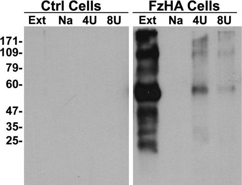
As an alternative approach to evaluate the specificity of Aβ-Fz interactions, Aβ40 was coupled to a gel matrix. The affinity matrix thus obtained was incubated with detergent-solubilized membranes from 293T cells that had been transfected with Fz5HA or with empty plasmids. The column was then eluted with increasingly denaturing buffers, and the eluates were analyzed by Western blotting with anti-HA. This showed that Fz5HA strongly bound to the immobilized Aβ in the gel and was eluted by 4 m urea (Fig. 4). Taken together, these results corroborate our hypothesis that Aβ binds with higher affinity to members of the Fz family that contain the cSP homologous sequence. They also show that Aβ binds to the extracellular CRD of Fz5 in both_in vitro_ solution assays and in cell membranes and that the cSP5 peptide blocks this interaction.
FIGURE 4.
Fz5HA binds with high affinity to immobilized Aβ40. Membrane extracts from 293T cells expressing Fz5HA (Fz5HA) or transfected with empty plasmid (mock) were incubated with the Aβ40 affinity gel as described under “Experimental Procedures.” The gel was sequentially eluted with 1 m NaCl (Na), 4 m urea (4U), and 8 m urea solutions (8U). Aliquots of the eluates were analyzed by SDS-PAGE and probed by Western immunoblotting with anti-HA. Most of the Fz5HA bound to Aβ40 was eluted with 4m urea. MW, molecular mass. Ext, total cell extract.
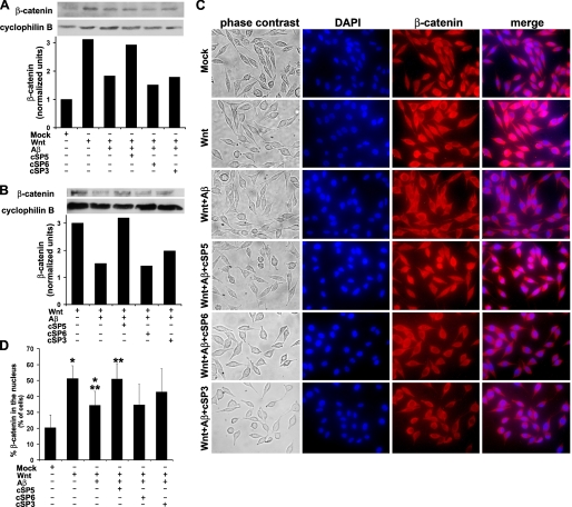
_cSP5 Blocks the Inhibition of Wnt/_β_-Catenin Signaling by A_β—Because the results described above indicated that Aβ binds to Fz at or in close proximity to the Wnt-binding site, we hypothesized that Aβ might compete with Wnt ligands for binding to Fz, thereby inhibiting downstream signaling via this pathway, including β-catenin stabilization and nuclear import. Indeed, we found that 500 nm Aβ40 significantly reduced (by ∼40%) total β-catenin levels in L-cells stimulated with Wnt3A (Fig. 5_A_) and reduced by ∼50% the amount of β-catenin in the nuclear fraction isolated from such cells (Fig. 5_B_). Remarkably, the addition of 1.5 μm cSP5 completely abolished the decrease in β-catenin levels induced by Aβ40 in L-cells. This indicates that Aβ40 binding to Fz is directly responsible for triggering a decrease in cellular levels of β-catenin.
FIGURE 5.
cSP5 blocks the inhibition of Wnt/β-catenin signaling by Aβ in L-cells. A, L-cells were incubated with 15% Wnt3A conditioned medium or 15% of control medium, 500 nm Aβ, and/or 1.5 μm cSP5, cSP3 or cSP6 for 9 h at 37°C. The cells were washed and lysed, and total (A) and nuclear (B) β-catenin levels were quantified by Western immunoblotting. The plots represent the means of two independent experiments, and the blot illustrates one of the experiments performed. C, L-cells incubated with Wnt, Aβ40, and/or cSP5, cSP3, or cSP6 as described in A were fixed and stained. The nuclei were stained with DAPI (blue). β-Catenin was detected using murine anti-β-catenin and anti-mouse Alexa 555 conjugate (red fluorescence). The images illustrate results from one of the experiments performed. D, nuclear localization of β-catenin was determined by co-localization of DAPI and β-catenin fluorescence in merged images. The plot represents the percentage of cells exhibiting nuclear β-catenin labeling (means ± S.D.) from four independent experiments, with five microscopic fields analyzed per condition in each experiment (∼1,200 cells counted per experimental condition).*, p < 0.00001; **, p < 0.0001.
These results prompted us to investigate the effects of Aβ40 and cSP5 on the nuclear translocation of β-catenin. As expected, stimulation of L-cells with Wnt3A markedly increased the nuclear localization of β-catenin (Fig. 5, C and_D_). In such Wnt-stimulated cells, treatment with Aβ40 decreased by ∼33% the presence of β-catenin in the nucleus (p < 0.00001). Interestingly, cSP5 completely blocked (p < 0.0001) the decrease in nuclear import of β-catenin induced by Aβ (Fig. 5, C and_D_).
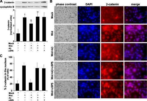
cSP3 and cSP6 did not significantly block the decrease in β-catenin levels promoted by Aβ, although cSP3 promoted a slight recovery in the levels of β-catenin in the nucleus (Fig. 5, A and_B_). Moreover, cSP3 (p = 0.06) and cSP6 (p = 0.93) did not significantly block the inhibition of β-catenin translocation to the nucleus promoted by Aβ (Fig. 5, C and_D_). To evaluate the effects of Aβ and cSP5 on Wnt/β-catenin signaling in a neuronal model, we performed the same experiments described in Fig. 5 using N2A cells (Fig. 6). The results showed that 500 nm Aβ40 significantly reduced (by ∼25%) β-catenin levels in N2A cells stimulated with Wnt3A (Fig. 6_A_). The addition of 1.5 μm cSP5 completely abolished the decrease in β-catenin level induced by Aβ40. As expected, stimulation of N2A cells with Wnt3A markedly increased the nuclear localization of β-catenin (Fig. 6_B_), and treatment with Aβ40 decreased by ∼70% the presence of β-catenin in the nucleus. cSP5 completely blocked the decrease in nuclear import of β-catenin induced by Aβ (Fig. 6,B and C). It is important to note that cSP5 had no effect on the increase in β-catenin levels promoted by Wnt (Fig. 6_A_), nor on β-catenin nuclear translocation (Fig. 6, B and C), indicating that cSP5 does not bind Wnt. Indeed, we found that biotinylated cSP5 does not bind to Wnt in vitro (supplemental Fig. S1).
FIGURE 6.
cSP5 blocks the inhibition of Wnt/β-catenin signaling by Aβ in neuronal differentiated N2A cells. A, differentiated N2A cells were incubated with 15% Wnt3A conditioned medium or 15% of control medium, 500 nm Aβ, and/or 1.5 μm cSP5 for 9 h at 37 °C. The cells were washed and lysed, and β-catenin levels were quantified by Western immunoblotting. The plot represents means ± S.D. of eight independent experiments, and the blot illustrates one of the experiments performed. *, p < 0.003;**, p < 0.008. B, cells were incubated with Wnt, Aβ40, and/or cSP5 as described in A, then fixed, and stained. The nuclei were stained with DAPI (blue). β-Catenin was detected using murine anti-β-catenin and anti-mouse Alexa 555 conjugate (red fluorescence). The images illustrate results from one of the experiments performed. C, nuclear localization of β-catenin was determined by co-localization of DAPI and β-catenin fluorescence in merged images. The plot represents the percentage of cells exhibiting nuclear β-catenin labeling (means ± S.D.) from three independent experiments, with five microscopic fields analyzed per condition in each experiment (∼1,200 cells counted per experimental condition).
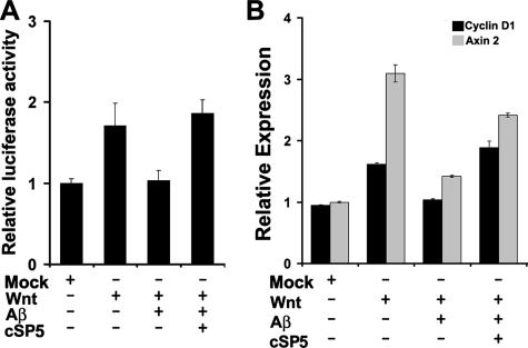
_cSP5 Rescues the Inhibition of the Expression of Wnt Target Genes by A_β—293T cells were incubated with Wnt and Aβ40 in the presence or absence of cSP5 and TCF-binding site reporter gene activity was measured (using the TOPFlash assay) in transient transfection experiments. As expected, Wnt significantly enhanced transcriptional activation of TCF/lymphoid enhancer factor-1. Importantly, Aβ40 inhibited stimulation by Wnt and cSP5 blocked the interference by Aβ (Fig. 7_A_).
FIGURE 7.
cSP5 rescues Wnt target gene transcription from inhibition by Aβ40. A, 293T cells were transfected with 0.1 μg of the TOPFlash or FOPFlash plasmids and with 0.025 μg of pGal and were incubated with 15% Wnt3A conditioned medium or 15% control medium, 500 nm Aβ, and/or 1.5 μm cSP5 for 9 h at 37°C. Reporter gene activities were assayed, normalized by pGal expression, and expressed relative to basal Wnt activity. B, differentiated N2A cells were incubated with Wnt3A, Aβ, and/or cSP5 as described for A. Expression levels of the housekeeping gene β-actin and of the Wnt target genes cyclin D1 and axin 2 were measured by RT-PCR. Cyclin D1 and axin 2 expression results are normalized by β-actin mRNA levels. Wnt-induced up-regulation of cyclin D1 and axin 2 expression was significantly blocked by Aβ, and cSP5 completely rescued Aβ inhibition.
To further substantiate the functional significance of the inhibition of Wnt/β-catenin signaling by Aβ in a neuronal model, N2A cells were incubated with Wnt, Aβ40, and/or cSP5, and expression of the Wnt target genes cyclin D1 and axin 2 was evaluated by RT-PCR (Fig. 7_B_). Consistent with the inhibition of nuclear translocation of β-catenin, Aβ treatment of the cells significantly reduced cyclin D1 (Fig. 7_B_, black bars) and axin 2 (Fig. 7_B_, gray bars) gene transcription, whereas cSP5 completely rescued the inhibition of cyclin D1 and almost completely rescued the inhibition of axin 2 expression by Aβ (Fig. 7_B_).
DISCUSSION
Aβ is thought to cause synaptic dysfunction and neuronal loss in AD. Applied to neurons in culture, Aβ induces an increase in levels of reactive oxygen species and hyperphosphorylation of Tau protein, which forms neurofibrillary tangles in AD brains (for recent examples, see Refs.31,32). However, the molecular events that determine the fate of neurons exposed to Aβ are unclear. Recent observations suggest that early memory loss in AD may be related to soluble Aβ-derived species and that formation of those species and/or their interaction with neurons may constitute primary targets for intervention in AD (1-3).
To find ligands for soluble Aβ that could provide insight into the mechanisms of Aβ toxicity, we used a phage display library of random heptapeptides. The display of random peptides on filamentous bacteriophage has allowed the identification of peptides that bind specifically to a variety of targets, including Aβ (25,33,34). Peptides thus identified have been shown to act as agonists or antagonists of various receptors and to mimic the binding sites of physiological receptors (25). Using this approach, we have now identified a cysteine-linked heptapeptide (denoted cSP5) that binds to soluble Aβ40/Aβ42 and is homologous to the extracellular cysteine-rich domain (FzCRD) present in several members of the Fz family of Wnt receptors.
The Fz domain homologous to cSP5 has been shown to be directly involved in Wnt binding (5). Insertion of a tripeptide (Gly-Ser-Gly) in the cSP5 homologous region of mouse Fz8 (between amino acid residues Ser81 and Pro82 or between Leu84 and Phe86) completely eliminates Wnt binding (5). Interestingly, among the 10 Fz family members that are expressed in humans and presented inFig. 1_A_, the SP peptide showed the lowest homology to Fz6. Fz6 is a member of the Fz family that does not transmit Wnt-induced β-catenin/TCF-dependent signaling when exposed to various Wnt ligands. In fact, Fz6 represses the Wnt pathway by transmitting an antagonistic signaling (30). Collectively, these results indicate that Aβ binds to several members of the Fz family that are homologous to cSP5 at or in close proximity to the Wnt-binding site.
Wnt signaling plays important roles in several aspects of neural circuit development, such as neuronal migration, axon path-finding, dendritic morphogenesis, and synaptic differentiation (for a review, see Ref.35). Moreover, synaptic activity causes the releaseofWnt3aatsynapsesinan_N_-methyl-d-aspartatereceptor-dependent manner, and inhibition of Wnt signaling impairs LTP (36). LTP is a classic paradigm for synaptic plasticity and a model for memory and learning, faculties that are selectively lost in early stage AD. Indeed, Aβ oligomers have been show to bind to dendritic spines in hippocampal neurons and inhibit LTP (37,39). Therefore, investigation of Aβ/Fz interactions may provide insight into cellular mechanisms involved in synaptic dysfunction and blockade of LTP.
Several intracellular binding partners for Wnt/Fz signaling have been implicated in AD. In accord with our data, all studies so far indicate that Aβ inhibits Wnt/Fz signaling. Aβ was shown to induce the expression of the Wnt antagonist Dickkopf-1 (DKK1), consistent with interference with normal Wnt signaling (40). Moreover, canonical Wnt signaling controls the kinase activity of GSK-3β, indicating that Wnt signaling could be a key link between two major hallmarks of AD, namely the brain accumulation of Aβ (which can interfere with Wnt signaling) (40) and neurofibrillary tangles formed by hyperphosphorylated Tau (a well known GSK-3β substrate) (16,17,40,41). However, no direct evidence of Aβ binding to a member of the Wnt/Fz signaling pathway had yet been presented.
Here we show, for the first time, that Fz5CRD binds to immobilized Aβ40 as well as to Aβ42, to soluble Aβ42 oligomers (ADDLs) and, to a lesser extent, to Aβ fibrils. In addition, Aβ40 co-immunoprecipitates with recombinant Fz5CRD and with Fz5HA present in the membranes of 293T cells transfected with Fz5HA. Moreover, we identified a peptide, denoted cSP5, that is homologous to amino acid residues 78-87 from Fz5 and almost completely blocks the binding of Fz5CRD or Fz5HA to Aβ40. Because cSP5 is homologous to a highly conserved domain of the human Fz family (Fig. 1_A_), our data strongly suggest that Aβ40 or Aβ42 are also capable of binding to other members of the Fz family that contain the conserved cSP5 domain, such as Fz4 (Fig. 1_C_). We propose that Aβ preferentially binds to members of the Fz family with higher homologies to cSP5, such as Fz5, Fz7, Fz2, and Fz8 (Fig. 1_A_). Interestingly, these members of the Fz family have been shown to be involved in transduction and intercellular transmission of polarity information during tissue morphogenesis and/or in differentiated tissues (42). Because the physiological functions of soluble Aβ40 have not yet been fully identified, our results may open new avenues for the study of the role of soluble Aβ40 in embryonic development.
Several lines of evidence show that β-catenin-mediated transcription plays an important role in neuronal viability (7). Previous studies indicated that presenilin-1 inherited mutations, which lead to particularly aggressive forms of AD, may affect the levels, trafficking, or phosphorylation state of cytosolic β-catenin (43). Moreover, exposure of hippocampal neurons to Wnt conditioned medium containing 5 μm fibrillar Aβ promoted a decrease in the levels of cytosolic β-catenin, significantly reducing the transcription of the Wnt target genes engrailed-1 (∼25%) and cyclin D1 (∼8%) (22,44). These observations are in agreement with our results, because we also observed a decrease in the levels of β-catenin and in transcription of cyclin D1 (∼36%) and axin 2 (∼55%) in cells incubated with Wnt and Aβ (Figs.5,6 and7). However, in contrast to the work of Garrido et al. (22), we used 500 nm of soluble Aβ40 instead of 5 μm of fibrillar Aβ42, suggesting that soluble Aβ40 species are mainly responsible for inhibition of the Wnt/Fz signaling pathway and, consequently, for the decrease in the levels of β-catenin and of Wnt target gene transcription. The incomplete functional blockade of Wnt/Fz signaling (Fig. 6_A_) by Aβ may result from the presence of endogenous Wnt in the medium and from the heterogeneity of Fz members expressed in N2A cells, with some members of the Fz family of receptors being less sensitive to inhibition by Aβ40.
In the present study, we also show that Aβ effects on β-catenin levels and on Wnt target gene transcription can be prevented by specifically inhibiting the binding of Aβ40 to Fz using micromolar concentrations of the synthetic peptide cSP5 (Figs.5,6 and7), which mimics the Aβ-binding site in Fz receptors. Taking into account that Aβ40-induced inhibition of Wnt/Fz signaling is completely rescued in the presence of cSP5 and that cSP5 by itself does not augment Wnt/Fz signaling, our results suggest that there is a common binding domain for Fz receptors and cSP5 on Aβ40 and Aβ42. However, we cannot completely rule out the possibility that other regions of the Fz receptor might also participate in interactions with Aβ. This issue clearly deserves deeper investigation, and cSP5 might be an important tool to understand the Wnt/Fz signaling function as well as Aβ toxicity. In this regard, it is important to note that cSP5 up to 10 μm is not toxic to cells (data not shown).
Similar to cSP5, cSP3 inhibits Aβ binding to Fz5 expressed by transfected 293T cells (Fig. 3_C_). However, cSP3 is not effective in blocking the effects of Aβ on β-catenin levels (Fig. 5). Significant differences between these two assays may explain this apparent discrepancy. First, the binding assay was performed for 2 h on ice in PBS buffer with 293T cells transfected exclusively with Fz5HA. On the other hand, β-catenin assays employed L-cells incubated for 9 h at 37°C in culture medium. Use of different cells and assay conditions may interfere with the interactions between cSP3 and Aβ and/or Aβ and Fz. Moreover, it is possible that, unlike cSP5, cSP3 does not effectively block the interaction of Aβ with other members of the Fz family (besides Fz5), which may be expressed by L-cells. Conceivably, the interaction between Aβ and cSP3 is weaker than with cSP5 and is disrupted under the more physiological conditions of the β-catenin assay, whereas the Aβ/cSP5 interaction seems to be more stable. Future studies aimed to characterize in more detail the interactions between Aβ and different members of the Fz family may shed light on such differences.
In conclusion, we present the first biochemical evidence of binding of Aβ to members of the Fz family, suggesting that the Aβ40-binding site in Fz co-localizes or is in close proximity to the Wnt-binding site. Moreover, we identified an Aβ ligand, the cSP5 peptide, that mimics the region comprising residues 78-87 of Fz5 and blocks the inhibitory effects of Aβ on Wnt/Fz function in L-cells and in N2A cells, restoring β-catenin levels and the transcription of Wnt target genes. The results presented here may provide valuable insight for the rational design of novel drugs that block the inhibition of Wnt/Fz signaling by Aβ.
Supplementary Material
[Supplemental Data]
*
This work was supported by grants from the Howard Hughes Medical Institute, Conselho Nacional de Desenvolvimento Científico e Tecnológico and Pronex-FAPERJ (to S. T. F.), Fundação de Amparo à Pesquisa do Estado do Rio de Janeiro (to M. H. M. and S. T. F.), the Third World Academy of Sciences (to M. H. M.), and Fundação de Amparo à Pesquisa do Estado de São Paulo (to M. A. J. and L. J.). The costs of publication of this article were defrayed in part by the payment of page charges. This article must therefore be hereby marked “_advertisement_” in accordance with 18 U.S.C. Section 1734 solely to indicate this fact.
The on-line version of this article (available athttp://www.jbc.org) contains supplemental Fig. S1.
Footnotes
2
The abbreviations used are: AD, Alzheimer disease; Aβ, amyloid-β peptide; Fz, Frizzled protein; FzCRD, extracellular cysteine-rich domain of Frizzled; DMEM, Dulbecco's modified Eagle's medium; PBS, phosphate-buffered saline; HA, hemaglutinin; GSK-3β, glycogen synthase kinase-3β; LTP, long term potentiation; BSA, bovine serum albumin; TCF, T-cell factor; ADDL, Aβ-derived diffusible ligand; DAPI, 4′,6′-diamino-2-phenylindole; RT, reverse transcription; EDAC, cyanide.
References
- 1.Klein, W. L. (2006) Alzheimer′s Dementia 260-272
- 2.Haass, C., and Selkoe, D. J. (2007) Nat. Rev. Mol. Cell Biol. 8 101-112 [DOI] [PubMed] [Google Scholar]
- 3.Ferreira, S. T., Vieira, M. N., and De Felice, F. G. (2007) IUBMB Life 59 332-345 [DOI] [PubMed] [Google Scholar]
- 4.Inestrosa, N. C., Varela-Nallar, L., Grabowski, C. P., and Colombres, M. (2007) IUBMB Life 59 316-321 [DOI] [PubMed] [Google Scholar]
- 5.Dann, C. E., Hsieh, J. C., Rattner, A., Sharma, D., Nathans, J., and Leahy, D. J. (2001) Nature 412 86-90 [DOI] [PubMed] [Google Scholar]
- 6.Gordon, M. D., and Nusse R. (2006) J. Biol. Chem. 281 22429-22433 [DOI] [PubMed] [Google Scholar]
- 7.Patapoutian, A., and Reichardt, L. F. (2000) Curr. Opin. Neurobiol. 10 392-399 [DOI] [PMC free article] [PubMed] [Google Scholar]
- 8.Lie, D. C., Colamarino, S. A., Song, H. J., Desire, L., Mira, H., Consiglio, A., Lein, E. S., Jessberger, S., Lansford, H., Dearie, A. R., and Gage, F. H. (2005) Nature 437 1370-1375 [DOI] [PubMed] [Google Scholar]
- 9.Speese, S. D., and Budnik, V. (2007) Trends Neurosci. 30 268-275 [DOI] [PMC free article] [PubMed] [Google Scholar]
- 10.Tamai, K., Semenov, M., Kato, Y., Spokony, R., Liu, C., Katsuyama, Y., Hess, F., Saint-Jeannet, J. P., and He, X. (2000) Nature 407 530-535 [DOI] [PubMed] [Google Scholar]
- 11.Aberle, H., Bauer, A., Stappert, J., Kispert, A., and Kemler, R. (1997) EMBO J. 16 3797-3804 [DOI] [PMC free article] [PubMed] [Google Scholar]
- 12.Salomon, D., Sacco, P. A., Roy, S. G., Simcha, I., Johnson, K. R., Wheelock, M. J., and Ben-Ze'ev, A. (1997) J. Cell Biol. 139 1325-1335 [DOI] [PMC free article] [PubMed] [Google Scholar]
- 13.He, X., Semenov, M., Tamai, K., and Zeng, X. (2004) Development 131 1663-1677 [DOI] [PubMed] [Google Scholar]
- 14.Luo, W., and Lin, S. C. (2004) Neurosignals 13 99-113 [DOI] [PubMed] [Google Scholar]
- 15.Nelson, W. J., and Nusse, R. (2004) Science 303 1483-1487 [DOI] [PMC free article] [PubMed] [Google Scholar]
- 16.De Ferrari, G. V., and Inestrosa, N. C. (2000) Brain Res. Rev. 33 1-12 [DOI] [PubMed] [Google Scholar]
- 17.Mudher, A., and Lovestone, S. (2002) Trends Neurosci. 25 22-26 [DOI] [PubMed] [Google Scholar]
- 18.Caricasole, A., Copani, A., Caruso, A., Caraci, F., Iacovelli, L., Sortino, M. A., Terstappen, G. C., and Nicoletti, F. (2003) Trends Pharmacol. Sci. 24 233-238 [DOI] [PubMed] [Google Scholar]
- 19.Salins, P., Shaweshc, S., Hea, Y., Dibrov, A., Kashour, T., Arthur, G., and Amaraa, F. (2007) Neuroscience Lett. 412 211-216 [DOI] [PubMed] [Google Scholar]
- 20.Levesque, G., Yu, G., Nishimura, M., Zhang, D. M., Levesque, L., Yu, H., Xu, D., Liang, Y., Rogaeva, E., Ikeda, M., Duthie, M., Murgolo, N., Wang, L., VanderVere, P., Bayne, M. L., Strader, C. D., Rommens, J. M., Fraser, P. E., and St George-Hyslop, P. (1999) J. Neurochem. 72 999-1008 [DOI] [PubMed] [Google Scholar]
- 21.Palacino, J. J., Murphy, M. P., Murayama, O., Iwasaki, K., Fujiwara, M., Takashima, A., Golde, T. E., and Wolozin, B. (2001) J. Biol. Chem. 276 38563-38569 [DOI] [PubMed] [Google Scholar]
- 22.Garrido, J. L., Godoy, J. A., Alvarez, A., Bronfman, M., and Inestrosa, N. C. (2002) FASEB J. 14 1982-1984 [DOI] [PubMed] [Google Scholar]
- 23.Baum, L., Hansen, L., Masliah, E., and Saitoh, T. (1996) Mol. Chem. Neuropathol. 29 253-261 [DOI] [PubMed] [Google Scholar]
- 24.Alvarez, A. R., Godoy, J. A., Mullendorff, K., Olivares, G. H., Bronfman, M., and Inestrosa, N. C. (2004) Exp. Cell Res. 297 186-196 [DOI] [PubMed] [Google Scholar]
- 25.Magdesian, M. H., Ulrich, H. U., Nery, A. A., Martins, A. H. B., Juliano, M. A., Juliano, L., and Ferreira, S. T. (2005) J. Biol. Chem. 280 31085-31090 [DOI] [PubMed] [Google Scholar]
- 26.Altschul, S. F., Madden, T. L., Schaffer, A. A., Zhang, J., Zhang, Z., Miller, W., and Lipman, D. J. (1997) Nucleic Acids Res. 25 3389-3402 [DOI] [PMC free article] [PubMed] [Google Scholar]
- 27.Lima, A. R., Juliano, L., and Juliano, M. A. (2004) Protein J. 23 287-294 [DOI] [PubMed] [Google Scholar]
- 28.Wigler, M., Pellicer, A., Silverstein, S., and Axel, R. (1978) Cell 14 725-731 [DOI] [PubMed] [Google Scholar]
- 29.Magdesian, M. H., Giordano, R., Ulrich, H., Juliano, M. A., Juliano, L., Schumacher, R. I., Colli, W., and Alves, M. J. (2001) J. Biol. Chem. 276 19382-19389 [DOI] [PubMed] [Google Scholar]
- 30.Golan, T., Yaniv, A., Bafico, A., Liu, G., and Gazit, A. (2004) J. Biol. Chem. 279 14879-14888 [DOI] [PubMed] [Google Scholar]
- 31.De Felice, F. G., Wu, D., Lambert, M. P., Fernandez, S. J., Velasco, P. T., Lacor, P. N., Bigio, E. H., Jerecic, J., Acton, P. J., Shughrue, P. J., Chen-Dodson, E., Kinney, G. G., and Klein, W. L. (2007) Neurobiol. Aging, in press [DOI] [PMC free article] [PubMed]
- 32.De Felice, F. G., Velasco, P. T., Lambert, M. P., Viola, K., Fernandez, S. J., Ferreira, S. T., and Klein, W. L. (2007) J. Biol. Chem. 282 11590-11601 [DOI] [PubMed] [Google Scholar]
- 33.Kang, C. K., Jayasinha, V., and Martin, P. T. (2003) Neurobiol. Dis. 14 146-156 [DOI] [PubMed] [Google Scholar]
- 34.Orner, B. P., Liu, L., Murphy, R. M., and Kiessling, L. L. (2006) J. Am. Chem. Soc. 128 11882-11889 [DOI] [PubMed] [Google Scholar]
- 35.Fradkin, L. G., Garriga, G., Salinas, P. C., Thomas, J. B., Yu, X., and Zou, Y. (2005) J. Neurosci. 25 10376-10378 [DOI] [PMC free article] [PubMed] [Google Scholar]
- 36.Chen, J., Park, C. S., and Tang, S. J. (2006) J. Biol. Chem. 281 11910-11916 [DOI] [PubMed] [Google Scholar]
- 37.Lambert, M. P., Barlow, A. K., Chromy, B. A., Edwards, C., Freed, R., Liosatos, M., Morgan, T. E., Rozovsky, I., Trommer, B., Viola, K. L., Wals, P., Zhang, C., Finch, C. E., Krafft, G. A., and Klein, W. L. (1998) Proc. Natl. Acad. Sci. U. S. A. 95 6448-6453 [DOI] [PMC free article] [PubMed] [Google Scholar]
- 38.Hsieh, J. C., Rattner, A., Smallwood, P. M., and Nathans, J. (1999) Proc. Natl. Acad. Sci. U. S. A. 96 3546-3551 [DOI] [PMC free article] [PubMed] [Google Scholar]
- 39.Walsh, D. M., Klyubin, I., Fadeeva, J. V., Cullen, W. K., Anwyl, R., Wolfe, M. S., Rowan, M. J., and Selkoe, D. J. (2002) Nature 416 535-539 [DOI] [PubMed] [Google Scholar]
- 40.Caricasole, A., Copani, A., Caraci, F., Aronica, E., Rozemuller, A. J., Caruso, A., Storto, M., Gaviraghi, G., Terstappen, G. C., and Nicoletti, F. (2004) J. Neurosci. 24 6021-6027 [DOI] [PMC free article] [PubMed] [Google Scholar]
- 41.Bhat, R. V., and Budd, S. L. (2002) Neurosignals 11 251-261 [DOI] [PubMed] [Google Scholar]
- 42.Seifert, J. R., and Mlodzik, M. (2007) Nat. Rev. Genet. 8 126-138 [DOI] [PubMed] [Google Scholar]
- 43.Nishimura, M., Yu, G., Levesque, G., Zhang, D. M., Ruel, L., Chen, F., Milman, P., Holmes, E., Liang, Y., Kawarai, T., Jo, E., Supala, A., Rogaeva, E., Xu, D. M., Janus, C., Levesque, L., Bi, Q., Duthie, M., Rozmahel, R., Mattila, K., Lannfelt, L., Westaway, D., Mount, H. T., Woodgett, J., and St George-Hyslop, P. (1999) Nat. Med. 5 164-169 [DOI] [PubMed] [Google Scholar]
- 44.Fuentealba, R. A., Farias, G., Scheu, J., Bronfman, M., Marzolo, M. P., and Inestrosa, N. C. (2004) Brain Res. Rev. 47 275-289 [DOI] [PubMed] [Google Scholar]
Associated Data
This section collects any data citations, data availability statements, or supplementary materials included in this article.
Supplementary Materials
[Supplemental Data]
