Medial temporal theta state before an event predicts episodic encoding success in humans (original) (raw)
Abstract
We report a human electrophysiological brain state that predicts successful memory for events before they occur. Using magnetoencephalographic recordings of brain activity during episodic memory encoding, we show that amplitudes of theta oscillations shortly preceding the onsets of words were higher for later-recalled than for later-forgotten words. Furthermore, single-trial analyses revealed that recall rate in all 24 participants tested increased as a function of increasing prestimulus theta amplitude. This positive correlation was independent of whether participants were preparing for semantic or phonemic stimulus processing, thus likely signifying a memory-related theta state rather than a preparatory task set. Source analysis located this theta state to the medial temporal lobe, a region known to be critical for encoding and recall. These findings provide insight into state-related aspects of memory formation in humans, and open a perspective for improving memory through theta-related brain states.
Keywords: hippocampus, magnetoencephalography, memory, oscillations, prestimulus
The neurocognitive processing that is instantiated by an event determines the longevity and quality of memory for that event. Studies using electrophysiological (1–3) and hemodynamic (4–9) techniques have shown that episodic memory—the ability to recollect an event and its spatiotemporal context—is associated with specific patterns of brain activity during the time the event is originally experienced. Recent observations that perception of a stimulus can be influenced by prestimulus brain activity (10, 11) raise the additional possibility that the brain state immediately preceding an event could predict later episodic memory. This possibility is supported by findings in rodents that the amplitude of hippocampal theta (3–8 Hz) oscillations, which are held to be important modulators of the induction of synaptic plasticity (12–14), is associated with enhanced learning in classical conditioning even before stimulus onset (15–17). Because the medial temporal lobe (MTL) and, in particular, the hippocampus are also critical for episodic memory encoding in humans (18–20), prestimulus mediotemporal theta states might be linked to effective episodic memory encoding.
For prestimulus brain activity to qualify as a general encoding-related state, it should be associated with encoding success independent of preparatory factors related to the cognitive control of particular task demands. A recent event-related potential (ERP) study of episodic memory showed that prestimulus, frontal-negative slow shifts predicted successful encoding of words 250 ms before the onset of word presentation (21). Critically, this prestimulus effect was observed only if the prestimulus task cue required a semantic judgment on the word and not when the task cue required an orthographic judgment (21). Therefore, this prestimulus ERP difference depended on a specific task set and is unlikely to reflect a general encoding-related state.
To examine whether prestimulus theta states predict episodic encoding success, we obtained whole-head magnetoencephalographic (MEG) recordings from 24 healthy young adults while they performed either semantic (pleasantness-rating) or phonemic (syllable-counting) judgments on visually presented words. The words were presented at a 2,750-ms rate in a series of 20-word study lists, with half the lists being processed semantically (deep study condition) and half phonemically (shallow study condition). Each list was followed by a distracter task to eliminate recency effects (i.e., recall advantages for later list words), then a free-recall test. Theta oscillations before and after word onsets at study were quantified by using single-trial wavelet transformations of unaveraged raw data. Thus, we were able to determine whether theta oscillations before word onset were related to later recall and whether this relationship was modulated by level of study processing (deep vs. shallow). We also localized the sources of the theta oscillations to assess whether they originated in the MTL.
Results
Behavioral Results.
At study, mean reaction times (RTs) in the deep study condition were 1,245 ms (SD, 198) for later-recalled and 1,246 ms (SD, 197) for later-forgotten words. The corresponding RTs in the shallow study condition were 1,221 ms (SD, 246) and 1,205 ms (SD, 225), respectively. A 2-way repeated-measures ANOVA on these RTs indicated no significant main effects of study condition (_F_1,23 = 0.85; P > 0.35) or of later-recalled versus later-forgotten status (_F_1,23 = 1.00; P > 0.30), and no significant interaction (_F_1,23 = 1.36; P > 0.25). Recall for the initial 3 list words (mean, 37.85%; SD, 12.74) was not significantly better than recall for the remaining 17 list words (mean, 34.65%; SD, 7.35; _t_23 = 1.62, P > 0.10). Thus, primacy effects were absent, probably because the study tasks discouraged selective rehearsal of the initial words. As typically observed, the deep study condition led to better recall (mean, 39.09%; SD, 8.54) than the shallow study condition (mean, 31.17%; SD, 8.46; _t_23 = 5.26; P < 0.001). The mean numbers of recalled words that entered MEG analysis were 69.7 (SD, 15.0) and 55.5 (SD, 15.4) in the deep and shallow study conditions, respectively, with the corresponding values for forgotten words being 108.8 (SD, 15.5) and 122.5 (SD, 15.1). Differences from 180 result from unclassifiable items (e.g., ambiguous oral responses) during free recall.
Theta Amplitude Changes Associated with Encoding Success.
All MEG analyses were performed on unbaselined raw theta amplitudes. To detect spatiotemporal patterns of theta amplitudes that predict later recall, epochs for later-recalled and later-forgotten words were initially collapsed across deep and shallow study conditions. First, a partial least squares (PLS) analysis (22, 23) was computed on theta amplitudes from −400 to 800 ms relative to word onset at study. PLS is a multivariate technique that examines the relationship between a set of design variables (here, frequency bands and experimental contrasts) and corresponding measures of brain activity (here, amplitude for each sensor, time point in the epoch, and participant). In this first analysis, theta amplitudes between 3 and 8 Hz were derived from a convolution of the raw signals with Morlet wavelets (22, 23) having ≈6 oscillation cycles and a frequency spacing of 0.25 Hz, resulting in 21 transforms. The high frequency resolution of these long wavelets permitted detection of the theta frequency most strongly associated with later recall.
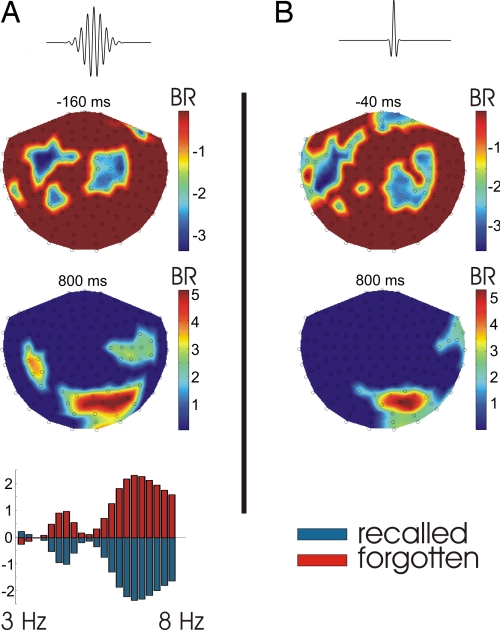
Fig. 1A shows that later-recalled words were associated with higher theta amplitudes than later-forgotten words between −250 and 50 ms at left frontotemporal and right frontal sensors, with the strongest difference being around 7 Hz. By contrast, later-forgotten words were associated with higher theta amplitudes than later-recalled words between 550 and 800 ms, primarily at right occipitotemporal sensors. To increase time resolution, we reanalyzed the data by using a short, 7-Hz wavelet having ≈1 oscillation cycle, after bandpass-filtering the raw data (eighth-order, zero-phase Butterworth, 5–9 Hz). The PLS results (Fig. 1B) show that the higher prestimulus theta amplitude for later-recalled words retained its topography but had a narrower temporal peak (−100 to −20 ms). The higher poststimulus theta amplitude for later-forgotten words retained both its topography and timing. Subsequent analyses are based on the transforms using the short 7-Hz wavelet.
Fig. 1.
Partial least squares analysis of theta amplitudes. The bottom plot in A shows the relative contribution of each frequency (between 3 Hz and 8 Hz) to the topographic maps shown at the top, indicating peak differences around 7 Hz. Blue regions in the maps denote higher theta amplitudes for later-recalled than later-forgotten words; red regions denote the converse. Maps display bootstrap ratios (BR) of the sensor saliences to their standard errors (analogous to z scores). One short, 7-Hz wavelet was used for B. The real parts of the wavelets used are shown at the top.
Time Course Analysis.
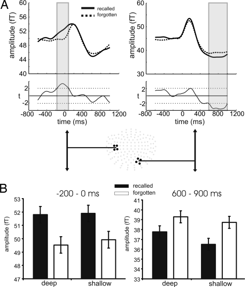
To elucidate the PLS results, sensor groups showing the strongest differences in PLS were selected, and theta amplitudes were averaged across sensors and submitted to t tests comparing later-recalled and later-forgotten words at each time point. Fig. 2A Left shows that as word onset approached, there was an increase in theta amplitude at left anterior temporal sensors for later-recalled words (solid line) but not for later-forgotten words (dashed line), leading to significant differences in amplitude starting at about −200 ms (hence referred to as prestimulus DM effect; difference due to later memory; ref. 2). Fig. 2A Right shows that the poststimulus amplitude difference between later-recalled and later-forgotten words at right occipitotemporal sensors (henceforth termed the late DM effect) resulted from a stronger amplitude decrease for later-recalled relative to later-forgotten words from 550 to 1,000 ms. We next addressed the question of whether the theta responses showed a constant phase relationship to stimulus onset (i.e., phase locking). Phase locking for later-recalled and later-forgotten words did not exceed chance level at the time of either the prestimulus or late DM effects. Thus, these effects reflected amplitude differences without significant phase locking, indicating that the phase of theta before word onset is not predictive of successful encoding.
Fig. 2.
Time course of theta amplitude responses. (A) Later-recalled words (solid lines) are associated with stronger prestimulus increases in theta amplitude than are later-forgotten words (dashed lines) at left anterior temporal sensors (Upper Left) and stronger poststimulus decreases in theta amplitude at right posterior occipitotemporal sensors (Upper Right). Amplitude is given in femtotesla (fT). Gray bars indicate significant differences at P < 0.05. (Lower) Graphs show the corresponding t values as a function of time, with the dotted lines indicating the significance threshold (df = 23, P < 0.05). (B) Effects of level of processing (deep vs. shallow) at study. The prestimulus increase and poststimulus decrease in theta amplitude for later-recalled words are independent of level of processing. Error bars denote SEM.
Correlation Between Prestimulus and Late DM Effects.
We investigated the relationship between the prestimulus and late DM effects by calculating the across-participants correlation between theta amplitude differences (later recalled minus later forgotten) in the prestimulus (−200 to 0 ms) and late (600–900 ms) time windows. The correlation was negative (i.e. larger prestimulus differences were associated with larger poststimulus differences) but not significant (R = −0.39; P = 0.062; 2-tailed).
Levels of Processing.
To test whether the prestimulus and late DM effects were responsive to prestimulus task demands, we examined whether level of processing (LOP) at study [semantic (deep) vs. phonemic (shallow)] interacted with these effects. A 2-way ANOVA on theta amplitudes for the left anterior temporal sensor group between −200 and 0 ms with the factors encoding success (later recalled vs. later forgotten) and LOP (deep vs. shallow) revealed a significant main effect of encoding success (_F_1,23 = 9.63; P < 0.005), but no significant main effect of LOP (_F_1,23 = 0.12; _P_ > 0.7) or interaction (_F_1,23 = 0.05; P > 0.8). A similar ANOVA on theta amplitudes for the right occipitotemporal sensor group between 600 and 900 ms revealed a significant main effect of encoding success (_F_1,23 = 17.18; P < 0.001), but no significant main effect of LOP (_F_1,23 = 2.73; _P_ > 0.10) or interaction (_F_1,23 = 1.18; P > 0.25). Thus, neither DM effect was significantly dependent on LOP at encoding. Fig. 2B shows the relevant theta amplitudes separated by LOP.
Recall Rate in Individual Participants Increases with Increasing Prestimulus Theta Amplitude.
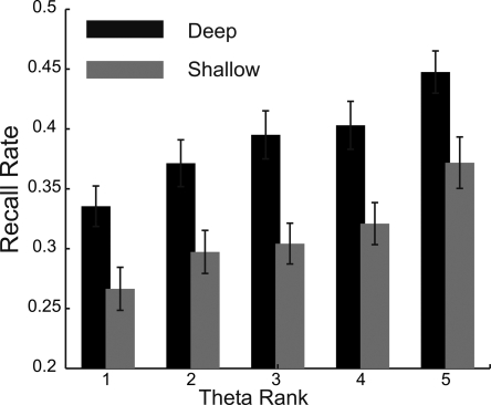
As shown above, prestimulus theta amplitude was, on average across participants, higher for later-recalled than for later-forgotten words. If prestimulus theta amplitude is indeed associated with successful encoding, the recall rate in individual participants should increase as a function of increased prestimulus theta amplitude. To investigate this possibility, we separated the trials during encoding for each participant into 5 equally sized bins of increasing theta amplitude. We then sorted the trials in every bin (or theta rank) according to their behavioral status (i.e., recalled vs. forgotten and deep vs. shallow). Thus, we were able to analyze the recall rate in individual participants as a function of LOP and 5 levels of prestimulus theta amplitude. Fig. 3 shows the recall rate across participants as a function of LOP (black vs. gray bars for deep vs. shallow, respectively) and prestimulus theta amplitude (theta rank). A 2-way ANOVA on recall rates with the factors LOP (deep vs. shallow) and theta rank (levels 1–5) showed both a main effect of LOP (_F_1,23 = 27.4; P < 0.001) and theta rank (_F_3.1,71.1 = 50.6; _P_ < 0.001). The lack of a significant interaction (_F_2.7,62.6 = 0.6; _P_ > 0.65) suggests that encoding success is modulated independently by the level of cognitive processing and the theta amplitude before stimulus onset. We fitted a first-order polynomial to the recall rates as a function of theta rank for each participant and tested whether the resulting slope was significantly different from zero across participants. The result was significant (_t_23 = 11.45, P < 0.001), indicating that recall rate increased as a function of increasing theta amplitude. Fig. S1 shows the recall rate modulation by theta amplitude of the individual participants. Remarkably, in all 24 participants the slope was greater than zero, and the highest theta rank was associated with a higher recall rate than the lowest theta rank. We also tested whether reaction times during study changed as a function of theta rank, which would suggest that theta amplitude before stimulus onset could exert its effect on encoding through general arousal or attentional mechanisms. A 1-way ANOVA on reaction times as a function of 5 theta ranks was not significant (_F_2.6,59.4 = 0.53; _P_ > 0.6). Additionally, we again fitted a first-order polynomial to the reaction times as a function of theta rank and found that the slopes were not significantly different from zero (_t_23 = −0.69, P > 0.45), indicating that the reaction times during study did not depend on prestimulus theta amplitude.
Fig. 3.
Recall rates across participants (y axis) as a function of LOP (black vs. gray) and theta amplitude (x axis; ranks 1 to 5).
Quantification of LOP and Theta-Related Recall Benefits.
To quantify the effect of prestimulus theta amplitude on later recall, we compared the recall rate modulation of LOP (deep vs. shallow) and theta amplitude (high vs. low, 50% of total trials each). High-theta trials were associated with an average recall rate benefit of 7.4% (SD, 5.0%) over low-theta trials. Deep processing was associated with an average recall benefit of 8.2% (SD, 7.5%) over shallow processing. If recall rate for items encoded under shallow and low-theta conditions is considered as baseline performance (mean, 27.4%; SD 8.4%), the recall benefit relative to this baseline of deep processing alone (under low-theta conditions) resulted in a performance increase of 27.8% (mean, 35.0%; SD 1%), whereas high-theta alone (under shallow encoding conditions) resulted in a performance increase of 24.8% (mean, 34.2%; SD 9.4%). Items encoded under both deep and high-theta conditions were associated with a performance increase of 56,8% (mean, 43.0%; SD 9.1%). A paired t test between the added recall benefits of deep processing and high theta alone and the recall benefit of deep processing under high-theta conditions was not significant (_t_23 = 0.59, P > 0.55). This suggests that LOP and theta-related recall benefits are not only statistically independent, but modulate encoding probability in an additive manner. Fig. S2 shows the distributions of recall benefits across participants separately for LOP and theta amplitude. Although both LOP and prestimulus theta amplitude robustly modulated performance in individual participants, we found no evidence that either contributed significantly to between-participant performance differences (SI Text).
Mechanisms of Prestimulus Theta Increase.
If the theta amplitude before stimulus onset plays a role in encoding of this stimulus, it is important to understand how it is modulated physiologically. One possibility is that theta amplitude fluctuates randomly over time, and words that are incidentally presented during high-theta states have a higher probability of being successfully encoded. The statistical difference in theta amplitude between later-recalled and later-forgotten words would then result from selective sampling of a random state. If this were the case, then the distribution of theta amplitudes (across all trials) should be the same during the time window of the statistically significant difference between later-recalled vs. later-forgotten words (subsequently called DM time window, −200 to 0 ms) as during a period in which theta amplitude is not related to encoding success. Alternatively, the distribution should be shifted to higher theta amplitudes during the DM time window if an active process is involved in generating higher theta amplitudes during some trials, with these trials in turn having a higher probability of being successfully encoded. The DM time window was indeed associated with a shift of the theta amplitude distributions toward higher values, as suggested by a paired t test on the median amplitude values of the individual participant distributions from the 2 time windows (_t_23 = 4.45, P < 0.001; see Fig. S3). Furthermore, later-forgotten items alone were also associated with a slight shift of the theta amplitude distributions toward higher values (_t_23 = 2.34, P < 0.05), which is incompatible with the idea of a random process underlying theta amplitude fluctuations.
Which factors could influence theta amplitude before stimulus onset? As outlined above, prestimulus theta amplitude was not modulated by LOP, nor was it related to reaction times during the study tasks. Another possibility is that prestimulus theta amplitude reflects the contextual influence of neighboring list items. It is well known that words in free-recall tasks tend to be recalled in groups; that is, a word has a higher probability of being successfully encoded if the preceding word was already successfully encoded (24). Theta amplitude before stimulus onset could thus reflect the context established by previous encoding success and help embed the following word in this context, thereby increasing the probability of successful encoding. We tested this possibility by comparing the recall rates and prestimulus theta amplitudes for words as a function of recall success of the 2 immediately preceding words. When neither of the 2 preceding words was recalled, the recall rate was 31.4% (SD, 7.6%). Recall rate increased to 36.9% (SD, 7.9%) when only the immediately preceding word was later recalled, and to 43.7% (SD, 11.1%) when both preceding words were later recalled. A 1-way ANOVA was significant (_F_1.9,44.2 = 18.8; P < 0.001). On the other hand, prestimulus theta amplitudes did not differ as a function of encoding success of the 2 previous words (_F_1.9,42.5 = 1.7; _P_ > 0.15; Fig. S4). Thus, increased prestimulus theta amplitude did not seem to be a reflection of contextual serial position effects that influence encoding.
Source Analysis of the Prestimulus DM Effect.
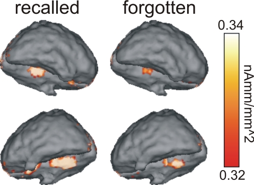
Theta field distributions were assessed by calculating between-sensor phase differences on a trial-by-trial basis for each participant for later-recalled and later-forgotten words. This approach was necessary because the prestimulus theta activity did not show above-chance phase locking, and averaging random phase values would lead to distorted field information (23). Inverse wavelet transformation (IWT) was used to bring the resulting phase information, together with the corresponding amplitude information, back to the time domain (see ref. 23 for more detail). Minimum-norm current–density reconstruction of the resulting theta waveforms revealed strong sources of theta activity bilaterally in the MTL between −100 and 0 ms, with later-recalled words being associated with stronger sources than later-forgotten words (Fig. 4).
Fig. 4.
Current density reconstructions of prestimulus theta amplitudes. Later-recalled words (Left) are associated with a stronger theta current source than are later-forgotten words (Right) in the medial temporal lobe. Upper shows the right hemisphere is in the foreground, with the brain tilted to expose the ventromedial surface of the left temporal lobe. Lower shows the reverse.
Discussion
Increased MTL theta oscillatory amplitude predicted successful memory for items even before they were presented. Remarkably, recall rate in all 24 participants increased as a function of prestimulus theta amplitude. The absence of a significant correlation between overall performance levels across participants and theta-related recall benefits suggests that theta oscillatory activity reflects a property of the MTL that exerts its effect on encoding across various levels of performance. Consistent with this notion is the absence of a significant interaction between theta- and LOP-related recall benefits. Although this finding underscores that prestimulus theta amplitude effectively modulates encoding across different levels of performance within individual participants, it also suggests that prestimulus theta amplitude does not reflect a qualitative difference in cognitive processing, and hence cannot be interpreted as a mere extension of the LOP effect of encoding (9, 25, 26) to the prestimulus time window.
Taken together with the recent finding that prestimulus ERP slow shifts correlate with encoding success (21), our findings suggest that episodic memory encoding can be enhanced by 2 different and potentially independent prestimulus phenomena. One is related to a semantic preparatory task set and contributes to improving encoding of words processed deeply (21). The other, reported here, is related to a task-independent brain state that improves encoding regardless of whether words are processed deeply or shallowly. Both phenomena provide evidence for the operation of item-independent encoding processes. Whereas poststimulus DM effects may be attributable to variation between to-be-remembered items in some characteristic that influences their later memorability, such as distinctiveness, no such hypothesis can be entertained for DM effects that occur before stimulus presentation.
It has previously been shown that the recall rate of a particular item is increased if the preceding list items were successfully encoded (24). Although we reproduced this behavioral phenomenon, it was not mirrored in higher prestimulus theta amplitudes (Fig. S4). Thus, prestimulus theta influences encoding for individual items, rather than reflecting an encoding state that is modulated across several list items, perhaps by promoting associations between successive items. Because prestimulus theta amplitude was also not significantly related to reaction times at study either within or across participants, the theta-related recall benefit does not appear to be due to an increase in general arousal, which should have been reflected in faster reaction times. It should be noted, however, that the lack of response time differences, even at an individual level, as demonstrated here, does not rule out the possibility that more specific attentional or preparatory factors could selectively influence MTL theta generation. For example, the intention to commit the upcoming stimulus to memory, or simply anticipating a novel stimulus, could influence the functional state of the MTL to promote successful encoding (27). Consistent with this proposal, our data suggest that increased prestimulus theta states are at least partly generated by an active process, rather than reflecting the coincidence of stimulus presentation with randomly fluctuating MTL theta amplitudes (Fig. S3). This implies that the prestimulus theta modulation of encoding should be reduced when to-be-encoded stimuli appear at unpredictable times.
One remaining possibility is that the prestimulus theta state represents contextual information about specific expectations formed by previous list items or by the experimental procedure, such as the expected position of a stimulus on the computer screen, its luminance, and timing. Several studies have now suggested an involvement of increased poststimulus theta amplitude during successful recollection (23, 28, 29), whereas we in the present study as well as others (30) found a decrease in poststimulus theta amplitude to be related to successful episodic encoding. If increased MTL theta amplitude is indeed a signature of recollection, it can be argued that the prestimulus DM effect reported here reflects the activation of a mnemonic context, in which the subsequently presented item can be embedded (31, 32). Preactivating such contextual information may thus promote recall and recollection by improving associative memory between episodic context and stimulus-related processing.
Several experiments have now shown that the phase of theta oscillations modulates the induction of synaptic plasticity in the rodent hippocampus (12, 14). Theta appears to reset its phase during the encoding of new stimuli (33), and it has been argued that this reset provides the basis for enhanced plasticity (13). These findings in rodents support the idea that the phase of theta oscillations provides a temporal separation of encoding and retrieval operations (34). The results we describe here differ from these accounts in 2 ways: First, an increase of theta amplitude, rather than a concentration of phase, was associated with encoding success. Second, because the theta amplitude increase preceded stimulus onset by 200 ms, it could not have reflected the period during which enhanced synaptic plasticity can support the encoding of stimulus information. However, it can be argued that theta needs to be present in the first place for its phase to influence encoding operations. Thus, the increased theta amplitude we observed before successful encoding may reflect a state of the MTL that is particularly conducive to the induction of plasticity. Evidence in support of this view comes from findings in rabbits that the learning rate in classical conditioning is higher when stimuli are presented in periods of high hippocampal theta (15–17).
Because theta-related encoding benefits appeared to be independent of LOP, the question arises as to how both contribute to encoding. A plausible neurocognitive interpretation comes from the effects of MTL damage (20). Patients with such damage are able to classify incoming information at different levels of perceptual, lexical, and semantic analysis, yet they show markedly impaired recall (25). This provides an explanation of why deep processing increases the probability of—but is not in itself sufficient for—encoding success (25): different levels of processing modify the cognitive structure of an episode independent of the MTL, but the actual submission of this episode to long-term memory critically depends on the integrity of the MTL. This possibility is supported by a recent animal model of schema learning: hippocampally lesioned animals were not impaired in their ability to recall schema information (knowledge about the spatial layout of food wells in an arena) learned by extensive training before the lesion, showing that using learned schemas does not depend on the integrity of the hippocampal formation, whereas the ability to integrate new information into existing schemata by 1-trial learning does (35). Theta activity in the MTL might promote plasticity without influencing the task-related cognitive structuring of an experience. Both MTL theta and deep processing would thus be required for encoding to be most successful. Indeed, we found that recall benefits during high-theta and deep-processing conditions amounted to the added recall benefits of high theta and deep processing alone (Fig. S2). We thus suggest that MTL theta reflects a fundamental network property that modulates episodic encoding independent of cognitive factors, like LOP or divided attention during study (36), and that therefore the benefits of deep processing and high theta are additive.
Our finding that MTL theta state predicts later memory, before to-be-remembered information is even perceived, suggests that memory might be improved by timing stimulus presentation to optimal theta states. A potential application of our finding might be that individuals could learn to optimize their MTL theta state via neurofeedback to modulate encoding success independent of item-specific content.
Materials and Methods
Participants.
Twenty-four young (age range 18–32 years; 14 female) native speakers of German, all right-handed, with normal or corrected-to-normal vision, participated in this MEG study in return for payment after giving informed consent. The study was carried out in accordance with the guidelines of the Ethics Commission of the University of Magdeburg Faculty of Medicine (Magdeburg, Germany). Present or past neurological or psychiatric disorders, or the use of any centrally acting drugs, as revealed by routine clinical screening, were contraindications for participation.
Behavioral Procedure.
To investigate the correlates of successful memory formation, theta signals during study of visually presented words were compared as a function of later recall versus later forgetting (6, 9). The experiment consisted of 18 successive encoding/retrieval blocks and participants were informed about the test being one of free recall. In each encoding phase, participants studied words at 1 of 2 levels of processing (LOP; deep vs. shallow processing). Each participant performed 9 deep and 9 shallow alternating study phases, with the starting study task being counterbalanced across participants. In the deep study task, participants were instructed to press a button with the right index finger if a word was pleasant, and a button with the right middle finger if a word was unpleasant (thus inducing semantic processing). In the shallow study task, they were instructed to press a button with the right index finger if a word had exactly 2 syllables and to press a button with the right middle finger for any other number of syllables (thus inducing phonemic processing).
Twenty German words were presented in each study list. Each study phase commenced with a cue stating either “pleasant or unpleasant” or “syllable counting,” which prompted the participant to perform either the deep or the shallow task on the ensuing word list. After presentation of a central fixation cross for 500 ms, each word was presented for 1,000 ms, again followed by a central fixation cross for 1,250 ms. Study lists were followed by a distracter task lasting ≈20 seconds and consisting of 4 moderately difficult arithmetic operations. Participants were asked to judge whether the result presented to them was correct or not and respond via button press. The distracter task ensured that recall was from long-term memory and not from working memory, and removed the recency effect (i.e., recall advantage for the last few words in the list) that would otherwise have occurred. After the distracter task, a cue (“please speak”) prompted participants to begin a 90-s free-recall test, in which they overtly recalled all studied words they could remember from the immediately preceding list in any order they wished. The oral responses were recorded and scored offline.
MEG Data Recording and Time–Frequency (Wavelet) Analysis.
The critical MEG recordings were made during study list presentation. MEG signals were recorded by using a 148-channel BTI Magnes 2500 whole-head magnetometer (Biomagnetic Technologies) with a digitization rate of 508 Hz. Epochs from 1,200 ms before stimulus onset to 2,000 ms thereafter were extracted for each studied word and sorted according to LOP and encoding success (later recalled vs. later forgotten). After artifact rejection, the data were analyzed by using continuous single-trial wavelet transforms, yielding information about the timing and frequency of neural oscillations present in the electromagnetic signals.
Statistical Analysis.
To detect spatiotemporal patterns of theta oscillatory activity separating later-recalled from later-forgotten words, task PLS analyses were applied to the theta amplitude data. Task PLS is a statistical technique similar to principal component or factor analysis, and it examines the relationship between a set of exogenous design variables (in this case, theta frequency bands and later-recalled vs. later-forgotten conditions) and corresponding multivariate measures of brain activity (in this case, mean amplitudes at each of the 148 sensors for each participant and each time point). For a more detailed description of PLS and its application to electrophysiological data, see previously published work (22, 23, 37). To elucidate the time course of the spatiotemporal differences in theta activity revealed by PLS, amplitudes for selected sensor groups were further analyzed by using paired t tests comparing conditions (later recalled vs. later forgotten) at each time point. Sensor groups were selected according to the amplitude differences revealed by the PLS analysis. To determine whether theta differences are observed solely as a function of encoding success or whether LOP affects the magnitudes of the theta responses, 2-way repeated-measure ANOVAs with the factors encoding success (later recalled vs. later forgotten) and level of processing (deep vs. shallow) were performed.
To test whether recall rate in single participants increased as a function of theta amplitude, we first took the average theta amplitude between −200 and 0 ms before stimulus onset for every trial (regardless of deep vs. shallow encoding status) and participant. Next, 8 sensors were selected for each participant that showed the largest difference in theta amplitude between later-recalled and later-forgotten words, and theta amplitude was averaged across these sensors (separately for each participant). The result yielded 1 value of theta amplitude per trial and participant. For each participant, the trials were then sorted as a function of increasing theta amplitude. Next, 5 bins were constructed for each participant that contained an equal number of trials. Finally, the trials in every bin were sorted according to their behavioral status (i.e., recalled vs. forgotten and deep vs. shallow; Fig. 3). Selecting 2 or 4 sensors and ranking theta amplitude into 10 bins yielded qualitatively similar results. For the performance comparisons with LOP, the data were separated into 2 bins consisting of high- and low-theta amplitudes, respectively (Fig. S2). Reported ANOVAs are Greenhouse–Geisser-corrected, where appropriate.
Source Analysis.
The phase of electromagnetic brain activity is important for obtaining reliable field information to reconstruct the cortical sources of this activity. Averaging random phase information between trials could lead to distorted field information, and hence inaccurate source reconstructions (23). Because we were interested in reconstructing the sources of non-phase-locked oscillatory activity (as indicated by the phase-alignment analyses), we assessed the theta field distributions for each participant and condition by calculating between-sensor phase differences on a trial-by-trial basis and then averaging across trials. The resulting phase information, together with the corresponding amplitude information, was submitted to inverse wavelet transformation (IWT). This approach yields theta oscillatory activity in the time domain that incorporates both absolute amplitude and reliable field information, regardless of the phase locking of the underlying activity, and was outlined previously in detail (23). The sources of IWT waveforms were localized independently for later-recalled and later-forgotten words after averaging across participants using Curry (Version 4; Neuroscan). Current density distributions were computed between −100 and 0 ms on triangle models restricted to the cortical surface of the MNI brain (Montreal Neurological Institute) using the linear least squares minimum-norm method (Curry User Guide, Version 4; ref. 38).
Acknowledgments.
We thank Michael Scholz for assistance with data analysis, Maja Fremuth for assistance with data acquisition, and 3 anonymous reviewers for helpful comments on a previous version of the manuscript. This work was supported by grants from the Deutsche Forschungsgemeinschaft (Kognitive Kontrolle von Gedaechtnis TP1 and TP3) and Bundesministerium für Bildung und Forschung (Center for Advanced Imaging).
Footnotes
The authors declare no conflict of interest.
References
- 1.Fernandez G, et al. Real-time tracking of memory formation in the human rhinal cortex and hippocampus. Science. 1999;285:1582–1585. doi: 10.1126/science.285.5433.1582. [DOI] [PubMed] [Google Scholar]
- 2.Paller KA, Kutas M, Mayes AR. Neural correlates of encoding in an incidental learning paradigm. Electroencephalogr Clin Neurophysiol. 1987;67:360–371. doi: 10.1016/0013-4694(87)90124-6. [DOI] [PubMed] [Google Scholar]
- 3.Schott B, Richardson-Klavehn A, Heinze HJ, Duzel E. Perceptual priming versus explicit memory: Dissociable neural correlates at encoding. J Cogn Neurosci. 2002;14:578–592. doi: 10.1162/08989290260045828. [DOI] [PubMed] [Google Scholar]
- 4.Brewer JB, Zhao Z, Desmond JE, Glover GH, Gabrieli JD. Making memories: Brain activity that predicts how well visual experience will be remembered. Science. 1998;281:1185–1187. doi: 10.1126/science.281.5380.1185. [DOI] [PubMed] [Google Scholar]
- 5.Buckner RL, Logan J, Donaldson DI, Wheeler ME. Cognitive neuroscience of episodic memory encoding. Acta Psychol (Amst) 2000;105:127–139. doi: 10.1016/s0001-6918(00)00057-3. [DOI] [PubMed] [Google Scholar]
- 6.Otten LJ, Henson RN, Rugg MD. State-related and item-related neural correlates of successful memory encoding. Nat Neurosci. 2002;5:1339–1344. doi: 10.1038/nn967. [DOI] [PubMed] [Google Scholar]
- 7.Reber PJ, et al. Neural correlates of successful encoding identified using functional magnetic resonance imaging. J Neurosci. 2002;22:9541–9548. doi: 10.1523/JNEUROSCI.22-21-09541.2002. [DOI] [PMC free article] [PubMed] [Google Scholar]
- 8.Schott BH, et al. Neuroanatomical dissociation of encoding processes related to priming and explicit memory. J Neurosci. 2006;26:792–800. doi: 10.1523/JNEUROSCI.2402-05.2006. [DOI] [PMC free article] [PubMed] [Google Scholar]
- 9.Wagner AD, et al. Building memories: Remembering and forgetting of verbal experiences as predicted by brain activity. Science. 1998;281:1188–1191. doi: 10.1126/science.281.5380.1188. [DOI] [PubMed] [Google Scholar]
- 10.Linkenkaer-Hansen K, Nikulin VV, Palva S, Ilmoniemi RJ, Palva JM. Prestimulus oscillations enhance psychophysical performance in humans. J Neurosci. 2004;24:10186–10190. doi: 10.1523/JNEUROSCI.2584-04.2004. [DOI] [PMC free article] [PubMed] [Google Scholar]
- 11.Super H, van der Togt C, Spekreijse H, Lamme VA. Internal state of monkey primary visual cortex (V1) predicts figure-ground perception. J Neurosci. 2003;23:3407–3414. doi: 10.1523/JNEUROSCI.23-08-03407.2003. [DOI] [PMC free article] [PubMed] [Google Scholar]
- 12.Hyman JM, Wyble BP, Goyal V, Rossi CA, Hasselmo ME. Stimulation in hippocampal region CA1 in behaving rats yields long-term potentiation when delivered to the peak of theta and long-term depression when delivered to the trough. J Neurosci. 2003;23:11725–11731. doi: 10.1523/JNEUROSCI.23-37-11725.2003. [DOI] [PMC free article] [PubMed] [Google Scholar]
- 13.McCartney H, Johnson AD, Weil ZM, Givens B. Theta reset produces optimal conditions for long-term potentiation. Hippocampus. 2004;14:684–687. doi: 10.1002/hipo.20019. [DOI] [PubMed] [Google Scholar]
- 14.Pavlides C, Greenstein YJ, Grudman M, Winson J. Long-term potentiation in the dentate gyrus is induced preferentially on the positive phase of theta-rhythm. Brain Res. 1988;439:383–387. doi: 10.1016/0006-8993(88)91499-0. [DOI] [PubMed] [Google Scholar]
- 15.Berry SD, Thompson RF. Prediction of learning rate from the hippocampal electroencephalogram. Science. 1978;200:1298–1300. doi: 10.1126/science.663612. [DOI] [PubMed] [Google Scholar]
- 16.Griffin AL, Asaka Y, Darling RD, Berry SD. Theta-contingent trial presentation accelerates learning rate and enhances hippocampal plasticity during trace eyeblink conditioning. Behav Neurosci. 2004;118:403–411. doi: 10.1037/0735-7044.118.2.403. [DOI] [PubMed] [Google Scholar]
- 17.Seager MA, Johnson LD, Chabot ES, Asaka Y, Berry SD. Oscillatory brain states and learning: Impact of hippocampal theta-contingent training. Proc Natl Acad Sci USA. 2002;99:1616–1620. doi: 10.1073/pnas.032662099. [DOI] [PMC free article] [PubMed] [Google Scholar]
- 18.Brown MW, Aggleton JP. Recognition memory: What are the roles of the perirhinal cortex and hippocampus? Nat Rev Neurosci. 2001;2:51–61. doi: 10.1038/35049064. [DOI] [PubMed] [Google Scholar]
- 19.Mishkin M, Vargha-Khadem F, Gadian DG. Amnesia and the organization of the hippocampal system. Hippocampus. 1998;8:212–216. doi: 10.1002/(SICI)1098-1063(1998)8:3<212::AID-HIPO4>3.0.CO;2-L. [DOI] [PubMed] [Google Scholar]
- 20.Squire LR, Stark CE, Clark RE. The medial temporal lobe. Annu Rev Neurosci. 2004;27:279–306. doi: 10.1146/annurev.neuro.27.070203.144130. [DOI] [PubMed] [Google Scholar]
- 21.Otten LJ, Quayle AH, Akram S, Ditewig TA, Rugg MD. Brain activity before an event predicts later recollection. Nat Neurosci. 2006;9:489–491. doi: 10.1038/nn1663. [DOI] [PubMed] [Google Scholar]
- 22.Duzel E, et al. A multivariate, spatiotemporal analysis of electromagnetic time-frequency data of recognition memory. Neuroimage. 2003;18:185–197. doi: 10.1016/s1053-8119(02)00031-9. [DOI] [PubMed] [Google Scholar]
- 23.Guderian S, Duzel E. Induced theta oscillations mediate large-scale synchrony with mediotemporal areas during recollection in humans. Hippocampus. 2005;15:901–912. doi: 10.1002/hipo.20125. [DOI] [PubMed] [Google Scholar]
- 24.Kahana MJ. Associative retrieval processes in free recall. Mem Cognit. 1996;24:103–109. doi: 10.3758/bf03197276. [DOI] [PubMed] [Google Scholar]
- 25.Craik FI. Levels of processing: Past, present, and future? Memory. 2002;10:305–318. doi: 10.1080/09658210244000135. [DOI] [PubMed] [Google Scholar]
- 26.Otten LJ, Henson RN, Rugg MD. Depth of processing effects on neural correlates of memory encoding: Relationship between findings from across- and within-task comparisons. Brain. 2001;124:399–412. doi: 10.1093/brain/124.2.399. [DOI] [PubMed] [Google Scholar]
- 27.Wittmann BC, Bunzeck N, Dolan RJ, Duzel E. Anticipation of novelty recruits reward system and hippocampus while promoting recollection. Neuroimage. 2007;38:194–202. doi: 10.1016/j.neuroimage.2007.06.038. [DOI] [PMC free article] [PubMed] [Google Scholar]
- 28.Klimesch W, et al. Theta synchronization during episodic retrieval: Neural correlates of conscious awareness. Brain Res Cogn Brain Res. 2001;12:33–38. doi: 10.1016/s0926-6410(01)00024-6. [DOI] [PubMed] [Google Scholar]
- 29.Osipova D, et al. Theta and gamma oscillations predict encoding and retrieval of declarative memory. J Neurosci. 2006;26:7523–7531. doi: 10.1523/JNEUROSCI.1948-06.2006. [DOI] [PMC free article] [PubMed] [Google Scholar]
- 30.Sederberg PB, et al. Hippocampal and neocortical gamma oscillations predict memory formation in humans. Cereb Cortex. 2007;17:1190–1196. doi: 10.1093/cercor/bhl030. [DOI] [PubMed] [Google Scholar]
- 31.Hasselmo ME, Eichenbaum H. Hippocampal mechanisms for the context-dependent retrieval of episodes. Neural Netw. 2005;18:1172–1190. doi: 10.1016/j.neunet.2005.08.007. [DOI] [PMC free article] [PubMed] [Google Scholar]
- 32.Lisman JE, Idiart MA. Storage of 7 +/- 2 short-term memories in oscillatory subcycles. Science. 1995;267:1512–1515. doi: 10.1126/science.7878473. [DOI] [PubMed] [Google Scholar]
- 33.Givens B. Stimulus-evoked resetting of the dentate theta rhythm: Relation to working memory. Neuroreport. 1996;8:159–163. doi: 10.1097/00001756-199612200-00032. [DOI] [PubMed] [Google Scholar]
- 34.Hasselmo ME, Bodelon C, Wyble BP. A proposed function for hippocampal theta rhythm: Separate phases of encoding and retrieval enhance reversal of prior learning. Neural Comput. 2002;14:793–817. doi: 10.1162/089976602317318965. [DOI] [PubMed] [Google Scholar]
- 35.Tse D, et al. Schemas and memory consolidation. Science. 2007;316:76–82. doi: 10.1126/science.1135935. [DOI] [PubMed] [Google Scholar]
- 36.Uncapher MR, Rugg MD. Effects of divided attention on FMRI correlates of memory encoding. J Cogn Neurosci. 2005;17:1923–1935. doi: 10.1162/089892905775008616. [DOI] [PubMed] [Google Scholar]
- 37.McIntosh AR, Lobaugh NJ. Partial least squares analysis of neuroimaging data: applications and advances. Neuroimage. 2004;23(Suppl 1):S250–S263. doi: 10.1016/j.neuroimage.2004.07.020. [DOI] [PubMed] [Google Scholar]
- 38.Fuchs M, Wagner M, Kohler T, Wischmann HA. Linear and nonlinear current density reconstructions. J Clin Neurophysiol. 1999;16:267–295. doi: 10.1097/00004691-199905000-00006. [DOI] [PubMed] [Google Scholar]


