Analysis of the Association between CIMP and BRAFV600E in Colorectal Cancer by DNA Methylation Profiling (original) (raw)
Abstract
A CpG island methylator phenotype (CIMP) is displayed by a distinct subset of colorectal cancers with a high frequency of DNA hypermethylation in a specific group of CpG islands. Recent studies have shown that an activating mutation of BRAF (BRAFV600E) is tightly associated with CIMP, raising the question of whether BRAFV600E plays a causal role in the development of CIMP or whether CIMP provides a favorable environment for the acquisition of BRAFV600E. We employed Illumina GoldenGate DNA methylation technology, which interrogates 1,505 CpG sites in 807 different genes, to further study this association. We first examined whether expression of BRAFV600E causes DNA hypermethylation by stably expressing BRAFV600E in the CIMP-negative, BRAF wild-type COLO 320DM colorectal cancer cell line. We determined 100 CIMP-associated CpG sites and examined changes in DNA methylation in eight stably transfected clones over multiple passages. We found that BRAFV600E is not sufficient to induce CIMP in our system. Secondly, considering the alternative possibility, we identified genes whose DNA hypermethylation was closely linked to BRAFV600E and CIMP in 235 primary colorectal tumors. Interestingly, genes that showed the most significant link include those that mediate various signaling pathways implicated in colorectal tumorigenesis, such as BMP3 and BMP6 (BMP signaling), EPHA3, KIT, and FLT1 (receptor tyrosine kinases) and SMO (Hedgehog signaling). Furthermore, we identified CIMP-dependent DNA hypermethylation of IGFBP7, which has been shown to mediate BRAFV600E-induced cellular senescence and apoptosis. Promoter DNA hypermethylation of IGFBP7 was associated with silencing of the gene. CIMP-specific inactivation of BRAFV600E-induced senescence and apoptosis pathways by IGFBP7 DNA hypermethylation might create a favorable context for the acquisition of BRAFV600E in CIMP+ colorectal cancer. Our data will be useful for future investigations toward understanding CIMP in colorectal cancer and gaining insights into the role of aberrant DNA hypermethylation in colorectal tumorigenesis.
Introduction
Aberrant DNA methylation at CpG islands has been widely observed in cancer. Promoter CpG island hypermethylation associated with inactivation of selected tumor suppressor genes appears to be critical in tumors from inception through to maintenance of the tumor phenotype [1]. Distinct subgroups of several types of human cancers have been proposed to have a CpG island methylator phenotype (CIMP) in which an exceptionally high frequency of cancer-specific DNA hypermethylation is found [2], [3]. Although this concept has been controversial [4], we have confirmed the existence of CIMP in colorectal cancer in a large-scale comprehensive study [5].
CIMP in colorectal cancer may arise through a distinct pathway originating in certain subtypes of serrated polyps [6] and is observed in approximately 15% of all colorectal cancer cases [5], [7]. Features associated with CIMP in colorectal cancer include gender (female), proximal location, and poorly differentiated or mucinous histology [3], [5], [7], [8]. Our study using a newly developed CIMP marker panel in colorectal cancers demonstrated that sporadic microsatellite instability (MSI+) occurs as a consequence of CIMP-associated MLH1 DNA hypermethylation [5]. Furthermore, we found a strong association of CIMP with the presence of an activated mutant form of BRAF (BRAFV600E) [5]. Both CIMP and BRAF mutations have been reported in the earliest stages of colorectal neoplasia: CIMP in apparently normal mucosa of patients predisposed to multiple serrated polyps [9] and BRAF mutations in aberrant crypt foci [10].
The RAS-RAF-MEK-ERK signaling pathway is frequently hyperactivated in colorectal cancer. KRAS mutations occur most frequently in 30–40% of all colorectal cancers [11] and BRAF mutations are present at a frequency of 5–22%, in which the constitutively activated BRAFV600E variant accounts for ∼90% of all the BRAF mutations [12]. Mutations in KRAS and BRAF are generally mutually exclusive, implying equivalent downstream effects in tumorigenesis [13]. However, recent studies have indicated that mutations of these genes might play distinct roles in tumor initiation and/or maintenance [10], [14].
The extremely tight association between BRAFV600E and CIMP raises the question of whether BRAFV600E plays a causal role in the development of CIMP or whether CIMP-associated promoter hypermethylation provides a favorable setting for the acquisition of BRAFV600E. In this study, we searched for possible molecular explanations for the association between BRAFV600E and CIMP using the Illumina GoldenGate DNA methylation platform, which examines the DNA methylation status of 1,505 CpG sites located at 807 genes. The GoldenGate DNA methylation assay has been widely used in various studies and is now a standard method for DNA methylation analysis [15]–[24]. Findings obtained from the commercially available “GoldenGate Methylation Cancer Panel I”, in particular, have been validated using various other techniques [15]–[17], [22], [23], making it a reliable source for DNA methylation measurements across 1,505 loci. We were not able to demonstrate a causal contribution of BRAFV600E to CIMP in our cell culture system. However, we identified genes whose DNA hypermethylation was significantly linked with BRAFV600E in primary colorectal tumors. Inactivation of these specific genes in the context of CIMP might drive the acquisition of BRAFV600E in CIMP+ colorectal tumors.
Results
Characterization of 21 Human Colorectal Cancer Cell Lines
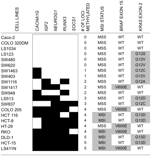
We first sought to determine whether expression of BRAFV600E would induce DNA hypermethylation at CpG sites associated with CIMP in an in vitro cell culture system. Since primary colonic epithelial cells were not readily available, we screened for colorectal cancer cell lines that do not have substantial DNA methylation at CIMP-defining loci and carry wild-type forms of both BRAF and KRAS. Such cell lines would serve as suitable systems for the introduction of BRAFV600E. We selected 21 colorectal cancer cell lines, characterized their DNA methylation profiles, and determined their BRAF and KRAS mutation status (Figure 1). We used MethyLight to assess the DNA methylation status of five CIMP-defining markers previously identified in our laboratory [5]. Using a PMR (percent of methylated reference) of ≥10 as a threshold for positive methylation, we identified six cell lines that lacked DNA methylation for all five CIMP-specific markers (Figure 1). To test our hypothesis, we initially chose the BRAF and KRAS wild-type Caco-2 and COLO 320DM cell lines for their ease in culturing and transfection. However, the study described below is limited to COLO 320DM cells, since we were not able to isolate any stably transfected Caco-2 clones that showed detectable level of BRAFV600E expression (data not shown).
Figure 1. Characteristics of 21 colorectal cancer cell lines.
MethyLight was used to assess the DNA methylation status of five CIMP-defining markers. A PMR of ≥10 was used as a threshold for positive methylation. Black boxes indicate PMR ≥10, and white boxes indicate PMR <10. The DNA methylation frequencies of the five CIMP markers increase from top to bottom. Microsatellite instability status for each cell line is listed as microsatellite stable (MSS) or harboring instability (MSI). The mutation status of BRAF exon 15 and KRAS exon 2 are listed.
Stable Transfection of BRAFV600E in COLO 320DM Cells
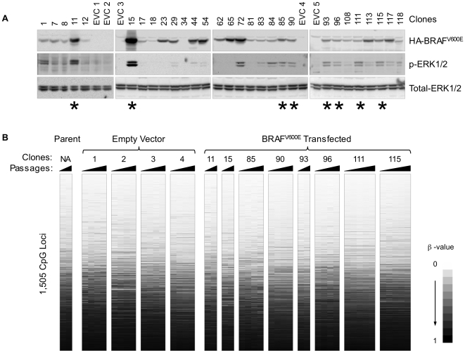
We transfected COLO 320DM cells with an HA-tagged BRAFV600E cDNA and isolated G418-resistant clones. The expression level of BRAFV600E was determined by western blotting using an antibody against the HA epitope (Figure 2A). The activity of BRAFV600E was confirmed by examining the activation of ERK1/2 using an antibody against phosphorylated ERK1/2 (Figure 2A). Eight stably transfected clones exhibiting high expression of BRAFV600E, as well as strong activation of ERK1/2, were individually grown in culture, and genomic DNA was isolated at various passages (between 2 and 27) from these clones. Four empty-vector transfected clones (EVCs) were grown in the same conditions and used as controls.
Figure 2. Selection of BRAFV600E stably-transfected clones and their Illumina GoldenGate DNA methylation profiles.
(A) Expression of BRAFV600E and ERK1/2 phosphorylation in stably transfected COLO 320DM cells. Blots were probed with the anti-HA antibodies for HA-BRAFV600E, anti-phospho-ERK1/2, and anti-ERK1. Asterisks indicate the eight BRAFV600E transfected clones that were subjected to DNA methylation analysis at various cell passages. (B) DNA methylation profiles of untransfected COLO 320DM cells, empty vector and BRAFV600E transfected COLO 320DM clones, as determined by the Illumina GoldenGate DNA methylation assay. The DNA methylation data were scored as β-values as previously defined [16]. Each row corresponds to an individual CpG locus and the data were sorted by average β-value across all samples. Each clone is ordered from left to right in increasing number of passages. EVC: empty-vector transfected clones.
DNA Methylation Analysis of the BRAFV600E Transfected COLO 320DM Clones
We next determined the DNA methylation status of 1,505 CpG sites located at 807 different genes in each of the eight BRAFV600E clones and four EVCs using the Illumina GoldenGate Methylation Cancer Panel 1 platform (Figure 2B). We found that the DNA methylation β-values across all 1,505 CpG sites in the BRAFV600E transfected clones (regardless of their BRAFV600E expression level) were very similar to those of empty-vector control clones and relatively stable over time. This suggests that there was no overall increase in DNA hypermethylation in BRAFV600E transfected clones in the CpG targets analyzed (Figure 2B).
We next determined whether the stable expression of BRAFV600E specifically increased the DNA methylation of only CIMP-associated markers in the 1,505 interrogated CpG sites. These sites were determined by screening 58 primary colorectal tumor samples using the Illumina GoldenGate DNA methylation platform (Dataset S1). The mutation status of BRAF and KRAS in these samples had been determined previously [5] (Table S1). Unsupervised two-dimensional cluster analysis of the DNA methylation β-values revealed a distinct cluster of 11 tumor samples, the majority of which contained BRAFV600E and showed frequent DNA methylation of known CIMP-associated markers, including CDKN2A, IGF2, and MLH1 (data not shown). We defined this subgroup as CIMP-positive tumors (Figure 3). We then identified a total of 100 CpG sites that have significantly higher levels of DNA methylation in CIMP-positive (CIMP+) versus CIMP-negative (CIMP−) tumors (P<0.001 after correction for multiple comparisons, see the Materials and Methods section) (Table S2). It should be noted that reactions for three of the MethyLight-based CIMP markers (CACNA1G, NEUROG1, and SOCS1) previously identified in our laboratory are not included in the Illumina GoldenGate Methylation Cancer Panel 1 platform. The RUNX3 Illumina GoldenGate reactions did not demonstrate CIMP-specific behavior. One possible explanation for this discrepancy could be that these reactions are designed around the transcription start site of RUNX3 isoform 1, whereas our CIMP-specific RUNX3 MethyLight reaction is designed at the promoter CpG island of the RUNX3 isoform 2 [5]. We saw no apparent difference in DNA methylation between BRAFV600E transfected clones and EVCs at these CIMP-associated CpG sites (Figure 3). Interestingly, we observed that the mean DNA methylation β-value of the 100 CIMP-specific loci increased as a function of cell passage (Figure 4A and 4B). However, this increase did not correlate with the levels of BRAFV600E expression and was also observed in cells transfected with the control vector (Figure 4B). This general increase in the mean β-value is specific for CIMP-associated loci, since the mean β-value from several sets of 100 randomly selected CpG sites did not show a similar trend (Figure 4C and 4D). Therefore, we concluded that, although CIMP-associated CpG islands may be prone to acquire DNA methylation in certain culture conditions, BRAFV600E does not specifically induce CIMP in COLO 320DM cells.
Figure 3. Illumina GoldenGate DNA methylation profiles of CIMP-associated loci.
CIMP+ tumors and the CIMP-associated loci in 58 primary tumor samples were defined as described in the Materials and Methods section. Each row corresponds to an individual locus of the 100 locus panel, and the data were sorted by the mean β-value of each locus over all 58 primary tumor samples. Each BRAFV600E transfected clone and EVC is ordered from left to right in increasing number of passages. Tumors with BRAF and KRAS mutations are indicated by a circle and a triangle, respectively. X: mutation status is not available. Each BRAFV600E transfected clone and EVC is ordered from left to right in increasing number of passages. “p” indicates the DNA methylation profiles of parent untransfected COLO 320DM cells.
Figure 4. Changes in DNA methylation levels over passages in BRAFV600E and EVC stably-transfected clones.
Black lines indicate BRAFV600E expressing clones and gray lines represent empty-vector transfected control clones. Each graphing point represents mean β-values across indicated CpG sites from the Illumina GoldenGate DNA methylation assay at various passages for each clone. (A) All 1,505 CpG loci from the Illumina GoldenGate assay. (B) Only 100 CIMP-associated loci are profiled. (C) One hundred randomly chosen CpG loci. (D) One hundred non-CIMP loci, which show mean β-values similar to the 100 CIMP-associated loci.
Identification of Genes That Are Significantly Methylated in Colorectal Tumors Harboring BRAFV600E
We also considered the alternative hypothesis that promoter methylation of specific gene targets provides a favorable setting for the acquisition of BRAF mutation in CIMP+ colorectal cancers. We previously identified the CIMP status and BRAF mutation status of 235 primary colorectal tumor samples [5]. We found BRAFV600E in 33 tumors (14.0%); 31 of these were classified as CIMP+ and only 2 as CIMP−. We performed the Illumina GoldenGate DNA methylation assay on these samples, and identified 60 genes, represented by 89 CpG sites, that are significantly methylated (P<0.001) in the 33 BRAFV600E-positive tumors (Table S3). These genes are candidates for CIMP-specific inactivation, which may closely synergize with the BRAFV600E to promote tumorigenesis.
To validate the data generated using the GoldenGate DNA methylation platform, we analyzed the DNA methylation status of four CIMP-specific genes (CALCA, EPHA3, KIT, and SLC5A8) on a subset of four CIMP-positive and 16 CIMP-negative tumors on the Illumina Infinium DNA methylation platform. These four genes were selected because both analytical platforms interrogate the DNA methylation status of the identical CpG dinucleotide. We then examined the concordance of DNA methylation at each of these loci between the two platforms, and found a high correlation coefficient in all cases (CALCA: 0.94, EPHA3: 0.95, KIT: 0.95, SLC5A8: 0.86), lending further support to our initial GoldenGate-based DNA methylation screen.
We confirmed the recently observed associations between DNA hypermethylation of BMP3 and MCC with CIMP+ and BRAFV600E in colorectal cancer [25], [26]. We also found CIMP-specific DNA hypermethylation of BMP6. The simultaneous epigenetic inactivation of BMP3 and BMP6 was shown to be associated with the activation of the RAS-RAF-MEK-ERK signaling pathway in non-small-cell lung cancer [27]. Moreover, we found an association of SLC5A8 and TIMP3 DNA methylation with BRAFV600E in our colorectal tumor samples, as had been previously reported in papillary thyroid carcinomas [28]. The functional consequence of DNA hypermethylation of such tumor suppressor genes linked with CIMP+ and BRAFV600E remains speculative [25]–[28].
Furthermore, we found that DNA methylation of SMO, a component of Hedgehog (Hh) signaling, was tightly linked to colorectal tumors with BRAFV600E (Table S3). Intriguingly, it has been recently reported that increased expression of SMO contributes to colorectal tumorigenesis [29]. However, Arimura et al. also showed that colorectal cancer cell lines harboring BRAFV600E, including COLO 205, HT-29 and RKO, did not appear to show expression of SMO [29]. Our data indicates that CIMP-specific promoter DNA hypermethylation might be involved in the repression of SMO in colorectal tumors carrying BRAFV600E (Table S3).
Promoter DNA Hypermethylation and Transcriptional Silencing of IGFBP7 in BRAF Mutant CIMP+ Colorectal Cancer
We identified the IGFBP7 promoter CpG island as a target for DNA methylation in colorectal tumors harboring BRAFV600E (P value = 3.1×10⁻⁹, Odds ratio = 12). BRAFV600E has been shown to induce cellular senescence [30]–[32]. Oncogene-induced senescence (OIS) has been recognized as an important tumor suppressor mechanism [33]. The underlying molecular mechanism of BRAFV600E-induced senescence and apoptosis has been elucidated in a recent study [34]. It has been demonstrated that expression of IGFBP7 is both necessary and sufficient to induce senescence and apoptosis mediated by BRAFV600E. Intriguingly, IGFBP7 was shown to be epigenetically silenced by CpG island promoter hypermethylation specifically in primary melanoma samples carrying BRAFV600E, indicating that loss of IGFBP7 expression is critical in the development of BRAFV600E-positive melanoma [34].
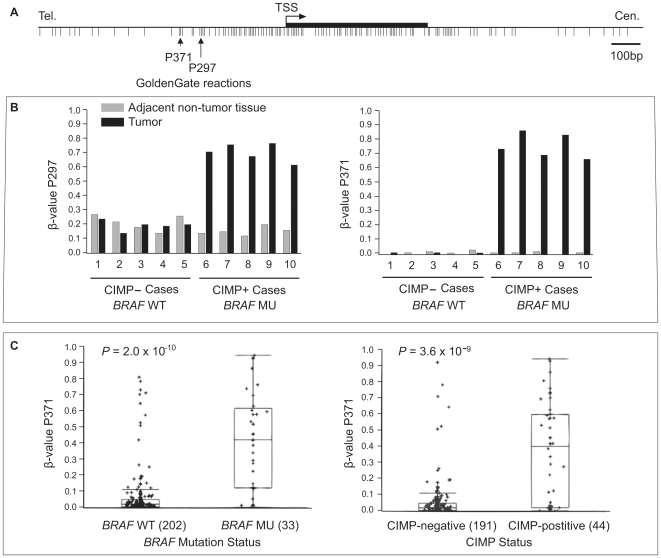
The Illumina GoldenGate Methylation Cancer Panel 1 platform contains two IGFBP7 probes (IGFBP7_P297_F and IGFBP7_P371_F) that interrogate the DNA methylation status of two distinct CpG dinucleotides in the IGFBP7 promoter CpG island (Figure 5A). We found that these two CpG sites in the IGFBP7 promoter are cancer-specifically methylated (Figure 5B) and strongly associated with both BRAFV600E (Wilcoxon rank-sum test, P value = 2.0×10⁻¹⁰) and CIMP (P value = 3.6×10⁻⁹) (Figure 5C). It has been reported that colorectal tumors with KRAS mutations also show DNA hypermethylation at CIMP-associated markers, albeit at a low frequency, and have high levels of DNA methylation of genes that undergo age-associated DNA hypermethylation. These tumors have been described as CIMP-low or CIMP2 [35], [36]. We did not find an association between IGFBP7 DNA hypermethylation and KRAS mutations when we excluded tumors with mutant BRAF (P value = 0.85). In agreement with these observations, we found that DNA hypermethylation of IGFBP7 is mostly present in colorectal cancer cell lines which harbor BRAFV600E and show frequent DNA methylation of the five-gene CIMP-specific marker panel previously described (Figure 6). Real-time RT-PCR analysis of colorectal cancer cell lines showed that IGFBP7 mRNA expression was inversely related to DNA hypermethylation, as cell lines with IGFBP7 hypermethylation showed little or no IGFBP7 gene expression (Figure 6). Among the CIMP− cells we examined, only COLO 320DM showed DNA hypermethylation of the IGFBP7 CpG island promoter with minimal level of expression. In retrospect, this unique characteristic of COLO 320DM cells compared to the other CIMP− cell lines might have enabled these cells to tolerate mutant BRAF overexpression, and may explain our difficulties in obtaining BRAFV600E expressing clones in other colorectal cancer cell line such as Caco-2.
Figure 5. IGFBP7 promoter DNA methylation in primary colorectal cancers.
(A) Genomic map of IGFBP7 promoter-associated CpG island, transcription start site (TSS) and exon 1 based on the UCSC genome browser (March 2006 assembly). The location of CpG sites interrogated by the Illumina GoldenGate DNA methylation assay is indicated by vertical arrows. (B) DNA methylation levels of the two CpG dinucelotides in the IGFBP7 promoter CpG island. β-values of each CpG site in 10 tumors (five CIMP− tumors with wild-type BRAF and five CIMP+ tumors with mutant BRAF, black bars) and adjacent non-tumor tissues (gray bars) are listed. (C) IGFBP7 promoter DNA methylation box plots of 235 human colorectal tumors stratified by BRAF mutation status (left) and CIMP+ status (right) at the IGFBP7 P371 locus. In the box plots, the ends of the box are the 25th and 75th quartiles. The line within the box identifies the median β-value. The whiskers above and below the box extend to at most 1.5 times the IQR. The CIMP status of each colorectal tumor sample is determined as described in the Materials and Methods section.
Figure 6. Analysis of DNA methylation and mRNA expression of IGFBP7 in colorectal cancer cell lines.
Quantitative real-time RT-PCR analysis of IGFBP7 expression. IGFBP7 expression levels are presented relative to PCNA expression. The error bars indicate the standard deviation of technical triplicate measurements. The number of methylated loci among the five CIMP markers and mutation status of BRAF and KRAS listed in Figure 1 are provided.
Discussion
CIMP in colorectal cancer provides a unique opportunity to study molecular mechanisms that lead to epigenetic changes in cancer and the contributions of these changes in the development of the disease [3], [37]. The distinct features found in CIMP are important clues in understanding this phenotype [3], [5], [7], [8]. Particularly striking is the extremely tight association between CIMP and BRAFV600E [5]. Mechanisms linking epigenetic (CIMP) and genetic (BRAF mutation) events and the temporal sequence in which these two events take place have attracted interest [37].
In this study, by using the high-throughput Illumina GoldenGate DNA methylation platform, we investigated the link between CIMP and BRAFV600E in colorectal cancer. We first tested whether expression of BRAFV600E causes DNA hypermethylation by stably expressing BRAFV600E in the CIMP-negative, BRAF wild-type COLO 320DM colorectal cancer cell line. We have examined DNA methylation changes in 100 CIMP-associated CpG sites, and found that BRAFV600E is not sufficient to induce DNA hypermethylation at these sites. One caveat of our system is that BRAFV600E could play a role in inducing DNA methylation only early in colorectal tumorigenesis, as BRAF mutations have been described at the earliest stage of tumor development [10], [38], [39]. It is possible that a unique set of genetic and/or epigenetic changes that occurred in COLO 320DM cells might have created an unfavorable environment for BRAFV600E to induce DNA hypermethylation. Experiments similar to those described above using Caco-2 cells, which also show CIMP– and carry _BRAF_-wild type, were not successful. We were not able to obtain any stably transfected clones that exhibited detectable levels of BRAFV600E (data not shown). Sustained BRAFV600E expression might be incompatible with Caco-2 cell proliferation due to cellular senescence or apoptosis induced by BRAFV600E. It is noteworthy that our RT-PCR analysis showed the robust expression of IGFBP7, a mediator of BRAFV600E-induced senescence or apoptosis, in Caco-2 cells in contrast to COLO 320DM cells.
Previously, we described CIMP-associated methylation of MLH1 as the underlying basis for mismatch repair deficiency (MSI+) in sporadic colorectal cancer [5]. Minoo et al. reported MLH1 DNA methylation upon stable transfection of BRAFV600E into the NCM460 cell line [40]. In our system, we did not detect such an increase in MLH1 DNA methylation (data not shown). Moreover, of the 33 BRAFV600E primary tumors we examined only 42% (14/33) showed MLH1 DNA hypermethylation. Therefore, BRAFV600E may affect DNA hypermethylation of MLH1 but only in certain circumstances. Interestingly, in the proposed serrated pathway to CIMP+ tumors, both BRAF mutations and CIMP+ have been observed in early precursor lesions, whereas MSI+ has not [6], [10], [41]. Thus, inactivation of MLH1 might occur at a later stage of tumor development. Minoo and colleagues observed the DNA hypermethylation of CDKN2A and 15 other CIMP-associated markers (IGFBP7 was not examined) in parent NCM460 cells, which limited their ability to study further the role of BRAFV600E inducing CIMP in their experimental system [40].
Intriguingly, we observed that the overall DNA methylation level of the CIMP-specific loci in our stably transfected cells increases as a function of cell passage. It is interesting to note that a selection drug in cultured cells has been described to result in changes in global chromatin structure [42], and a similar process may be associated with our observations here.
In addition, we found relatively large inter-clonal (among different clones) variation in DNA methylation levels in our transfection experiments (Figures 2B and 3), with an average R2 correlation calculated based on four EVCs of 0.88±0.01 (± s.d.). Our average intra-clonal (within clones at different passages) R2 correlation is 0.97±0.01 and the R2 correlation between technical replicates in Illumina GoldenGate DNA methylation analysis is 0.98±0.02 [16]. Consequently, we found some large differences in DNA methylation at several loci even among control clones (Figures 2B and 3). This emphasizes the importance of using multiple clones for this type of studies.
Alternatively, the strong association between CIMP and BRAFV600E might arise if CIMP specifically provides a favorable cellular context for BRAFV600E to promote tumorigenesis. In the second set of experiments, we determined genes whose DNA hypermethylation was tightly linked to BRAFV600E and CIMP+ in colorectal cancer. Intriguingly, we observed CIMP-dependent DNA hypermethylation and transcriptional inactivation of IGFBP7, which has been shown to mediate BRAFV600E-induced cellular senescence and apoptosis [34]. BRAFV600E has been shown to induce cellular senescence in cultured and primary human cells [30], [31], as well as mouse model [32]. Oncogene-induced senescence (OIS) has been recognized as an important tumor suppressor mechanism [33]. In order for BRAFV600E to promote its oncogenic effects, additional cooperative events are required to bypass senescence [33]. Recently, the molecular basis of BRAFV600E-induced senescence and apoptosis has been studied in detail. Wajapeyee et al. identified IGFBP7 as a mediator of BRAFV600E-induced senescence in human primary fibroblasts using a genome-wide shRNA screen. Their subsequent findings suggest that IGFBP7 expression is both necessary and sufficient to induce senescence and apoptosis in human primary melanocytes and melanoma, respectively. Moreover, they observed loss of IGFBP7 in primary BRAFV600E-positive melanoma samples and concluded that silencing of IGFBP7 expression is a critical step in the development of a melanoma harboring BRAFV600E [34].
Promoter-associated CpG island DNA hypermethylation of IGFBP7 has been reported in human colorectal cancer cell lines as well. The DNA methylation inhibitor 5-aza-2′-deoxycytidine has been shown to restore IGFBP7 expression in colorectal cancer cell lines, indicating that the DNA hypermethylation plays a major role in silencing of this gene in colorectal cancer [43]. However, its association with BRAF mutation and CIMP+ status in human colorectal cancers has not been explored. In this study, we found that IGFBP7 DNA hypermethylation is tumor-specific and tightly associated with colorectal tumors carrying BRAFV600E and exhibiting CIMP. Moreover, we found that IGFBP7 DNA hypermethylation is associated with loss of expression in CIMP+ colorectal cancer cell lines. CIMP-specific inactivation of BRAFV600E-induced senescence and apoptosis pathway by IGFBP7 DNA hypermethylation might create a favorable context for the acquisition of BRAFV600E in CIMP+ colorectal cancer.
Importantly, IGFBP7 DNA hypermethylation was not observed in all of the BRAF mutant colorectal tumors. Lin et al. examined the DNA sequence of the promoter and exonic regions of IGFBP7 in ten colorectal cancer cell lines. They did not find mutations associated with inactivation of IGFBP7 in their cell lines [43]. However, an increasing number of genes have recently been reported to be involved in OIS, and cooperation of multiple different signals appears to be critical for OIS [44]. It is therefore possible that CIMP-associated DNA hypermethylation events may impair OIS by affecting other components of the OIS signaling pathway in colorectal cancer.
Additional genes that showed CIMP-specific DNA hypermethylation include those that mediate various signaling pathways implicated in colorectal tumorigenesis (Table S3). The functional consequence of CIMP-specific DNA hypermethylation of such genes will be the subject of future investigations. We found that both BMP3 and BMP6 are targeted for CIMP-specific DNA hypermethylation and are strongly linked with BRAFV600E. Disruption of the BMP signaling pathway has been proposed to play a role in colorectal tumorigenesis [25]. Concurrent epigenetic inactivation of BMP3 and BMP6 was shown to be associated with the hyperactivation of the RAS-RAF-MEK-ERK signaling pathway in non-small-cell lung cancer [27]. Furthermore, receptor tyrosine kinases (RTKs) such as EPHA3, KIT, and FLT1 also showed CIMP-associated DNA hypermethylation (Table S3). Somatic mutations or overexpression of these genes has been implicated in colorectal tumorigenesis, which may involve the activation of the RAS-RAF-MEK-ERK signaling [45]–[50]. The potential inactivation of these genes in CIMP may lead to the development of tumors dependent on oncogenic BRAF-driven hyperactivation of the RAS-RAF-MEK-ERK signaling pathway. Furthermore, we also found that DNA methylation of SMO and HHIP were closely linked to colorectal tumors with BRAFV600E (Table S3). SMO and HHIP are involved in the regulation of the Hedgehog (Hh) signaling pathway. It has been demonstrated that elevated expression of SMO might contribute to colorectal tumorigenesis through activation of the Wnt signaling pathway in a mouse model and colorectal cancer cell lines [29]. Notably, it appeared that the expression of SMO was silenced in colorectal cancer cell lines harboring BRAFV600E [29]. Our data in primary colorectal tumors indicate that the CIMP-specific promoter DNA hypermethylation may result in a different effect of the Hedgehog (Hh) signaling pathway on colorectal tumorigenesis (Table S3).
Our data will be a useful resource for future investigations toward understanding CIMP and the role of aberrant DNA hypermethylation in colorectal tumorigenesis. The inactivation of a senescence pathway by IGFBP7 DNA hypermethylation in CIMP+ tumors may provide a permissive environment for the acquisition of BRAFV600E, thus providing a possible explanation for the link between BRAFV600E and CIMP in colorectal cancer.
Materials and Methods
Ethics Statement
This study was conducted according to the principles expressed in the Declaration of Helsinki. The study was approved by the Institutional Review Board of the Royal Brisbane Hospital Human Research Ethics Committee, the Bancroft Centre Ethics Committee and the University of Southern California. All patients provided written informed consent for the collection of samples and subsequent analysis. DNA from these patients was also analyzed in a previous publication [5].
Cell Culture and Genomic DNA Isolation
Colorectal cancer cell lines were obtained from American Type Culture Collection (Manassas, VA, USA). COLO 320DM cells were grown in DMEM supplemented with 10% FBS, 1 mM glutamine. An empty vector and an HA-tagged BRAFV600E cDNA clone (pMEV-HA, pMEV-BRAF-V599E, Biomyx Technology, San Diego, CA, USA) were transfected into COLO 320DM cells using Lipofectamine 2000 (Invitrogen, Carlsbad, CA, USA). G418 (Sigma-Aldrich, St. Louis, MO, USA) (1 mg/ml) was added 48 hours after transfection and resistant clones were randomly isolated and expanded. Stably expressing clones were maintained in 500 µg/ml of G418. Genomic DNA from each clone was isolated as previously described [51].
MethyLight Analysis of Five CIMP-Specific Markers in Colorectal Cancer Cell Lines
Genomic DNA was treated with sodium bisulfite and subsequently analyzed by MethyLight as previously described [5], [52]. The primer and probe sequences for the MethyLight reactions were described previously [5]. The results of MethyLight analyses were scored as PMR (Percent of Methylated Reference) values as previously defined [5].
Mutation Analysis and MSI Status of Colorectal Cancer Cell Lines
Primer sequences and PCR conditions for direct sequencing of BRAF at codon 600 in exon 15 and at codons 12 and 13 of KRAS in exon 2 were reported previously [13]. The MSI status of each cell line was based on the Sanger Institute Cancer Genome Project (http://www.sanger.ac.uk/) and based on a previous study [53].
Western Blot Analysis
Whole cell extracts were prepared from each resistant clone at the first passage using CelLytic M Cell Lysis Reagent (Sigma-Aldrich). Equal amounts of protein from whole cell extracts were separated on gradient (4–20%) polyacrylamide gels (Invitrogen) and then transferred to polyvinylidene difluoride (PVDF) membranes (Bio-Rad, Hercules, CA, USA). Blots were probed with the anti-HA antibodies (Roche, Indianapolis, IN, USA) for HA-BRAFV600E, anti-phospho-ERK1/2 (Cell Signaling, Beverly, MA, USA), and anti-ERK1 (Santa Cruz Biotechnology, Santa Cruz, CA, USA) followed by incubation with species specific horseradish peroxidase-conjugated secondary antibodies (Santa Cruz). Proteins were visualized using SuperSignal West Pico Chemiluminescent Substrate (Pierce, Rockford, IL, USA).
Primary Colorectal Tissue Samples
Primary colorectal tissue samples were collected and DNA was extracted as previously described [5]. A 58 sample set included five CIMP+ tumors, five CIMP– tumors and 48 randomly selected tumors, as indicated previously [5]. A 235 sample set included the same 48 randomly selected samples described above along with an additional 187 randomly collected tumors described previously [5]. CIMP status and mutation status of BRAF and KRAS for each tumor sample was previously determined [5]. BRAF mutations and KRAS mutations were found in 15% (33/235) and 33% (74/221) of the colorectal tumor samples, respectively. The KRAS mutation status of 14 tumor samples was not available. BRAF mutations and KRAS mutations were mutually exclusive [5].
DNA Methylation Analysis by the Illumina GoldenGate and Infinium DNA Methylation Platforms
Genomic DNA was bisulfite converted using the EZ-96 DNA Methylation Kit (ZYMO Research, Orange, CA, USA) according to manufacturer's protocol. Illumina GoldenGate DNA methylation analyses were performed as previously described [16] at the USC Epigenome Center Core Facility. Target sequences for the assay and detailed information on each interrogated CpG site and the associated gene on the “GoldenGate Methylation Cancer Panel 1” are described at www.illumina.com. The Illumina Infinium DNA methylation assay was performed following manufacturer's specifications. Detailed information on each interrogated CpG site and the associated gene on the Infinium BeadArray is available at www.illumina.com.
Identification of CIMP-Associated DNA Methylation Markers Using 58 Primary Colorectal Tumor Samples
For the hierarchical cluster analysis on 58 primary tumor samples shown in Figure 3, we used the β-values obtained from 1,421 reactions (84 X-linked reactions were omitted). We used Euclidian distance and Ward's linkage method to perform the clustering (JMP 6.0 software, SAS Institute, Cary, NC, USA). In order to identify CIMP-associated CpG sites, we performed a _t_-test on the difference in the β-value between the CIMP-positive group (11 tumors) and CIMP-negative group (47 tumors). We selected 100 CpG sites with P<0.001 after a correction for multiple-comparison [45] and mean |Δβ|>0.17, the estimated error in β [16].
Quantitative Real-Time RT-PCR
Total RNA from colorectal cancer cell lines were isolated using RNeasy Mini Kit (QIAGEN GmbH, Hilden, Germany). Reverse transcription reaction was performed using the SuperScript® III First-Strand Synthesis kit (Invitrogen). Quantitative real-time PCR was performed with primers and probe purchased from Applied Biosystems (Assay ID Hs00266026_m1) (Foster City, CA, USA). The raw expression values were normalized to those of PCNA.
Supporting Information
Dataset S1
Raw β-values obtained from the Illumina GoldenGate DNA methylation assay on 58 primary colorectal tumor samples. Samples are labeled with internal IDs.
(1.72 MB XLS)
Table S1
Characteristics of the 58 Primary colorectal tumor samples.
(0.02 MB XLS)
Table S2
One hundred (100) CpG sites that have significantly higher levels of DNA methylation in CIMP-positive versus CIMP-negative tumors. P values and difference in mean β-values between CIMP-positive tumors and CIMP-negative tumors are also included.
(0.02 MB XLS)
Table S3
The Illumina GoldenGate DNA methylation loci specifically methylated in colorectal tumors harboring BRAFV600E. We performed the Illumina GoldenGate DNA methylation assay on 235 primary colorectal tumor samples, whose CIMP status and BRAF mutation status have been determined previously [5]. We dichotomized the DNA methylation β-value (methylated or unmethylated) for each locus. The dichotomization threshold was chosen for each locus using the mean β-value + 3SD (standard deviations) from ten normal mucosal samples. The table lists 89 Illumina GoldenGate DNA methylation targets (of 60 genes) selected with P<0.001 (Fisher's exact test) after Bonferroni correction for multiple comparisons. Target CpG sites are sorted based on their P values.
(0.03 MB XLS)
Footnotes
Competing Interests: Peter Laird is a consultant for Epigenomics AG and for Celgene Corporation. The other authors disclosed no potential competing interest. This work was not supported by either Epigenomics AG or Celgene Corporation. These companies had no role in study design, data collection and analysis, decision to publish, or preparation of the manuscript.
Funding: National Institutes of Health (NIH) grants R01 CA118699-02 (P.W. Laird), R01 CA075090-09 (P.W. Laird). The funders had no role in study design, data collection and analysis, decision to publish, or preparation of the manuscript.
References
- 1.Jones PA, Baylin SB. The epigenomics of cancer. Cell. 2007;128:683–692. doi: 10.1016/j.cell.2007.01.029. [DOI] [PMC free article] [PubMed] [Google Scholar]
- 2.Toyota M, Ahuja N, Ohe-Toyota M, Herman JG, Baylin SB, et al. CpG island methylator phenotype in colorectal cancer. Proc Natl Acad Sci U S A. 1999;96:8681–8686. doi: 10.1073/pnas.96.15.8681. [DOI] [PMC free article] [PubMed] [Google Scholar]
- 3.Issa JP. CpG island methylator phenotype in cancer. Nat Rev Cancer. 2004;4:988–993. doi: 10.1038/nrc1507. [DOI] [PubMed] [Google Scholar]
- 4.Yamashita K, Dai T, Dai Y, Yamamoto F, Perucho M. Genetics supersedes epigenetics in colon cancer phenotype. Cancer Cell. 2003;4:121–131. doi: 10.1016/s1535-6108(03)00190-9. [DOI] [PubMed] [Google Scholar]
- 5.Weisenberger DJ, Siegmund KD, Campan M, Young J, Long TI, et al. CpG island methylator phenotype underlies sporadic microsatellite instability and is tightly associated with BRAF mutation in colorectal cancer. Nat Genet. 2006;38:787–793. doi: 10.1038/ng1834. [DOI] [PubMed] [Google Scholar]
- 6.O'Brien MJ. Hyperplastic and serrated polyps of the colorectum. Gastroenterol Clin North Am. 2007;36:947–68, viii. doi: 10.1016/j.gtc.2007.08.007. [DOI] [PubMed] [Google Scholar]
- 7.Samowitz WS, Albertsen H, Herrick J, Levin TR, Sweeney C, et al. Evaluation of a large, population-based sample supports a CpG island methylator phenotype in colon cancer. Gastroenterology. 2005;129:837–845. doi: 10.1053/j.gastro.2005.06.020. [DOI] [PubMed] [Google Scholar]
- 8.Kambara T, Simms LA, Whitehall VL, Spring KJ, Wynter CV et al. BRAF mutation is associated with DNA methylation in serrated polyps and cancers of the colorectum. Gut. 2004;53:1137–1144. doi: 10.1136/gut.2003.037671. [DOI] [PMC free article] [PubMed] [Google Scholar]
- 9.Minoo P, Baker K, Goswami R, Chong G, Foulkes WD, et al. Extensive DNA methylation in normal colorectal mucosa in hyperplastic polyposis. Gut. 2006;55:1467–1474. doi: 10.1136/gut.2005.082859. [DOI] [PMC free article] [PubMed] [Google Scholar]
- 10.Rosenberg DW, Yang S, Pleau DC, Greenspan EJ, Stevens RG, et al. Mutations in BRAF and KRAS differentially distinguish serrated versus non-serrated hyperplastic aberrant crypt foci in humans. Cancer Res. 2007;67:3551–3554. doi: 10.1158/0008-5472.CAN-07-0343. [DOI] [PubMed] [Google Scholar]
- 11.Oliveira C, Westra JL, Arango D, Ollikainen M, Domingo E, et al. Distinct patterns of KRAS mutations in colorectal carcinomas according to germline mismatch repair defects and hMLH1 methylation status. Hum Mol Genet. 2004;13:2303–2311. doi: 10.1093/hmg/ddh238. [DOI] [PubMed] [Google Scholar]
- 12.Garnett MJ, Marais R. Guilty as charged: B-RAF is a human oncogene. Cancer Cell. 2004;6:313–319. doi: 10.1016/j.ccr.2004.09.022. [DOI] [PubMed] [Google Scholar]
- 13.Davies H, Bignell GR, Cox C, Stephens P, Edkins S, et al. Mutations of the BRAF gene in human cancer. Nature. 2002;417:949–954. doi: 10.1038/nature00766. [DOI] [PubMed] [Google Scholar]
- 14.Haigis KM, Wistuba II, Kurie JM. Lung premalignancy induced by mutant B-Raf, what is thy fate? To senesce or not to senesce, that is the question. Genes Dev. 2007;21:361–366. doi: 10.1101/gad.1532107. [DOI] [PubMed] [Google Scholar]
- 15.Bibikova M, Chudin E, Wu B, Zhou L, Garcia EW, et al. Human embryonic stem cells have a unique epigenetic signature. Genome Res. 2006;16:1075–1083. doi: 10.1101/gr.5319906. [DOI] [PMC free article] [PubMed] [Google Scholar]
- 16.Bibikova M, Lin Z, Zhou L, Chudin E, Garcia EW, et al. High-throughput DNA methylation profiling using universal bead arrays. Genome Res. 2006;16:383–393. doi: 10.1101/gr.4410706. [DOI] [PMC free article] [PubMed] [Google Scholar]
- 17.Ladd-Acosta C, Pevsner J, Sabunciyan S, Yolken RH, Webster MJ, et al. DNA methylation signatures within the human brain. Am J Hum Genet. 2007;81:1304–1315. doi: 10.1086/524110. [DOI] [PMC free article] [PubMed] [Google Scholar]
- 18.Houseman EA, Christensen BC, Yeh RF, Marsit CJ, Karagas MR, et al. Model-based clustering of DNA methylation array data: a recursive-partitioning algorithm for high-dimensional data arising as a mixture of beta distributions. BMC Bioinformatics. 2008;9:365. doi: 10.1186/1471-2105-9-365. [DOI] [PMC free article] [PubMed] [Google Scholar]
- 19.TCGA-Consortium Comprehensive genomic characterization defines human glioblastoma genes and core pathways. Nature. 2008;455:1061–1068. doi: 10.1038/nature07385. [DOI] [PMC free article] [PubMed] [Google Scholar]
- 20.Martinez R, Martin-Subero JI, Rohde V, Kirsch M, Alaminos M, et al. A microarray-based DNA methylation study of glioblastoma multiforme. Epigenetics. 2009;4:255–264. doi: 10.4161/epi.9130. [DOI] [PubMed] [Google Scholar]
- 21.Bibikova M, Fan JB. GoldenGate(R) Assay for DNA Methylation Profiling. Methods Mol Biol. 2009;507:149–163. doi: 10.1007/978-1-59745-522-0_12. [DOI] [PubMed] [Google Scholar]
- 22.Byun HM, Siegmund KD, Pan F, Weisenberger DJ, Kanel G, et al. Epigenetic profiling of somatic tissues from human autopsy specimens identifies tissue- and individual-specific DNA methylation patterns. Hum Mol Genet. 2009;18:4808–4817. doi: 10.1093/hmg/ddp445. [DOI] [PMC free article] [PubMed] [Google Scholar]
- 23.Christensen BC, Houseman EA, Marsit CJ, Zheng S, Wrensch MR, et al. Aging and environmental exposures alter tissue-specific DNA methylation dependent upon CpG island context. PLoS Genet. 2009;5:e1000602. doi: 10.1371/journal.pgen.1000602. [DOI] [PMC free article] [PubMed] [Google Scholar]
- 24.Katari S, Turan N, Bibikova M, Erinle O, Chalian R, et al. DNA methylation and gene expression differences in children conceived in vitro or in vivo. Hum Mol Genet. 2009;18:3769–3778. doi: 10.1093/hmg/ddp319. [DOI] [PMC free article] [PubMed] [Google Scholar]
- 25.Loh K, Chia JA, Greco S, Cozzi SJ, Buttenshaw RL, et al. Bone morphogenic protein 3 inactivation is an early and frequent event in colorectal cancer development. Genes Chromosomes Cancer. 2008;47:449–460. doi: 10.1002/gcc.20552. [DOI] [PubMed] [Google Scholar]
- 26.Kohonen-Corish MR, Sigglekow ND, Susanto J, Chapuis PH, Bokey EL, et al. Promoter methylation of the mutated in colorectal cancer gene is a frequent early event in colorectal cancer. Oncogene. 2007;26:4435–4441. doi: 10.1038/sj.onc.1210210. [DOI] [PubMed] [Google Scholar]
- 27.Kraunz KS, Nelson HH, Liu M, Wiencke JK, Kelsey KT. Interaction between the bone morphogenetic proteins and Ras/MAP-kinase signalling pathways in lung cancer. Br J Cancer. 2005;93:949–952. doi: 10.1038/sj.bjc.6602790. [DOI] [PMC free article] [PubMed] [Google Scholar]
- 28.Xing M. BRAF mutation in papillary thyroid cancer: pathogenic role, molecular bases, and clinical implications. Endocr Rev. 2007;28:742–762. doi: 10.1210/er.2007-0007. [DOI] [PubMed] [Google Scholar]
- 29.Arimura S, Matsunaga A, Kitamura T, Aoki K, Aoki M, et al. Reduced level of smoothened suppresses intestinal tumorigenesis by down-regulation of Wnt signaling. Gastroenterology. 2009;137:629–638. doi: 10.1053/j.gastro.2009.04.059. [DOI] [PubMed] [Google Scholar]
- 30.Zhu J, Woods D, McMahon M, Bishop JM. Senescence of human fibroblasts induced by oncogenic Raf. Genes Dev. 1998;12:2997–3007. doi: 10.1101/gad.12.19.2997. [DOI] [PMC free article] [PubMed] [Google Scholar]
- 31.Michaloglou C, Vredeveld LC, Soengas MS, Denoyelle C, Kuilman T, et al. BRAFE600-associated senescence-like cell cycle arrest of human naevi. Nature. 2005;436:720–724. doi: 10.1038/nature03890. [DOI] [PubMed] [Google Scholar]
- 32.Dankort D, Filenova E, Collado M, Serrano M, Jones K, et al. A new mouse model to explore the initiation, progression, and therapy of BRAFV600E-induced lung tumors. Genes Dev. 2007;21:379–384. doi: 10.1101/gad.1516407. [DOI] [PMC free article] [PubMed] [Google Scholar]
- 33.Mooi WJ, Peeper DS. Oncogene-induced cell senescence–halting on the road to cancer. N Engl J Med. 2006;355:1037–1046. doi: 10.1056/NEJMra062285. [DOI] [PubMed] [Google Scholar]
- 34.Wajapeyee N, Serra RW, Zhu X, Mahalingam M, Green MR. Oncogenic BRAF induces senescence and apoptosis through pathways mediated by the secreted protein IGFBP7. Cell. 2008;132:363–374. doi: 10.1016/j.cell.2007.12.032. [DOI] [PMC free article] [PubMed] [Google Scholar]
- 35.Ogino S, Kawasaki T, Kirkner GJ, Loda M, Fuchs CS. CpG island methylator phenotype-low (CIMP-low) in colorectal cancer: possible associations with male sex and KRAS mutations. J Mol Diagn. 2006;8:582–588. doi: 10.2353/jmoldx.2006.060082. [DOI] [PMC free article] [PubMed] [Google Scholar]
- 36.Shen L, Toyota M, Kondo Y, Lin E, Zhang L, et al. Integrated genetic and epigenetic analysis identifies three different subclasses of colon cancer. Proc Natl Acad Sci U S A. 2007;104:18654–18659. doi: 10.1073/pnas.0704652104. [DOI] [PMC free article] [PubMed] [Google Scholar]
- 37.Schuebel K, Chen W, Baylin SB. CIMPle origin for promoter hypermethylation in colorectal cancer? Nat Genet. 2006;38:738–740. doi: 10.1038/ng0706-738. [DOI] [PubMed] [Google Scholar]
- 38.Beach R, Chan AO, Wu TT, White JA, Morris JS, et al. BRAF mutations in aberrant crypt foci and hyperplastic polyposis. Am J Pathol. 2005;166:1069–1075. doi: 10.1016/S0002-9440(10)62327-9. [DOI] [PMC free article] [PubMed] [Google Scholar]
- 39.O'Brien MJ, Yang S, Mack C, Xu H, Huang CS, et al. Comparison of microsatellite instability, CpG island methylation phenotype, BRAF and KRAS status in serrated polyps and traditional adenomas indicates separate pathways to distinct colorectal carcinoma end points. Am J Surg Pathol. 2006;30:1491–1501. doi: 10.1097/01.pas.0000213313.36306.85. [DOI] [PubMed] [Google Scholar]
- 40.Minoo P, Moyer M, Jass J. Role of BRAF-V600E in the serrated pathway of colorectal tumourigenesis. J Pathol. 2007;212:124–133. doi: 10.1002/path.2160. [DOI] [PubMed] [Google Scholar]
- 41.Velho S, Moutinho C, Cirnes L, Albuquerque C, Hamelin R, et al. BRAF, KRAS and PIK3CA mutations in colorectal serrated polyps and cancer: primary or secondary genetic events in colorectal carcinogenesis? BMC Cancer. 2008;8:255. doi: 10.1186/1471-2407-8-255. [DOI] [PMC free article] [PubMed] [Google Scholar]
- 42.Muthuswami R, Mesner LD, Wang D, Hill DA, Imbalzano AN, et al. Phosphoaminoglycosides inhibit SWI2/SNF2 family DNA-dependent molecular motor domains. Biochemistry. 2000;39:4358–4365. doi: 10.1021/bi992503r. [DOI] [PubMed] [Google Scholar]
- 43.Lin J, Lai M, Huang Q, Ma Y, Cui J, et al. Methylation patterns of IGFBP7 in colon cancer cell lines are associated with levels of gene expression. J Pathol. 2007;212:83–90. doi: 10.1002/path.2144. [DOI] [PubMed] [Google Scholar]
- 44.Cichowski K, Hahn WC. Unexpected pieces to the senescence puzzle. Cell. 2008;133:958–961. doi: 10.1016/j.cell.2008.05.027. [DOI] [PMC free article] [PubMed] [Google Scholar]
- 45.Bardelli A, Parsons DW, Silliman N, Ptak J, Szabo S, et al. Mutational analysis of the tyrosine kinome in colorectal cancers. Science. 2003;300:949. doi: 10.1126/science.1082596. [DOI] [PubMed] [Google Scholar]
- 46.Bates RC, Goldsmith JD, Bachelder RE, Brown C, Shibuya M, et al. Flt-1-dependent survival characterizes the epithelial-mesenchymal transition of colonic organoids. Curr Biol. 2003;13:1721–1727. doi: 10.1016/j.cub.2003.09.002. [DOI] [PubMed] [Google Scholar]
- 47.Noren NK, Pasquale EB. Eph receptor-ephrin bidirectional signals that target Ras and Rho proteins. Cell Signal. 2004;16:655–666. doi: 10.1016/j.cellsig.2003.10.006. [DOI] [PubMed] [Google Scholar]
- 48.Graells J, Vinyals A, Figueras A, Llorens A, Moreno A, et al. Overproduction of VEGF concomitantly expressed with its receptors promotes growth and survival of melanoma cells through MAPK and PI3K signaling. J Invest Dermatol. 2004;123:1151–1161. doi: 10.1111/j.0022-202X.2004.23460.x. [DOI] [PubMed] [Google Scholar]
- 49.Bellone G, Smirne C, Carbone A, Buffolino A, Scirelli T, et al. KIT/stem cell factor expression in premalignant and malignant lesions of the colon mucosa in relationship to disease progression and outcomes. Int J Oncol. 2006;29:851–859. [PubMed] [Google Scholar]
- 50.Wood LD, Parsons DW, Jones S, Lin J, Sjoblom T, et al. The genomic landscapes of human breast and colorectal cancers. Science. 2007;318:1108–1113. doi: 10.1126/science.1145720. [DOI] [PubMed] [Google Scholar]
- 51.Laird PW, Zijderveld A, Linders K, Rudnicki MA, Jaenisch R, et al. Simplified mammalian DNA isolation procedure. Nucleic Acids Res. 1991;19:4293. doi: 10.1093/nar/19.15.4293. [DOI] [PMC free article] [PubMed] [Google Scholar]
- 52.Weisenberger DJ, Campan M, Long TI, Kim M, Woods C, et al. Analysis of repetitive element DNA methylation by MethyLight. Nucleic Acids Res. 2005;33:6823–6836. doi: 10.1093/nar/gki987. [DOI] [PMC free article] [PubMed] [Google Scholar]
- 53.Suter CM, Norrie M, Ku SL, Cheong KF, Tomlinson I, et al. CpG island methylation is a common finding in colorectal cancer cell lines. Br J Cancer. 2003;88:413–419. doi: 10.1038/sj.bjc.6600699. [DOI] [PMC free article] [PubMed] [Google Scholar]
Associated Data
This section collects any data citations, data availability statements, or supplementary materials included in this article.
Supplementary Materials
Dataset S1
Raw β-values obtained from the Illumina GoldenGate DNA methylation assay on 58 primary colorectal tumor samples. Samples are labeled with internal IDs.
(1.72 MB XLS)
Table S1
Characteristics of the 58 Primary colorectal tumor samples.
(0.02 MB XLS)
Table S2
One hundred (100) CpG sites that have significantly higher levels of DNA methylation in CIMP-positive versus CIMP-negative tumors. P values and difference in mean β-values between CIMP-positive tumors and CIMP-negative tumors are also included.
(0.02 MB XLS)
Table S3
The Illumina GoldenGate DNA methylation loci specifically methylated in colorectal tumors harboring BRAFV600E. We performed the Illumina GoldenGate DNA methylation assay on 235 primary colorectal tumor samples, whose CIMP status and BRAF mutation status have been determined previously [5]. We dichotomized the DNA methylation β-value (methylated or unmethylated) for each locus. The dichotomization threshold was chosen for each locus using the mean β-value + 3SD (standard deviations) from ten normal mucosal samples. The table lists 89 Illumina GoldenGate DNA methylation targets (of 60 genes) selected with P<0.001 (Fisher's exact test) after Bonferroni correction for multiple comparisons. Target CpG sites are sorted based on their P values.
(0.03 MB XLS)


