Inducible Foxp3+ regulatory T-cell development by a commensal bacterium of the intestinal microbiota (original) (raw)
Abstract
To maintain intestinal health, the immune system must faithfully respond to antigens from pathogenic microbes while limiting reactions to self-molecules. The gastrointestinal tract represents a unique challenge to the immune system, as it is permanently colonized by a diverse amalgam of bacterial phylotypes producing multitudes of foreign microbial products. Evidence from human and animal studies indicates that inflammatory bowel disease results from uncontrolled inflammation to the intestinal microbiota. However, molecular mechanisms that actively promote mucosal tolerance to the microbiota remain unknown. We report herein that a prominent human commensal, Bacteroides fragilis, directs the development of Foxp3+ regulatory T cells (Tregs) with a unique “inducible” genetic signature. Monocolonization of germ-free animals with B. fragilis increases the suppressive capacity of Tregs and induces anti-inflammatory cytokine production exclusively from Foxp3+ T cells in the gut. We show that the immunomodulatory molecule, polysaccharide A (PSA), of B. fragilis mediates the conversion of CD4+ T cells into Foxp3+ Treg cells that produce IL-10 during commensal colonization. Functional Foxp3+ Treg cells are also produced by PSA during intestinal inflammation, and Toll-like receptor 2 signaling is required for both Treg induction and IL-10 expression. Most significantly, we show that PSA is not only able to prevent, but also cure experimental colitis in animals. Our results therefore demonstrate that B. fragilis co-opts the Treg lineage differentiation pathway in the gut to actively induce mucosal tolerance.
Keywords: Bacteroides fragilis, inflammatory bowel disease, Toll-like receptor, polysaccharide A, interleukin 10
Bacteria colonize almost every environmentally exposed surface of the human body, with the greatest quantity and diversity found in the lower gastrointestinal tract (1). One-hundred trillion bacteria representing hundreds of species live in homeostasis with the intestinal immune system. Maintenance of homeostasis toward the intestinal microbial milieu is critical to the preservation of health (2). Defects in mucosal tolerance lead to disorders, such as inflammatory bowel disease (IBD); Crohn disease and ulcerative colitis are idiopathic human syndromes marked by unrestrained gastrointestinal inflammation. In the United States alone, over 1 million people suffer from IBD, making it a serious social and medical problem. Additionally, the overall rates of diagnosis are increasing and the mean age of onset is decreasing. Although the causes for IBD remain enigmatic, interactions between host genetics, the immune system, and the intestinal microbiota appear to be central to the development and severity of disease. A growing body of evidence suggests that a breakdown in immune tolerance to the microbiota is a critical event in the onset and progression of IBD. Indeed, IBD patients have increased reactivity to antigens of the normal microbiota and antibiotics offer some therapeutic benefits. Furthermore, germ-free animals do not develop experimental colitis and the microbial community of the gut is significantly altered in IBD patients (2, 3). The microbiota and the mucosal immune system appear to have forged an intricate evolutionary relationship with important implications for human health, and disturbances in our partnership with symbiotic bacteria may be a risk factor for IBD.
Intestinal immune health is achieved, in part, through the development of opposing arms of the adaptive immune response. Induction of a proinflammatory T-cell response by microorganisms can elicit development of either T-helper-1 (Th1), Th2, or Th17 cells (4). Proinflammatory T-cell responses are suppressed by the action of various subsets of regulatory T cells (Tregs). Naturally occurring Tregs emerge from the thymus and are marked by expression of CD4, CD25, and the transcription factor Foxp3 (Forkhead Box transcription factor for Treg differentiation) (5). More recent studies have provided evidence that other populations of Tregs exist that have variable expression of Foxp3 and secrete anti-inflammatory cytokines, such as IL-10 (6).These Treg subsets, often referred to as “inducible Tregs,” are not found in the thymic environment but, rather, seem to be induced in peripheral tissues, such as the gut. Suppression of Th1 and Th17 cell responses by Tregs prevents inflammation in experimental models of colitis (7, 8). Emerging studies have highlighted a key role for the commensal microbiota in the development of intestinal Th17 cells. Germ-free mice display very low numbers of Th17 cells within the lamina propria that is restored upon colonization with a complex microbial consortium (9–11) or even specific bacterial species (12, 13). Studies have also shown that germ-free animals are altered in Treg proportions of the gut (10, 14). Thus, the microbiota plays an active role in the development and function of both pro- and anti-inflammatory T-cell pathways. However, microbial molecules that coordinate the Treg/Th17 axis remain to be described.
Polysaccharide A (PSA), produced by the human symbiont Bacteroides fragilis, is able to protect animals from intestinal inflammation by suppressing IL-17 production (15). We report here that colonization of germ-free animals with B. fragilis mediates the development of inducible Tregs with a unique genetic signature. CD4+Foxp3+ Tregs that produce IL-10 differentiate in the gut, but not in systemic compartments in response to PSA. Natural Treg subsets are not affected by B. fragilis colonization. PSA engenders mucosal tolerance by promoting the differentiation of functional Treg cells during homeostasis and intestinal inflammation. Toll-like receptor 2 (TLR2) signaling is required for induction of Foxp3+ Tregs and IL-10 production by PSA, and TLR2−/− animals treated with PSA are not protected from colitis. Finally, we show that PSA is capable of reversing experimental colitis in animals, thus representing a potentially unique and natural therapy for human IBD. Our findings support recent speculation that Foxp3+ Treg cells respond to commensal microbial antigens (16, 17), and define the cellular and molecular pathway by which PSA functions to control intestinal inflammatory disease.
Results
Bacteroides fragilis Colonization Elicits Mucosal Tolerance.
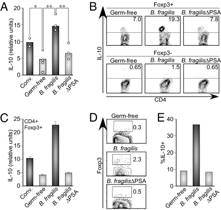
The microbiota has profound influences on the development and function of the immune system (2). Colonization of germ-free animals with B. fragilis represents a model system for the study of immune-bacterial symbiosis. Because recent reports have revealed a critical role for Treg-produced IL-10 during maintenance of intestinal homeostasis, we sought to understand how B. fragilis colonization directly affects Treg development (18). Germ-free C57BL/6 mice were lethally irradiated and reconstituted with bone marrow from Foxp3-GFP mice. Mice were either left germ-free or monoassociated with WT B. fragilis or a strain deleted of PSA (_B. fragilis_ΔPSA) (Fig. S1). Consistent with recent reports (9, 10), the percentage of Treg cells (CD4+Foxp3+) residing within either the mesenteric lymph node (MLN) or colon did not differ significantly between groups (Fig. S2 A and B), suggesting that the microbiota is not a requirement for the presence of naturally occurring Tregs cells. Remarkably, production of IL-10 within the intestine is deficient in the absence of the microbiota (compare conventional with germ-free mice); moreover, B. fragilis colonization restores the production of IL-10 within the colon in a PSA-dependent manner (Fig. 1_A_). Monoassociation of germ-free animals with B. fragilis results in a 2-fold increase in the percentage of IL-10–producing Foxp3+ Tregs within the colon (Fig. S3_A_). The induction of IL-10 from Foxp3+ cells by B. fragilis completely requires PSA, as Tregs in _B. fragilis_ΔPSA-colonized animals have similar IL-10 levels as germ-free mice (Fig. 1_B_). Colonization with B. fragilis directs IL-10 production almost exclusively from Foxp3+ (and not Foxp3−) T cells (Fig. 1_B_). Compared with conventionally-colonized animals, purified CD4+Foxp3+ Tregs from germ-free mice display lower expression of IL-10, and the Treg-associated cytokine TGF-β2 (Fig. 1_C_ and Fig. S3_B_). B. fragilis monoassociation restores expression of these anti-inflammatory genes, a phenotype that is completely dependent on PSA production. Microbial colonization does not affect all Treg-associated genes, as CD25 expression does not change in all groups tested (Fig. S3_B_). Thus, homeostatic microbial colonization by B. fragilis has a dramatic impact on the programming of CD4+Foxp3+ Tregs in the colon.
Fig. 1.
Bacteroides fragilis colonization elicits tolerant T cell responses in the intestine. (A) Germ-free C57BL/6 mice were reconstituted with Foxp3-GFP bone marrow and colonized with bacteria as indicated (Conv. refers to specific pathogen-free conventional mice). RNA was extracted from the colons of mice and IL-10 measured by qRT-PCR. Each symbol indicates an individual mouse. *P < 0.05; **_P_ < 0.01. These data are representative of two independent trials with at least three mice in each group. (_B_) Lamina propria lymphocytes were assessed for IL-10 from CD4+Foxp3+ and CD4+Foxp3− subsets by flow cytometry (FC). These data are representative of three independent trials. (_C_) Cells from the MLNs of mice were purified to isolate CD4+Foxp3-GFP T cells. RNA was extracted and the IL-10 transcript levels analyzed by qRT-PCR. Cell populations were >99% pure. Error bars indicate SD from samples run in triplicate from the same experiment. These data are representative of two independent trials with at least four mice per group. (D and E) Germ-free C57BL/6 Rag−/− animals were sublethally irradiated and CD4+Foxp3− T cells transferred from Foxp3-GFP donor mice. Cell purity was always >99%. Animals were either left germ-free or colonized with indicated bacterial strains. Foxp3 was analyzed by GFP expression in D and IL-10 in E. (E) The percentage of IL-10+ cells within the CD4+Foxp3+ converted subset. These data are representative of two independent trials with at least three mice in each group.
We wanted to examine whether PSA is able to convert Foxp3+ T cells from Foxp3− precursors. CD4+Foxp3− T cells were purified from Foxp3-GFP animals (19) and adoptively transferred into germ-free Rag−/− mice. Groups of animals were either left germ-free or colonized with WT or _B. fragilis_ΔPSA bacteria. Three weeks posttransfer, CD4+ T cells were analyzed for the expression of Foxp3 and IL-10. Fig. 1_D_ shows that although germ-free animals have few CD4+ T cells that expressed Foxp3, animals colonized with WT B. fragilis contained significant levels of converted Foxp3+ Tregs (statistical analysis in Fig. S3_C_). Conversion to Foxp3 expression was dependent on PSA, as _B. fragilis_ΔPSA-colonized animals contained cell levels comparable to germ-free animals (Fig. 1_D_). Remarkably, Foxp3+ T cells from _B. fragilis_-colonized animals acquired IL-10 expression (Fig. 1_E_). These results identify a unique bacterial molecule of the intestinal microbiota that is able to promote lineage differentiation of Foxp3+ Treg cells.
PSA Promotes Inducible Foxp3+ Tregs with Suppressive Activity.
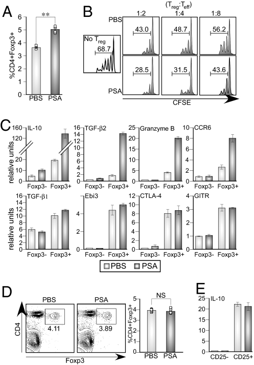
We orally administered PSA to mice and monitored the CD4+CD25+Foxp3+ population of Tregs in the MLN. Mice treated with PSA displayed increased percentages of CD4+Foxp3+ T cells compared with control mice (Fig. 2_A_). One of the primary functions of Treg cells is to suppress the activation and proliferation of inflammatory effector T cells (Teffs). The suppressive properties of Tregs following PSA administration were determined by the addition of varying ratios of CD4+CD25+ T cells purified from MLNs to naive responder cells. Tregs isolated from PSA-treated animals have increased suppressive capacity compared with PBS-treated control animals (28.5% proliferating responder cells vs. 43.0% at a 1:2 Treg:Teff ratio) (Fig. 2_B_). These findings demonstrate that PSA induces the differentiation of functional Foxp3+ Tregs with enhanced suppressive activity.
Fig. 2.
PSA is sufficient to induce functional Foxp3+ Tregs with an inducible phenotype. (A) Foxp3-GFP mice were orally gavaged with purified PSA, MLNs harvested, and cells stained for CD4 and CD25. Each symbol represents the percent of CD4+ Foxp3-GFP+ cells from a single mouse. Results are representative of three independent trials with four mice per group. **P < 0.01. (_B_) CD4+CD25+ cells were purified from the MLNs of mice treated with either PBS or PSA, as indicated in _A_, and incubated with CFSE-pulsed CD4+CD25− effector T cells in an in vitro suppression assay. Numbers indicate the percentage of cells undergoing at least one cellular division at three different ratios of effector T cells (Teff) and Tregs. These data are representative of two independent trials. (_C_) Foxp3-GFP mice were orally treated with purified PSA. MLNs were extracted and CD4+Foxp3+ or the CD4+Foxp3− T cells were purified based on ±GFP expression (purity >99%). RNA was extracted and used for qRT-PCR. These data are representative of three independent experiments. Light bars indicated cells derived from PBS treated mice and dark bars from PSA-treated mice. (D) TLR2−/− mice were orally treated with PSA. MLNs were extracted and analyzed for the percentage of CD4+Foxp3+ cells. Each symbol in the bar graph represents the percentage of CD4+Foxp3+ from an individual mouse. NS, not significant. Results are representative of three independent trials with at least three mice per group. (E) CD4+CD25hi+ and CD4+CD25− T cell populations were FACS purified from MLNs of PSA-treated TLR2−/− mice. IL-10 levels were analyzed by qRT-PCR. Light and dark bars indicate IL-10 levels in PBS or PSA treated animals, respectively. Error bars represent the SD from samples of the same experiment run in triplicate. Results are representative of two independent trials with four mice per group.
Various subsets of Tregs exist within the Foxp3+ and Foxp3− T-cell populations; therefore, we analyzed the expression of Treg-associated genes to understand how PSA affects the development of Foxp3+ Tregs. Foxp3-GFP mice were gavaged with purified PSA (or PBS control), and RNA was extracted from either CD4+Foxp3− or CD4+Foxp3+ T cells from the MLNs following cell purification. As expected, gene expression in Foxp3− and Foxp3+ T-cell subsets differed dramatically and included higher basal levels of IL-10, TGF-β2, GITR (glucocorticoid-induced TNF-related protein), ICOS, CTLA-4, and Ebi3 (subunit of IL- 35) in Foxp3+ T cells (Fig. 2_C_ and Fig. S4). Remarkably, PSA induces over 8-fold increased levels of IL-10 from CD4+Foxp3+ Tregs than that expressed in PBS-treated cells, and had virtually no impact on CD4+Foxp3− T cells. Accordingly, although TGF-β2 expression in CD4+Foxp3− T cells was not altered, PSA elicited significant induction of TGF-β2 from Foxp3+ Treg cells. Although Treg subsets that do not express Foxp3 have been described based on IL-10 and TGF-β production (Tr1 and Th3 cells, respectively) (20), PSA's effects are restricted to the Foxp3+ Treg population. PSA treatment also significantly increases the transcription of granzyme B, perforin, and CCR6 (a chemokine receptor associated with the migration of Treg cells) from Foxp3+ Tregs (Fig. 2_C_ and Fig. S4). It is important to note that PSA does not globally impact all Treg-derived cytokines, as expression of TGF-β1 and Ebi3 are not altered, demonstrating specificity for a distinct Treg profile. Furthermore, production of the natural Treg-associated surface receptors CTLA-4, GITR, and ICOS are not changed among Foxp3+ cells in response to PSA treatment (Fig. 2_C_ and Fig. S4). Taken together, these data reveal that PSA activates inducible Foxp3+ Tregs and identifies a “PSA-specific” gene expression program within Foxp3+ Treg cells.
We have previously reported that colonization of germ-free animals with PSA-producing bacteria induces production of the Th1 cytokine IFN-γ among splenic CD4+ T cells (21). To further investigate the lineage differentiation of immune cells directed by PSA, we examined animals for IFN-γ expression among Foxp3+ T cells. Intriguingly, IFN-γ expression in the MLNs is found exclusively in the Foxp3− subset and is not affected by microbial colonization (Fig. S5). Furthermore, splenic CD4+ T cells produce IFN-γ only from Foxp3−/IL-10− T cells that is dependent on PSA expression (Fig. S6). These results demonstrate that there is a compartmental difference in the ability of PSA to induce a Th1 profile in the spleen, although promoting a tolerogenic immune environment in the gut consisting of CD4+Foxp3+IFN-γ− Treg cells.
PSA Activity Requires TLR2 Signaling.
Many microbial products are sensed by pattern recognition receptors, such as TLRs. Although historically believed to induce inflammation, a series of studies now show that TLR signaling can also promote anti-inflammatory responses (reviewed in ref. 22). PSA has been shown to coordinate cytokine production from innate immune cells through TLR2 signaling (23); however, a role for TLR2 in Treg development remains uncertain. To understand the mechanism by which PSA promotes Tregs, TLR2-deficient animals were orally treated with PSA and analyzed for CD4+Foxp3+ T-cell development. In contrast to the Treg expansion seen in WT animals (see Fig. 2_A_), we observed no difference in the percentage of CD4+Foxp3+ T cells in TLR2−/− mice treated with PSA compared with PBS controls (Fig. 2_D_). Additionally, induction of IL-10 by Tregs (CD4+CD25+) in response to PSA is lost in the absence of TLR2 expression (Fig. 2_E_). Our findings are entirely consistent with reports that TLR2−/− mice have defects in Foxp3+ Treg cells (24, 25). Although further work is needed to fully understand how innate immune signaling contributes to Treg lineage differentiation, PSA-mediated Treg development is a TLR2-dependent mechanism.
PSA Expands Functional Foxp3+ Tregs During Protection from Experimental Colitis.
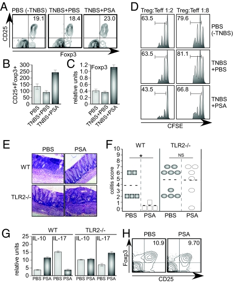
Activation of innate and adaptive immune cells and secretion of inflammatory cytokines are not found under steady state conditions within the intestine. We next wished to examine Foxp3+ Treg cell development during intestinal disease. TNBS (2,4,6-trinitrobenzene sulfonic acid) treatment of animals results in gut inflammation, which activates T-cell responses; animals lose a significant amount of weight and display marked thickening of the colon, with lymphocyte infiltration and concomitant epithelial hyperplasia (26). As previously reported, disease was not evident in TNBS-treated animals that were treated with purified PSA (15). Vehicle treated (PBS; −TNBS) and PBS treated TNBS animals (TNBS+PBS) had a similar percentage of Foxp3+ Treg cells within the CD4+CD25+ T cells of the MLN (Fig. 3_A_). Consistent with PSA's anti-inflammatory properties, mice given PSA reproducibly had a 5 to 10% increase in the percentage of Foxp3+ cells within the CD4+CD25+ compartment of the MLNs (Fig. 3_A_). Additionally, the absolute number of CD4+CD25+Foxp3+ cells in the MLNs was significantly higher in PSA-treated mice compared with PBS or noncolitic animals (Fig. 3_B_). PSA expansion of the Foxp3+ Treg population is specific, as the percentage of B cells in the MLNs did not differ between PBS and PSA fed mice (Fig. S7). Consistent with an increase in the percentage of Foxp3+ cells in PSA-treated mice, there was an increase in the expression of the foxp3 transcript in MLNs (Fig. 3_C_). Furthermore, we also found that the Foxp3 expression was increased on a per cell basis in CD4+CD25+ cells during PSA-mediated protection from colitis (Fig. S8), demonstrating that PSA up-regulates proportional and cell-intrinsic Foxp3 expression. The suppressive capacity of Tregs during PSA-mediated protection from intestinal inflammation was determined by in vitro suppression assays. As expected, proliferation was partially suppressed (proliferation of effector cells in the absence of Tregs was > than 90%) when Tregs from vehicle (PBS; −TNBS) or PBS-treated colitic mice (TNBS+PBS) were added to the culture (Fig. 3_D_). Notably however, Tregs isolated from the MLNs of PSA fed mice (TNBS+PSA) suppressed T-cell proliferation to a significantly higher level than cells from untreated animals (43.5% proliferating responder cells vs. 63.5% at a 1:2 Treg:Teff ratio), demonstrating Tregs from animals protected from colitis by PSA have increased functional suppressive activity.
Fig. 3.
PSA expands functional Foxp3+ Tregs during protection from experimental colitis. (A) BALB/c mice were treated with PSA or PBS during TNBS induced colitis and analyzed for the percentage of CD25+Foxp3+ cells within the CD4+ population of the MLN. These data are representative of three independent trials with four mice per group. (B) Mice were treated as in A, MLN cells were counted, and absolute numbers of CD4+CD25+Foxp3+ cells determined (×103). Numbers represent the average of four mice with error bars indicting SD, and are representative of three independent trials. (C) RNA was extracted from the MLNs of mice treated with PSA and foxp3 transcript expression is shown, normalized to β-actin expression in the total lymph node. Numbers represent the average of four mice, with error bars indicating SD, and are representative of three independent trials. (D) CD4+CD25+ cells were purified from the MLNs of colitic PBS or PSA-treated mice and incubated with CFSE-pulsed CD4+CD25− responder cells in an in vitro suppression assay. Numbers indicate the percentage of cells undergoing at least one cellular division at two different ratios of Teffs and Tregs. These data are representative of two independent trials. (E) C57BL/6 WT or TLR2−/− animals were gavaged with purified PSA (or PBS) before TNBS administration. Colons were fixed, sectioned, and H&E stained. Representative histological sections are shown. (F) Colitis scores show that PSA protects WT but not TLR2−/− animals from experimental colitis. Each symbol represents a separate animal, and data are from two independent trails. *P < 0.05. NS, not significant. (G) RNA was extracted from the MLNs of indicated mice either treated with PBS or PSA, and the expression levels of IL-10 and IL-17A were assayed by qRT-PCR. Relative units are represented as transcript levels relative to expression of a housekeeping gene (L32). Results are representative of three independent trials with four mice per group; error bars indicate SD. (H) Percentages of CD4+CD25+Foxp3+ T cells in the MLNs of TLR2−/− mice treated with PBS or PSA during TNBS induced colitis. Plots are gated on CD4+ T cells. Results are representative of three independent trials with four mice per group.
PSA Protection from Intestinal Inflammation Requires TLR2.
Multiple studies have implicated TLR signaling by innate immune cells during inflammatory disease (27). Furthermore, TLR2 signaling has been shown to have significant effects on Treg expansion and function (24, 25). We are therefore in a unique position to examine how TLR2 responses to a natural microbial ligand affect intestinal inflammatory disease. WT or TLR2−/− animals were orally treated with PSA or PBS before induction of TNBS colitis. WT animals had ulcerative lesions, lymphocyte infiltration, and thickening of the intestinal mucosa following administration of TNBS (PBS treatment); WT animals treated with PSA were protected from disease (Fig. 3_E_). In contrast, TLR2−/− mice treated with PSA had marked inflammation including thickening of the mucosa, infiltrating leukocytes, and crypt loss (Fig. 3_E_). Scoring by a blinded pathologist revealed that TLR2−/− animals are resistant to PSA-mediated protection, as no differences in colitis scores were observed for TLR2-deficient animals (Fig. 3_F_). As previously reported, WT animals were protected from colitis by PSA (Fig. 3_F_) (15). We next determined the impact of TLR2 signaling on cytokine induction by PSA in animals. IL-10 expression was induced in response to PSA during protection from colitis in WT animals (Fig. 3_G_); however, in the absence of TLR2 signaling, IL-10 was no longer up-regulated in PSA-treated animals. Suppression of the proinflammatory cytokine IL-17 by PSA was evident in WT animals; intriguingly, IL-17 levels were actually increased in TLR2−/− mice given PSA compared with PBS consistent with no protection from colitis in the absence of TLR2 (Fig. 3_G_). To examine a role for TLR2 in coordination of Treg responses by PSA, we determined the proportions of CD4+Foxp3+ Tregs in TLR2−/− animals treated with PSA during TNBS colitis. As shown in Fig. 3_H_, there are no differences in the percentage of Tregs in TLR2−/− animals in response to PSA. These results demonstrate that TLR2 signaling by PSA is required for Treg induction and provide a molecular mechanism for protection from experimental colitis.
PSA Treatment Cures Animals with Experimental Colitis.
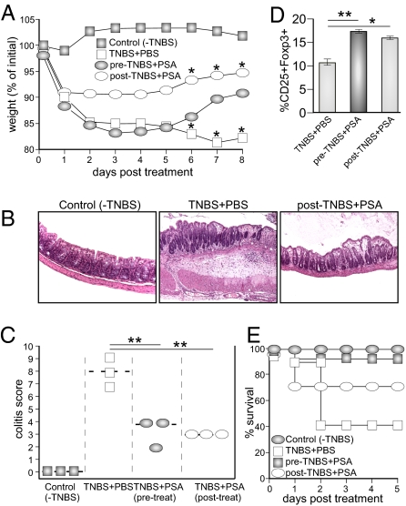
Prophylactic therapies for IBD are unsuitable, as there are no diagnostics that accurately predict disease development and patients must be treated after onset of symptoms. We thus sought to investigate the possibility that PSA would be efficacious for established colitis, potentiating its application as a treatment for established IBD. Animals were induced for TNBS colitis, and groups were treated with PBS before disease induction (TNBS+PBS) or PSA, either before disease induction (pre-TNBS+PSA) or following rectal TNBS administration (post-TNBS+PSA). PBS-treated TNBS animals lost a significant amount of weight (approximately 20% weight loss) by 8 d (Fig. 4_A_). PSA treatment 1 d following onset of intestinal inflammation corrected weight loss equal to or better than PSA treatment before disease; treatment 2 d after colitis induction also provided protection (Fig. S9). Animals with colitis exhibited severe disease 5 d post-TNBS treatment, which was not seen in animals treated with PSA post-TNBS (Fig. 4_B_). Using a standard scoring system, animals treated with TNBS showed a high degree of disease (Fig. 4_C_). However, both pre- and posttreatment with PSA significantly prevented the development of colitis similarly. Animals administered PSA following the onset of disease showed increased IL-10 and Foxp3 expression in gut tissues (Fig. S9), and displayed a significant decrease in IL-17 levels during both protection and cure of colitis (Fig. S10). Accordingly, the percentage of CD4+CD25+Foxp3+ subset was significantly increased in PSA-treated animals compared with diseased animals (Fig. 4_D_). Rectal administration of high doses of TNBS causes a severe intestinal immune response that leads to mortality of animals. Remarkably, animals treated with PSA, even after the commencement of disease, were dramatically rescued from death compared with control treated animals (Fig. 4_E_). Together our studies reveal that gut bacteria produce molecules that coordinate the development of inducible Foxp3+ Tregs, and immunomodulation of Tregs by PSA may represent a unique approach to engendering mucosal tolerance as a therapy for intestinal inflammatory diseases.
Fig. 4.
PSA treatment cures animals with colitis. (A) BALB/c mice were either pretreated with PSA before administration of 1% TNBS (pre) or orally treated with PSA 24 h after the administration of TNBS (post). Mice were weighed daily for 8 d. *P < 0.05 at the indicated time points. (B and C) Colons were extracted from groups of mice treated as in A, fixed, sectioned, and stained with H&E to determine disease severity 5 d post-TNBS administration. Representative sections are shown in B. Colitis scores are shown in C. Data are representative of three independent experiments, P values determined by a two-tailed Mann-Whitney U test. **P < 0.01. (D) MLNs from indicated mice were extracted and cells stained with antibodies to detect for CD4, CD25, and Foxp3. Numbers indicate the percentage of CD25+ Foxp3+ cells within CD4+ population of cells. Results are representative of three independent trials with four mice per group; error bars indicate SD. (E) BALB/c mice were treated with PSA as indicated in A and rectally administered 2% TNBS. Number of animals: 8 mice in the control group, 7 in the pre-TNBS PSA group, and 5 in the post-TNBS PSA groups, and 11 in the TNBS+PBS group. Data in E are the combination of two independent experiments.
Discussion
Tregs represent an important immune mechanism for inducing tolerance to antigens (both self and foreign) to prevent autoimmunity and unwanted inflammation. Although there has been speculation regarding a role for the microbiota in the generation of various Treg subsets (17), identification of Treg-inducing molecular signals from gut bacteria have not been described previously. Here we identify that B. fragilis produces a microbial molecule that can coordinate the development of an inducible population of CD4+Foxp3+ Treg cells. PSA of B. fragilis directs a specific gene-expression profile in functionally suppressive Tregs. Notably, PSA induces IL-10 and TGF-β2 responses from Foxp3+ Tregs and not from Foxp3− T-cell subsets. Moreover, levels of natural Treg markers such as GITR, CD25, and CTLA-4 are not affected by PSA. We also report that germ-free mice have a reduction in the levels of IL-10 expression within the Foxp3+ compartment that is restored upon colonization with B. fragilis. PSA can convert Foxp3− T cells into a Foxp3+ Tregs that express IL-10. Foxp3+ Treg induction and IL-10 production in response to PSA require expression of TLR2. Accordingly, TLR2-deficient animals are not protected from experimental colitis by PSA. Although further work is required to understand the nature of TLR signaling, these results provide a molecular mechanism for PSA's anti-inflammatory properties.
Numerous human and animal studies now indicate that IBD results from a loss of tolerance to commensal bacteria; we show that PSA directs the development of Tregs during protection and cure of experimental colitis. These findings are consistent with studies that show inducible IL-10 production by Foxp3+ T cells is important for mediating tolerance at mucosal surfaces and preventing intestinal inflammation (18). Accordingly, PSA represents a unique example of a commensal bacterial molecule that directs the development and function of inducible Tregs during health and disease. We propose that gut bacteria are able to modulate peripheral Treg functions as a mechanism to engender mucosal tolerance. Collectively, a growing body of evidence now indicates that the intricate interplay between commensal bacteria and regulation of the immune system has profound influences on human health. Conceivably, harnessing the immunomodulatory potential of PSA may lead to natural biological therapies for IBD based on novel pathways for Treg induction.
Materials and Methods
Animals and Bacteria.
Eight- to 10-wk-old specific pathogen-free BALB/c and C57BL/6 mice were purchased from Taconic Farms. TLR2−/− animals were purchased from Jackson Laboratories. Mice were colonized with strains of B. fragilis NCTC9343 and verified for proper colonization by plating under aerobic and anaerobic conditions and PCR of fecal samples with primers specific for B. fragilis and 16S rRNA bacterial primers. PSA was purified as previously described for all treatment experiments (15). All animals were cared for by approved Institutional Animal Care and Use Committee guidelines from the California Institute of Technology.
In Vitro Suppression Assay.
Either CD4+CD25+ or CD4+Foxp3+ cells were used as a source of Tregs. CD4+CD25− cells were pulsed with 1 μL of a 5 μM CFSE stock for 10 min at 37 °C. CFSE labeled cells were washed in PBS twice and immediately used. 1 × 10⁵ mitomycin C (Sigma)-treated CD4-depleted splenocytes were mixed with CFSE-pulsed CD4+CD25− (or Foxp3−) responder cells. Indicated dilutions of CD4+CD25+ Treg cells were titrated in and 1 μg/mL of anti-CD3 was added in a round bottom 96-well plate. Cultures were incubated for 3 to 5 d and then analyzed by flow cytometry.
Experimental Colitis.
Eight-week-old BALB/c mice were purchased from Taconic Farms. For prophylactic studies, animals were pretreated with 50 μg of PSA every other day for 6 d before administration of TNBS and every other day after TNBS induction until necropsy. For curing studies, animals were treated with a single dose of 50 μg of PSA 1 or 2 d following TNBS and every other day until necropsy. See SI Materials and Methods for details.
Statistics.
Differences between data sets were analyzed by Mann-Whitney U test or student's t test where applicable, and analyzed using GraphPad Prism 5.0 or Microsoft Excel software.
Supplementary Material
Supporting Information
Acknowledgments
We thank members of the Mazmanian laboratory for their critical review of the manuscript and Dr. Talal Chatila (University of California Los Angeles) for the kind gift of the Foxp3-GFP mice, Rochelle A. Diamond for help with cell sorting, and Dr. Gregory W. Lawson (University of California Los Angeles) for providing expert pathology analysis. J.L.R. is a Merck Fellow of the Jane Coffin Child's Memorial Fund. S.K.M. is a Searle Scholar. Work in the laboratory of the authors is supported by funding from the National Institutes of Health (DK 078938), the Damon Runyon Cancer Research Foundation, and the Crohn's and Colitis Foundation of America (to S.K.M.).
Footnotes
The authors declare no conflict of interest.
References
- 1.Turnbaugh PJ, et al. The human microbiome project. Nature. 2007;449:804–810. doi: 10.1038/nature06244. [DOI] [PMC free article] [PubMed] [Google Scholar]
- 2.Round JL, Mazmanian SK. The gut microbiota shapes intestinal immune responses during health and disease. Nat Rev Immunol. 2009;9:313–323. doi: 10.1038/nri2515. [DOI] [PMC free article] [PubMed] [Google Scholar]
- 3.Frank DN, et al. Molecular-phylogenetic characterization of microbial community imbalances in human inflammatory bowel diseases. Proc Natl Acad Sci USA. 2007;104:13780–13785. doi: 10.1073/pnas.0706625104. [DOI] [PMC free article] [PubMed] [Google Scholar]
- 4.Dong C. Diversification of T-helper-cell lineages: Finding the family root of IL-17-producing cells. Nat Rev Immunol. 2006;6:329–333. doi: 10.1038/nri1807. [DOI] [PubMed] [Google Scholar]
- 5.Fontenot JD, et al. Regulatory T cell lineage specification by the forkhead transcription factor foxp3. Immunity. 2005;22:329–341. doi: 10.1016/j.immuni.2005.01.016. [DOI] [PubMed] [Google Scholar]
- 6.Feuerer M, Hill JA, Mathis D, Benoist C. Foxp3+ regulatory T cells: Differentiation, specification, subphenotypes. Nat Immunol. 2009;10:689–695. doi: 10.1038/ni.1760. [DOI] [PubMed] [Google Scholar]
- 7.Kullberg MC, et al. IL-23 plays a key role in Helicobacter hepaticus-induced T cell-dependent colitis. J Exp Med. 2006;203:2485–2494. doi: 10.1084/jem.20061082. [DOI] [PMC free article] [PubMed] [Google Scholar]
- 8.Coombes JL, Robinson NJ, Maloy KJ, Uhlig HH, Powrie F. Regulatory T cells and intestinal homeostasis. Immunol Rev. 2005;204(1):184–194. doi: 10.1111/j.0105-2896.2005.00250.x. [DOI] [PubMed] [Google Scholar]
- 9.Atarashi K, et al. ATP drives lamina propria T(H)17 cell differentiation. Nature. 2008;455:808–812. doi: 10.1038/nature07240. [DOI] [PubMed] [Google Scholar]
- 10.Ivanov II, et al. Specific microbiota direct the differentiation of IL-17-producing T-helper cells in the mucosa of the small intestine. Cell Host Microbe. 2008;4:337–349. doi: 10.1016/j.chom.2008.09.009. [DOI] [PMC free article] [PubMed] [Google Scholar]
- 11.Niess JH, Leithäuser F, Adler G, Reimann J. Commensal gut flora drives the expansion of proinflammatory CD4 T cells in the colonic lamina propria under normal and inflammatory conditions. J Immunol. 2008;180:559–568. doi: 10.4049/jimmunol.180.1.559. [DOI] [PubMed] [Google Scholar]
- 12.Gaboriau-Routhiau V, et al. The key role of segmented filamentous bacteria in the coordinated maturation of gut helper T cell responses. Immunity. 2009;31:677–689. doi: 10.1016/j.immuni.2009.08.020. [DOI] [PubMed] [Google Scholar]
- 13.Ivanov II, et al. Induction of intestinal Th17 cells by segmented filamentous bacteria. Cell. 2009;139:485–498. doi: 10.1016/j.cell.2009.09.033. [DOI] [PMC free article] [PubMed] [Google Scholar]
- 14.Strauch UG, et al. Influence of intestinal bacteria on induction of regulatory T cells: Lessons from a transfer model of colitis. Gut. 2005;54:1546–1552. doi: 10.1136/gut.2004.059451. [DOI] [PMC free article] [PubMed] [Google Scholar]
- 15.Mazmanian SK, Round JL, Kasper DL. A microbial symbiosis factor prevents intestinal inflammatory disease. Nature. 2008;453:620–625. doi: 10.1038/nature07008. [DOI] [PubMed] [Google Scholar]
- 16.Maynard CL, Weaver CT. Diversity in the contribution of interleukin-10 to T-cell-mediated immune regulation. Immunol Rev. 2008;226:219–233. doi: 10.1111/j.1600-065X.2008.00711.x. [DOI] [PMC free article] [PubMed] [Google Scholar]
- 17.Belkaid Y, Tarbell K. Regulatory T cells in the control of host-microorganism interactions (*) Annu Rev Immunol. 2009;27:551–589. doi: 10.1146/annurev.immunol.021908.132723. [DOI] [PubMed] [Google Scholar]
- 18.Rubtsov YP, et al. Regulatory T cell-derived interleukin-10 limits inflammation at environmental interfaces. Immunity. 2008;28:546–558. doi: 10.1016/j.immuni.2008.02.017. [DOI] [PubMed] [Google Scholar]
- 19.Lin W, et al. Regulatory T cell development in the absence of functional Foxp3. Nat Immunol. 2007;8:359–368. doi: 10.1038/ni1445. [DOI] [PubMed] [Google Scholar]
- 20.O'Garra A, Vieira PL, Vieira P, Goldfeld AE. IL-10-producing and naturally occurring CD4+ Tregs: Limiting collateral damage. J Clin Invest. 2004;114:1372–1378. doi: 10.1172/JCI23215. [DOI] [PMC free article] [PubMed] [Google Scholar]
- 21.Mazmanian SK, Liu CH, Tzianabos AO, Kasper DL. An immunomodulatory molecule of symbiotic bacteria directs maturation of the host immune system. Cell. 2005;122(1):107–118. doi: 10.1016/j.cell.2005.05.007. [DOI] [PubMed] [Google Scholar]
- 22.van Maren WW, Jacobs JF, de Vries IJ, Nierkens S, Adema GJ. Toll-like receptor signalling on Tregs: To suppress or not to suppress? Immunology. 2008;124:445–452. doi: 10.1111/j.1365-2567.2008.02871.x. [DOI] [PMC free article] [PubMed] [Google Scholar]
- 23.Wang Q, et al. A bacterial carbohydrate links innate and adaptive responses through Toll-like receptor 2. J Exp Med. 2006;203:2853–2863. doi: 10.1084/jem.20062008. [DOI] [PMC free article] [PubMed] [Google Scholar]
- 24.Sutmuller RP, et al. Toll-like receptor 2 controls expansion and function of regulatory T cells. J Clin Invest. 2006;116:485–494. doi: 10.1172/JCI25439. [DOI] [PMC free article] [PubMed] [Google Scholar]
- 25.Liu H, Komai-Koma M, Xu D, Liew FY. Toll-like receptor 2 signaling modulates the functions of CD4+ CD25+ regulatory T cells. Proc Natl Acad Sci USA. 2006;103:7048–7053. doi: 10.1073/pnas.0601554103. [DOI] [PMC free article] [PubMed] [Google Scholar]
- 26.Neurath M, Fuss I, Strober W. TNBS-colitis. Int Rev Immunol. 2000;19(1):51–62. doi: 10.3109/08830180009048389. [DOI] [PubMed] [Google Scholar]
- 27.Strober W. The multifaceted influence of the mucosal microflora on mucosal dendritic cell responses. Immunity. 2009;31:377–388. doi: 10.1016/j.immuni.2009.09.001. [DOI] [PubMed] [Google Scholar]
Associated Data
This section collects any data citations, data availability statements, or supplementary materials included in this article.
Supplementary Materials
Supporting Information


