Autocrine VEGF signaling is required for vascular homeostasis (original) (raw)
. Author manuscript; available in PMC: 2010 Dec 28.
SUMMARY
The Vascular Endothelial Growth Factor (VEGF) signaling pathway is essential for the emergence, differentiation and morphogenesis of the cardiovascular system. Moreover, VEGF is required for neoangiogenesis in multiple pathological settings in the adult. However, its participation in the homeostatic control of the vasculature has not been fully explored. Here we showed that in the absence of any pathological insult, autocrine VEGF is required for the homeostasis of blood vessels in the adult. Genetic deletion of VEGF specifically in the endothelial lineage leads to progressive endothelial degeneration, microhemorrhagic events, development of intravascular thrombosis and sudden death in 65% of mutant mice by 25 weeks of age. The phenotype was manifested without detectable changes in the levels of circulating VEGF or in levels of total VEGF mRNA in all of the organs examined, indicating that paracrine VEGF could not compensate for the absence of endothelial VEGF. We further showed that wild-type, but not VEGF null endothelial cells were able to phosphorylate VEGFR2 in the absence of exogenous VEGF. Activation of the receptor in wild-type cells was suppressed by small molecule antagonists, but not by extracellular blockade of VEGF. Together these results revealed a novel, cell-autonomous activation of the VEGF signaling pathway that holds strong significance to vascular homeostasis, but it is dispensable for the angiogenic cascade.
Keywords: angiogenesis, blood vessels, capillaries, endothelial cells
INTRODUCTION
Genetic ablation experiments in mice have demonstrated that VEGF is critical to the differentiation of endothelial cells and morphogenesis of the vascular system during development. In fact, a single VEGF allele is insufficient to establish a proper vascular network and mice die early in embryogenesis (Carmeliet et al., 1996; Ferrara et al., 1996). Excess VEGF (by only 2 fold) also results in embryonic lethality re-stating the significance of VEGF gene dosage during development (Miquerol et al., 2000). In the adult, ample experimental evidence has unequivocally shown that VEGF is required for pathological growth of vessels in many conditions including inflammation, retinopathies and arthritis (Bates et al., 2003; Dvorak, 2005; Dvorak, 2006; Ferrara et al., 2007; Gitler et al., 2004; Hicklin and Ellis, 2005). Furthermore, the dependency of tumor expansion on neovascularization has served to propel investigations aimed at suppressing VEGF signaling by targeting either the ligand or its receptors (Ferrara and Kerbel, 2005).
VEGF acts through two high affinity receptor tyrosine kinases, VEGFR-1/flt-1 and VEGFR2/KDR/flk-l; both are expressed on normal vascular endothelial cells (Mustonen and Alitalo, 1995) and are up-regulated during angiogenesis (Brown et al., 1997; Dvorak et al., 1999). VEGFR-2 is thought to mediate most of the angiogenic functions attributed to VEGF, whereas the role of VEGFR-1 signaling is less clear, and generally considered to be a decoy receptor, at least during development (Zeng et al., 2001). Most of the VEGF isoforms also bind to a third, non-tyrosine kinase receptor on vascular endothelium, neuropilin-1 (Soker et al., 1998). Neuropilin contributes to the sum of pro-angiogenic functions mediated by VEGFR2, and it might also participate in endothelial guidance and vascular patterning (Gerhardt et al., 2004).
Notably, as a pro-angiogenic cytokine, the significance of VEGF is unquestionable. However this is not its only function. VEGF was originally discovered for its effects on vascular permeability (Dvorak et al., 1979; Senger et al., 1983). Subdermal injection of VEGF results in microvascular hyperpermeability which leads to clotting of extravasated plasma fibrinogen and edema (Senger et al., 1983). More recently, VEGF has also been shown to be required for maintenance of the differentiated phenotype in endothelial cells. In particular, the presence of endothelial fenestrations in the pancreatic islands and glomeruli is dependent on constitutive signaling through VEGF (Eremina et al., 2003; Kamba et al., 2006). However, relevance of VEGF in non-pathological adult tissues remains largely unknown, despite its biological and therapeutic significance. In this study we investigated the functional relevance of endothelial-derived VEGF in vivo.
RESULTS
VEGF is expressed by endothelial cells in vivo
VEGF traditionally has been recognized as a paracrine factor in both developmental and pathological settings. Although expression of VEGF has been found in endothelial cultures under hypoxia (Namiki et al., 1995), the synthesis of VEGF by endothelial cells in vivo has not been reported until recently. Even then, its relevance has remained questionable due to its spotty expression (Maharaj et al., 2006). We expanded those evaluations to include nine organs at three ages in two independent reporter models (Damert et al., 2002; Miquerol et al., 2000) by X-gal staining. These studies demonstrated frequent VEGF promoter activity in the endothelium of large and small vessels. Expression was noted in normal adult mice (Figure 1A, arrows), suggesting the presence of an autocrine signaling loop in adult non-pathological vessels.
Figure 1. VEGF Expression by Endothelial Cells and Generation of VEGFECKO Mice.
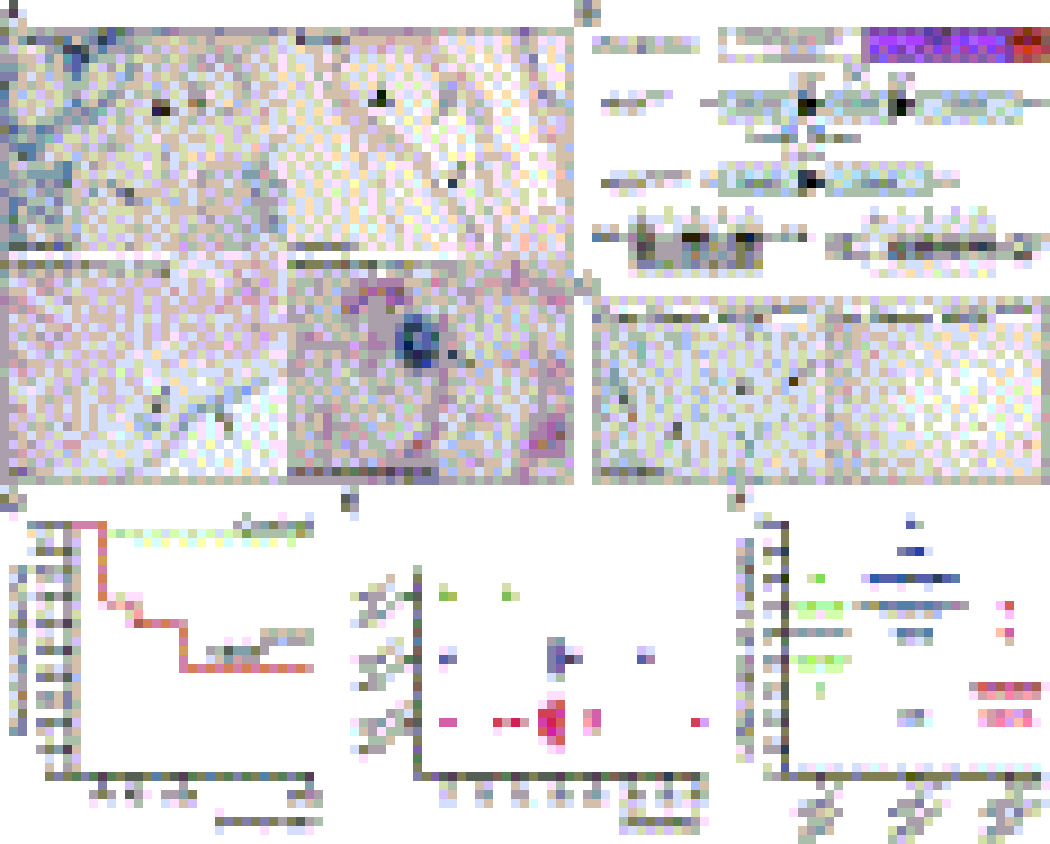
(A) Expression of LacZ reporter (blue) driven by the VEGF promoter in non-pathological adult tissues. Note promoter activity in endothelial cells (arrows). In larger vessels: positive (arrows) and negative (arrowhead) endothelial cells are observed.
(B) Schematic diagram of the transgenic mice used to generate VEGFECKO mice (top). Genotyping of tail DNA. A band of 850 indicates the presence of the Cre transgene. Only animals from lanes 2 and 4 harbored the Cre-recombinase transgene (bottom, left). A 100-bp band indicates wild type (wt) allele and the 150-bp corresponds to the floxed VEGF allele (flox) (bottom, right).
(C) Endothelial cells isolated from Cre+/Rosa+/VEGFlox/lox (VEGFECKO) and Cre-/Rosa+/VEGFlox/lox mice stained for β-gal. Arrows point to endothelial cells expressing Cre and arrowheads to cells with no Cre expression (approximately 95% of the isolated cells were positive).
(D) Survival analysis. Mortality rate of newborn VEGFECKO at P0 was calculated by predicted mendelian distribution and expected litter size. Survival rate of VEGFECKO at six months was 34.7%.
(E) Mortality of adult cohorts.
(F) Litter size variability. Wt × Wt, 6 ± 1.33 (n = 10); Wt/KO × Wt/KO, 7.15 ± 1.76 (n = 20); KO/KO, 4.0 ± 1.30 (n = 11).
Generation and analysis of VE-Cadherin-Cre/VEGFlox/lox mice (VEGFECKO)
To elucidate the biological significance of VEGF expression by vascular endothelial cells in vivo, VE-Cadherin-Cre (VE-CAD-Cre) transgenic mice (Alva et al., 2006) were crossed to VEGFlox/lox mice (Gerber et al., 1999) to generate VE-CAD-Cre/VEGFlox/+ mice. These were further crossed with VEGFlox/lox to obtain VE-Cadherin-Cre/VEGFlox/lox mice (referred to as VEGFECKO) (Figure 1B). Specificity of Cre excision was assessed by crossing VEGFECKO mice to the Rosa26R reporter (Soriano, 1999) line followed by X-gal staining of endothelial cells isolated from wild type and VEGFECKO mice (Figure 1C). Using this approach, we found that more than 95% of the cells in culture showed Cre activity and lacked VEGF transcripts (as per RT-PCR results). As a control, X-gal staining was performed on endothelial cells isolated from mice without Cre transgene (Figure 1C).
We genotyped 261 mice resulting from 35 litters of crosses between VE-Cadherin-Cre/VEGFlox/+ mice. Analysis of the resulting genotypes revealed a lower Mendelian frequency for VEGFECKO mice (18.7% shown vs. 25% expected, STable 1). Evaluation of the viability profile for VEGFECKO mice showed that 31.6% died in utero, 11.5% of newborn mice died between P0 and P5, and 26.1% died between 4 weeks and 6 months (Figure 1D). A cohort of mice was followed for 6 months to ascertain whether lethality occurred at a particular stage. We found an average lethality rate of 23.8% in adult VEGFECKO mice with a peak between 20 and 25 weeks (Figure 1E and STable 2). Additional crosses (between: VE-CAD-Cre/VEGF+/+, VE-CAD-Cre/VEGFlox/+ and VEGFECKO) and genotype analyses of the resulting mice at 4-weeks showed consistent small size litters for VEGFECKO crosses (Figure 1F) and stressed the contribution of endothelial VEGF to vascular development.
STable 1.
Genotyping analysis of 4-week old mice
| Genotype (at 4 weeks) | Number | % | Expected % of mice |
|---|---|---|---|
| Cre+, VEGF+/+ | 71 | 27.3% | 25% |
| Cre+, VEGFlox/+ | 141 | 54.0% | 50% |
| Cre+, VEGFlox/lox | 49 | 18.7% | 25% |
STable 2.
Mortality rate of adult VEGFECKO mice
| 10/05 | 11/05 | 12/05 | 1/06 | 2/06 | 3/06 | |
|---|---|---|---|---|---|---|
| Total # (VEGFECKO) | 18 | 21 | 34 | 41 | 74 | 83 |
| Death | 4 | 7 | 8 | 10 | 13 | 17 |
| % Death | 22.2 | 33.3 | 24.5 | 24.4 | 17.8 | 20.5 |
Mice in the cohort which survived the embryonic window of lethality were indistinguishable from control littermates. VEGFECKO mice between 8 and 24 weeks did not show any external pathological manifestations that could aid in predicting sudden death. After 30 weeks, however, VEGFECKO mice frequently exhibited signs of fatigue, waste, and became lethargic.
Systemic vascular pathologies in VEGFECKO mice
Evaluation of VEGFECKO mice and littermate controls between 40 and 50 weeks of age revealed organ failure associated with systemic vascular pathologies. Hemorrhages (Figures 2Ag, i, l), intestinal perforations (Figures S1Bg), and signs of multiple micro-infarcts were frequently noted in older mice after macroscopic inspection (Figure 2Aj). In addition, older mice exhibited occasional neointimal hyperplasia and frequent disruption in the elastic lamina of larger vessels (Figure S1A).
Figure 2. Systemic Vascular Pathologies VEGFECKO mice.
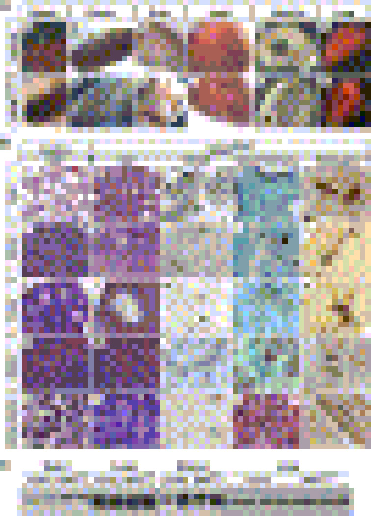
(A) Macroscopic analyses. a–f, Control. g–l, VEGFECKO. VEGFECKO organs of older mice show hemorrhage (arrows in g, i and l), tortuous and dilated vessels (arrows in h), areas of suggestive collagen accumulation in the heart indicative of microinfarcts (arrows, j) and intestinal perforations (arrows, k) (also see histology in Fig. S1).
(B) Histological sections of organs from younger mice. a–e, lung. Lung in VEGFECKO mice shows chronic inflammation (b), fibrosis (d) and excess of intravascular fibrin(ogen) deposits (e). PECAM staining reveals ruptured endothelial cells (arrowhead, c) and collapsed lumen (arrow, c). f–j, uterus. k–o, ovary. VEGFECKO ovary shows significantly enlarged vessels surrounding mature follicles (l) compared to wild type ovary (k). p–t, spleen. Asterisk indicates fibrosis. u–y, bone marrow. Fibrin(ogen) staining reveals clotting from VEGFECKO organs (arrows, e, o, t and y). Hemosiderin deposits were indicated by arrows in h, i, m, n, r, s, w and x. Bar, 100µm.
(C) VEGFR2 protein levels from VEGFECKO and control at 25 weeks were determined by immunoblots (same amount of protein was loaded per well). Slight differences in the uterus correlate with estrous cycle.
To gain further insight into the defects that preceded these outcomes, histological analysis was performed in mice between 8 and 20 weeks, a time when no differences were noted between control and VEGFECKO mice (Figure 2B). Evaluation of lungs from VEGFECKO mice showed a chronic inflammatory state even in young mice (Figure 2Bb), endothelial cell rupture (Figure 2Bc), vascular constriction (Figure 2Bc) and significant fibrosis (Figure 2Bd). Fibrinogen accumulation was found in several organs including lung (Figure 2Be), uterus (Figure 2Bj), ovary (Figure 2Bo), spleen (Figure 2Bt) and bone marrow (Figure 2By). In addition, hemosiderin deposits were detected in most organs examined (Figures 2Bh, m, n, r, s, w and x,) indicating the previous occurrence of hemorrhagic events. Together, these results indicate that endothelial VEGF is required for the systemic stability of the vasculature. These defects occurred in the absence of alterations in VEGFR2 protein levels, as confirmed by Western blot analysis (Figure 2C).
Systemic endothelial apoptosis in VEGFECKO mice
We postulated that endothelial apoptosis in VEGFECKO mice could be a possible cause of the multiple hemorrhagic and thrombotic events. Indeed, endothelial cells positive for cleaved caspase-3 were frequently observed in VEGFECKO mice and the percentage increased with age (Figure 3A), this was in sharp contrast to control littermates. Ultrastructural examination of endothelial cells in VEGFECKO also revealed typical morphological changes associated with apoptotic cell death. These included membrane blebbing (Figure 3Bb), cell shrinkage (Figure 3Bc), cell rupture (Figure 3Bd), nuclear condensation (Figure 3Bd) and exposure of cytosolic components (Figure 3Bd). Since apoptotic endothelial cells are procoagulant (Blann, 2003a; Blann, 2003b; Choy et al., 2001), these results explain the development of thromboembolitic events and cardiac ischemic episodes (platelet adhesion, thrombi, accumulation of von-Willebrand Factor (vWF) and fibrinogen deposits) that resulted in the sudden death of a cohort of animals.
Figure 3. Evidence of Endothelial Cell Apoptosis in VEGFECKO Mice.
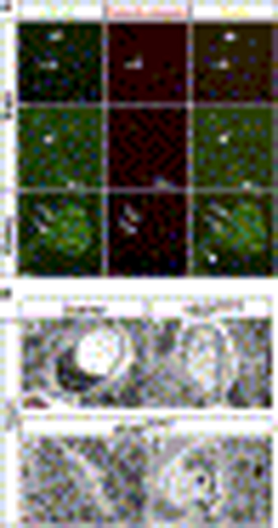
(A) Endothelial expression of cleaved caspase-3 in VEGFECKO mice. Representative sections double-stained for PECAM (green) and cleaved caspase-3 (red) are shown. Arrows point to vessels with active caspase-3. Arrowheads indicate caspase-3 negative vessels. Bar, 100µm.
(B) Electron microscopic evidence of endothelial cell apoptosis in heart sections. Endothelial cells of VEGFECKO heart (b–d) shows morphological characteristic of apoptotic cells, including cytoplasmic swelling (b–c, arrow), nuclear condensation (d, arrow) and the exposure of cytosolic components (d, asterisk). Also note mitochondria swelling in d (arrowhead). Bar, 3.5µm
Micro-infarctions and intravascular thrombosis in the coronary vessels of VEGFECKOmice
Prior 24 weeks, VEGFECKO mice experienced sudden death. Therefore, we suspected cardiac pathology or strokes. Macroscopic analysis of the heart from adult VEGFECKO mice showed possible signs of multiple micro-infarctions in VEGFECKO mice, revealed as accumulated fibrotic areas (Figure 2Aj) that were not found in littermate controls (Figure 2Ad). Myocardial infarction (MI) results from the rapid development of cellular necrosis caused by a critical imbalance between the oxygen supply and demand of the myocardium. The most common cause of MI is narrowing of the epicardial blood vessels due to atheromatous plaques. Plaque rupture with subsequent exposure of the basement membrane results in thrombus formation, plaque development and acute reduction of blood supply to a portion of the myocardium (Kawai, 1994; van Gaal et al., 2006). Histological evaluation of heart sections from VEGFECKO mice and control littermates revealed the presence of intravascular thrombi (Figure 4Ab, asterisks), platelet adhesion to the endothelial wall (Figure 4Ad, arrows) and disrupted endothelial lining (Figure 4Ac, arrows), but the absence of any sign of atherosclerosis. Accumulation of both vWF (Figures 4Ae–h) and fibrinogen (Figures 4Ai–l) were also found by immunocytochemistry. The thrombotic phenotype was noted in most animals and its frequency increased with age. Together, these results indicate that endothelial-derived VEGF is essential to the regulation of the anti-thrombotic properties of the vessel wall.
Figure 4. Intravascular Thrombosis in VEGFECKO Mice Affect Cardiac Physiology.
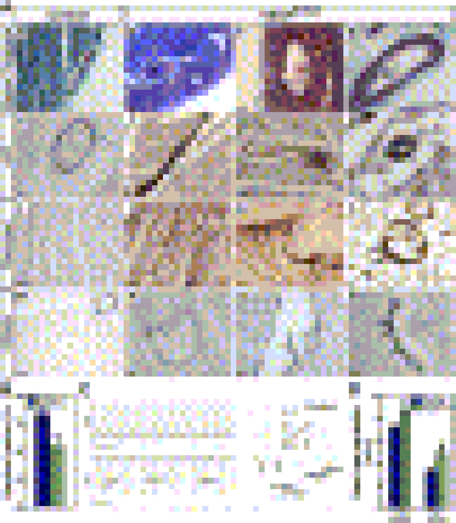
(A) a–d, Frequent intravascular thrombi (b, asterisks), detachment of endothelial lining (c, arrow) and platelet adhesion (d, arrows) in VEGFECKO hearts. (a–d, Gomori trichrome staining). e–h, von-Willebrand Factor (vWF) staining shows accumulation in the vascular wall of mutant mice (arrows). i–l. Fibrinogen staining was also increased (arrows). m–p, PECAM staining shows detachment of endothelial cells (n, o, arrows) and collapse of vascular wall (p, arrows). Bars: a–c, f, g, k, m, p= 35µm; d, e, h, l, n, o, p= 10µm.
(B) VEGFECKO mice (green) have reduced ejection fraction compared to control littermates (blue) (means ± SD., n=11 ea, P<0.001).
(C) ECG traces obtained from wild type (top) and VEGFECKO (bottom) mice. The left-right arrows in VEGFECKO (bottom) mice indicate the variability in heart rate and amplitude. Time bar = 0.5 sec. The right panels show expanded ECG traces with the defections annotated. Time bar = 0.5 sec.
(D) Increased heart rate variability from wild type (black) to VEGFECKO (green) mice during both diurnal cycles (mean ± SD., n=3 ea, P<0.001).
To further explore the relationship between the cardiac phenotype and the nature of the cellular insults imposed on the endothelium, tissue sections were examined after immunostaining with a pan-endothelial marker. PECAM staining of hearts from VEGFECKO mice showed ruptured and discontinued endothelial lining (Figures 4Am–p, arrows).
The intravascular thrombotic phenotype may also relate to alterations in the hematopoietic compartment. Indeed VEGF has been shown to regulate hematopoietic progenitors (Gerber et al., 2002), and unfortunately, the constitutive VE-Cadherin Cre model is penetrant in approximately 60% of all hematopoietic lineages (Alva et al., 2006). To ascertain whether VEGF deletion from bone marrow cells contributed to the observed phenotype, we examined the constituency of circulating and bone marrow cells by flow cytometry (Figure S2). No significant differences were noted in blood counts (Figure S2A, B) or in the frequency of myeloid (Gr-1/Mac-1), erythroid (Ter-119/CD71), megakaryocytic (CD71/CD41) and B-cell (B220) lineages between control littermates and VEGFECKO mice (Figure S2C, D). Surprisingly, we also failed to detect differences in apoptosis when Cre+ cells were compared to Cre- cells in the bone marrow of VEGFECKO mice (Figure S3). To further ascertain the possible contribution of bone marrow cells in the thrombotic phenotype, we performed a series of bone marrow transplantation experiments in which VEGFECKO bone marrow cells were introduced into lethally irradiated wild-type mice, and vice versa. Both recipient mice survived and showed equivalent levels of bone marrow incorporation (Figure S4). However, only VEGFECKO recipients of wild-type bone marrow developed vascular rupture, intra-vascular thrombosis and hemorrhagic events (Figure S5); in contrast wild-type recipients of VEGFECKO marrow did not develop vascular pathology. Combined, these results suggest that the phenotype observed was endothelial cell specific and cell-autonomous.
VEGFECKO mice have significant cardiac dysfunction
To ascertain the relationship between cellular abnormalities in VEGFECKO mice and the sudden-death phenotype, we examined several aspects of cardiac physiology. Wild-type and VEGFECKO male mice (n=11 each) were first evaluated for left ventricular dimensions and contractile function via echocardiography. The VEGFECKO mice demonstrated lower contractile function in comparison to their wild-type littermates. As shown in Figure 4B, their ejection fraction was reduced by 19% while other indices of ventricular performance showed similar dysfunction; fractional shortening dropped from 37.4 ± 1.3% to 28.8 ± 1.5% (P<0.001; wild type vs. VEGFECKO, mean ± SEM) and the velocity of circumferential fiber shortening (Vcf) was reduced from 6.28 ± 0.28 to 4.59 ± 0.26 (lengths/s) (P<0.001). VEGFECKO hearts were dilated with greater end diastolic dimensions (EDD: 4.07 ± 0.08 mm to 4.37 ± 0.06 mm, wild type vs. VEGFECKO) and end systolic dimensions (ESD: 2.55 ± 0.09 to 3.12 ± 0.1mm; P<0.001) without changes in wall thickness.
With evidence for ventricular dysfunction and vascular clots, the electrocardiograms (ECG), body temperatures and cage activity were evaluated via telemetry in awake, free-roaming male mice (n=3 each) and one female VEGFECKO mouse. There were no significant differences in cage activity or body temperature between the groups at the same time during the day. All wild-type mice showed normal mouse waveforms exhibiting P, QRS, Tri and T waves (Figure 4C, top waveforms) (London, 2001). In contrast, VEGFECKO mice displayed abnormal ECG waveforms (Figure 4C, lower traces) mixed in with relatively normal waveforms. Abnormal waveforms could have P waves going either positive or negative, large negative R waves, minimal or missing Tri waves and normal T waves. Though the PR and QT intervals were normal in both groups, the QRS duration increased from 12 ms in wild-type mice to 16–22 ms in the abnormal QRS complexes of the VEGFECKO mice. Two of the VEGFECKO mice had abnormal waveforms over 25% of the time during the 4 months of the recording period while the other two had fewer episodes (1–2% range). Interestingly, the frequency of abnormal waves increased as the mice became older.
Heart rates were lower in VEGFECKO as compared to wild-type mice. Figure S6A (baseline) shows the difference during the light (resting) circadian cycle. During the dark (active) cycle, heart rates increased in both groups, but remained lower in the VEGFECKO mice (584 ± 15 vs. 537 ± 1 0, wild type vs. VEGFECKO, P<0.05). Additionally, the coefficient of variance in heart rate (% CV) was significantly (P<0.001) greater in the VEGFECKO mice, indicating more variability in the heart rate (Figure 4D). Such variability can be seen in the abnormal VEGFECKO mouse waveform (Figure 4C, bottom, left-right arrows). We also exposed mice to exercise-induced stress and found that the VEGFECKO group performed more poorly than controls (Figures S6A, B).
Because of the variation in heart rate, we examine whether Cre-mediated deletion affected the cardiac conduction system. X-gal staining in Purkinje fibers (identified by Periodic Acid Schiff (PAS)) demonstrated that Cre-activity was confined to the endothelial component of the heart (Figure S7).
Autocrine VEGF signaling promotes endothelial survival
Further mechanistic analysis of the VEGFECKO phenotype required a detailed systemic assessment of VEGF. Levels of VEGF in plasma and serum were indistinguishable between VEGFECKO and littermate controls (Figure 5A). Furthermore, evaluation of VEGF transcripts in whole organs by real time PCR showed no differences between the groups except for the lung, where VEGFECKO mice had a statistically significant increase, likely due to the chronic inflammatory state (Figure 5B). These findings indicate that endothelial VEGF is not a major source of circulating or total VEGF levels in tissues and that the systemic endothelial dysfunction of VEGFECKO mice was not rescued by unaltered levels of paracrine VEGF. Additional comparisons between VEGFECKO and control mice showed equivalent capillary density (Figure 5C), angiogenic response in matrigel plugs (Figure 5D), frequency of endothelial fenestrations (Figures 5E and F), and vascular permeability (Figure 5G).
Figure 5. VEGF Levels, Vessel Density, Endothelial Fenestrations and Vessel Permeability in VEGFECKO and Control Mice.
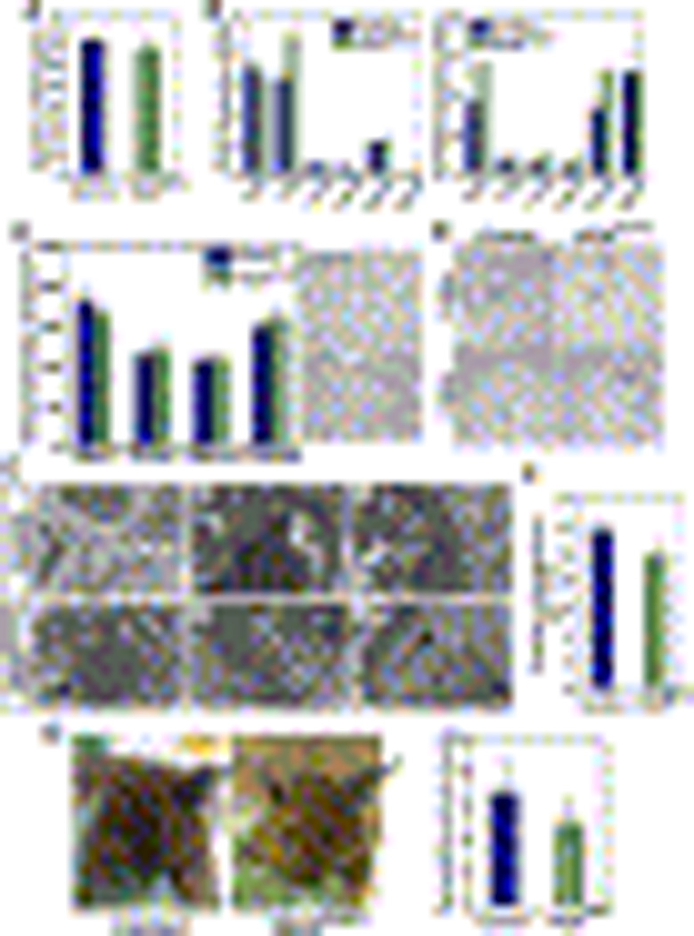
(A) Serum VEGF levels from wild type (blue) and VEGFECKO (green) mice were determined by ELISA.
(B) Organs from wild type (blue) and VEGFECKO (green) mice were analyzed for VEGF (left) and GAPDH (right) transcripts by real-time RT-PCR. Relative RNA units (RRU) were normalized to β-actin levels and calculated from standard curves.
(C) Assessment of vascular density. Data shown are means (n=4) ± S.D (left). Representative PECAM-stained sections of uterus is shown on the right.
(D) Induction of angiogenesis in wild type and VEGFECKO using Matrigel plugs containing VEGF. Arrows indicate neovessels invading the Matrigel plug.
(E) Ultrastructural analyses of glomerular capillaries. Arrows show fenestrations in control and VEGFECKO mice. Note the swelling of VEGFECKO endothelium (asterisk in d and closed triangle in f), yet these cells still retain fenestrations (arrows).
(F) Quantification of fenestrations in control and VEGFECKO mice. Glomerular fenestrations from wild type (blue) and VEGFECKO (green) mice were determined using ultrastructural information obtained from 4 mice. Data shown are means ± SD. Evaluation of P value indicates that the difference is statistically insignificant.
(G) Vascular permeability responses. (left) Photographs of control and VEGFECKO mice injected with Evans Blue followed by application of vehicle or mustard oil (arrow) in the ear. (right) Quantification of extravasated Evans Blue dye from wild type (blue) and VEGFECKO (green) mice. Data are presented as ratio to control. P value was not significant.
To gain further insight into the cell-autonomous functions of VEGF, we isolated endothelial cells from VEGFECKO and control mice. After several isolations, it became clear that the VEGFECKO endothelium either proliferated with slower kinetics, or died more frequently. While this was more evident after a week in culture, the difference was statistically significant as early as 72 hrs post-culture (Figure 6A). Stress, such as serum starvation, exacerbated differences between control and VEGFECKO endothelial cells (Figure 6B). Consistent with the findings in vivo, assessment of active caspase levels upon exposure to CoCl2 (to simulate hypoxia) showed a four-fold increased in VEGFECKO endothelial cells in comparison to control (Figure 6C). This was correlated with a significant difference in endothelial cell survival (Figure 6D). Furthermore, addition of exogenous VEGF did not rescue for the increased cell death exhibited by VEGFECKO endothelial cells (Figure 6D). These findings uncovered an important role for autocrine VEGF signaling in survival mechanisms following hypoxia-mediated stress for which paracrine sources of VEGF cannot compensate.
Figure 6. Endothelial VEGF Mediates Survival in a Cell-Autonomous Manner.
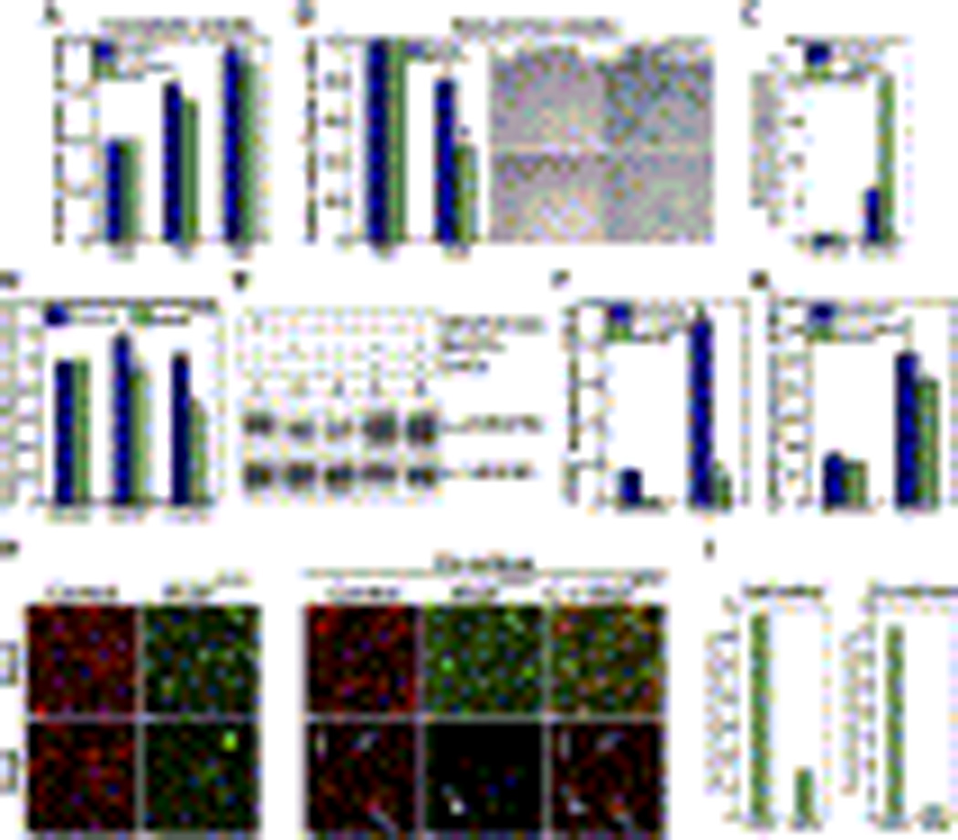
(A–B) Endothelial cells from control (blue) and VEGFECKO (green) mice were cultured in the presence (A) or absence (B) of serum and viable cells were counted at indicated times. Endothelial cells were stained for x-gal and nuclear fast red (right panels, B). Note the difference in cell number between control and VEGFECKO cells.
(C) Endothelial cells from control (blue) and VEGFECKO (green) mice were cultured in the presence of CoCl2, active caspases were fluorescence-labeled and quantified by fluorometer.
(D) Endothelial cells from control (blue) and VEGFECKO (green) mice were cultured in the presence of recombinant hVEGF165 or CoCl2 and viable cells assessed after 48hs.
(E) HUVECs were cultured under hypoxia for 24 h and analyzed for VEGFR2 phosphorylation (pVEGFR2, top) and VEGFR2 total levels (bottom). Lanes: 1, VEGF (100 ng/ml) for 5 min; 2–3, normoxia in the presence (2) and absence (3) of sodium orthovanadate (Na3VO4) for 24 h; 4–5, CoCl2 (hypoxia mimetic) in the presence of Na3VO4 for 24 h.
(F–G) Endothelial cells from control (blue) and VEGFECKO (green) mice were cultured in the presence of CoCl2 (100 µM) for 24 h and transcript levels of VEGF (F) and GAPDH (G) were determined by real-time RT-PCR. Relative RNA units (RRU) were normalized to β-actin. Data shown are means ± SD.
(H) Endothelial cells differentially labeled (Cy3 for wild type and BODIFY green for VEGFECKO) were cultured alone (left) or combined (right). Photographs were taken after 2 days and 5 days. Arrows indicate control cells (red). Arrowhead points to VEGFECKO cells (green).
(I) Quantification of cell survival following co-culture. Fluorescence labeled cells were counted at indicated times. Data are presented as percentage to control.
If these are correct, one should be able to detect activation of VEGF receptors in the absence of exogenous VEGF. Indeed, phosphorylation of VEGFR2 was consistently found in human endothelial cells (Figure 6E). Levels of VEGFR2 phosphorylation significantly increased when the cells were exposed to CoCl2 (Figure 6E). To assess whether hypoxia triggered VEGF increase, endothelial cells were treated with CoCl2 and the levels of VEGF transcripts were measured by real-time PCR. Wild-type endothelial cells upregulated VEGF transcripts by 5.3-fold under hypoxia, whereas VEGFECKO endothelial cells showed only minimal induction (Figure 6F) (consistent with the 95% endothelial VEGF excision previously shown (Figure 1C)). Notably, under normoxia, wild-type endothelial cells had 6.4 times more VEGF transcripts than VEGFECKO endothelial cells, which further confirms expression of VEGF by endothelial cells and its up-regulation following hypoxic stress (Figure 6F). Interestingly, the same VEGFECKO endothelial cells were able to respond to hypoxia by increasing GAPDH to levels similar to control (Figure 6G), indicating that these cells were not only viable, but also fully capable of responding to hypoxic stress.
To ascertain whether the effect of VEGF was paracrine (i.e., secreted and acting in adjacent cells) or autocrine, we performed individual and co-culture experiments with wild-type and VEGFECKO endothelial cells. Under both situations, VEGFECKO endothelial cells displayed a greater degree of cell death (Figures 6H, I). These findings demonstrate that the effect of VEGF was indeed cell-autonomous, as the wild-type cells were not able to rescue VEGFECKO endothelial cells.
Further exploration of the autocrine VEGF signaling pathway was performed with extracellular inhibitors of VEGF (Avastin) and intracellular suppressors of VEGFR2 activation (SU4312). In the absence of exogenous VEGF, Avastin failed to block phosphorylation of VEGFR2, whereas SU4312 substantially neutralized the activity of endogenous VEGF by blocking VEGFR2 phosphorylation (Figure 7A). These results indicate that the phosphorylation of VEGFR2 is likely to occur intracellularly and in the absence of VEGF secretion. Controls for both Avastin (Figure 7B) and SU4312 (Figure 7C) were shown to block VEGFR2 phosphorylation mediated by exogenous VEGF. The possibility of VEGFR2 autophosphorylation in the absence of VEGF was ruled out by evaluation of receptor activation in endothelial cells from VEGFECKO mice. In these cells, exogenous VEGF elicited significant VEGFR2 phosphorylation; whereas no phosphorylation was observed in the absence of exogenous growth factor (Figure 7D). Finally, intracellular, but not extracellular suppression of VEGFR2 was cytotoxic, as HUVECs cultured in the presence of SU4312 were unable to survive under serum-free conditions (Figure 7E).
Figure 7. Autocrine VEGF Signaling in Endothelial Cells.
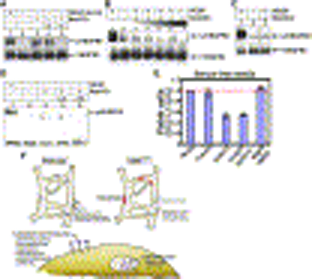
(A) HUVECs were cultured under normoxia for 24 h and in the presence of the indicated compounds and analyzed for VEGFR2 phosphorylation (top) and VEGFR2 total levels (bottom). Lanes: 1, 100 ng/ml VEGF for 5 min; 2, media; 3, Na3VO4; 4–5, Avastin (10 µg/ml, 4) or SU4312 (0.4 µM, 5) in the presence of Na3VO4.
(B) HUVECs were exposed to VEGF (100 ng/ml, pre-incubated with Avastin at 37°C for 2 h) for 5 min. Phosphorylation of VEGFR2 (top) and VEGFR2 (bottom) are shown. Lanes: 1, VEGF; 2, media; 3–6, VEGF pre-incubated with 1 ug/ml Avastin (3), 5 ug/ml Avastin (4), 10 ug/ml Avastin (5) and 100 ug/ml Avastin (6); 7, Avastin (1 ug/ml).
(C) HUVECs incubated with VEGF (100 ng/ml, lane 1) or vehicle (lane 2). In lane 3, HUVECs were pre-incubated with SU4312 at 37°C for 2 h and then exposed to VEGF (100 ng/mlPhosphorylation of VEGFR2 (top) and total levels of VEGFR2 (bottom) are shown.
(D) Endothelial cells from VEGFECKO were cultured under normoxia for 24 h and subjected to the indicated treatments. Lysates were analyzed for VEGFR2 phosphorylation. Lanes: 1, VEGF (100 ng/ml) for 5 min; 2, media; 3, Na3VO4; 4–5, Avastin (10 µg/ml, 4) or SU4312 (0.4 µM, 5) in the presence of Na3VO4.
(E) HUVECs were cultured in the absence of serum with indicated VEGF or VEGF signaling blockers.
(F) Schematic representation of VEGF function in endothelial homeostasis. Stress induced by radiation, reactive-oxygen species and hypoxia triggers activation of VEGFR2 by both autocrine and intracrine VEGF sources supporting endothelial survival. In the absence of intracrine VEGF, some endothelial cells undergo apoptosis resulting in hemorrhage (smaller vessels), and / or exposure of the underlying basement membrane with subsequent development of thrombi (larger vessels). In the bottom, the scheme shows an endothelial cell in which paracrine and intra/autocrine activation is taking place.
DISCUSSION
While the contribution of VEGF to the induction of vascular growth has long been accepted, our data indicate that the function of this growth factor expands its classical, effect as the main mediator of developmental and pathological angiogenesis. Here we show that in vivo autocrine VEGF signaling is required for endothelial cell survival under non-pathological conditions in a manner that is cell-autonomous.
One of the surprises reveled by these experiments was that removal of endothelial VEGF could not be sufficiently compensated for by VEGF secreted from adjacent cell types. Evaluation of circulating VEGF protein and transcripts levels from several organs did not show any significant difference between control and VEGFECKO mice, indicating that endothelial VEGF does not sum to a detectable proportion of the total growth factor present in any given organ. Interestingly, while endothelial VEGF constitutes a minor proportion of the total VEGF, it holds high functional significance. A second important conclusion drawn from our studies is that autocrine VEGF does not contribute to the angiogenic response, as vascular density and patterning was virtually identical between wild-type and VEGFECKO mice. Clearly, the majority of the experimental data published so far supports the notion that the progression of angiogenesis requires exogenous presentation of VEGF. Indeed, removal of VEGF from cells other than endothelial has repeatedly resulted in the reduction of vascular growth (Gerber et al., 1999), a phenotype that is not shared by the VEGFECKO mice. Vascular density and pattern was not affected by removal of endothelial VEGF. Also, reduction in size or developmental retardation as a consequence of general VEGF decrease was never noted in VEGFECKO mice. The nature of the defects in our model appears to be restricted to endothelial viability. Together these findings indicate that cell-autonomous signaling triggers a response that does not fully overlap with the events initiated by paracrine activation. Thus, paracrine VEGF signaling is essential for the angiogenic cascade, proliferation, survival, permeability responses and endothelial differentiation. In contrast, autocrine VEGF signaling only conveys survival signals (Figure 7F). Interestingly, both paracrine and autocrine activation are mediated by the main initiating receptor (VEGFR2). It should be stressed that our data does not exclude paracrine VEGF signaling in survival functions. Instead, our findings indicate that provision of survival signals in a paracrine mode alone is insufficient.
The concept of an autocrine signaling loop for VEGF has been previously shown in bone-marrow endothelial cell progenitors (Gerber et al., 2002). The experiments here would indicate that the functional significance of cell-autonomous signaling is broader than anticipated and it impacts fully differentiated, normal endothelial cells as part of a homeostatic program. Thus, we propose a model in which survival signals are required to support viability of the endothelium under normal conditions. These survival signals are likely triggered by stress situations, such as irradiation, hypoxia and reactive oxygen species. Activation of VEGFR2 by endogenous VEGF sources enables endothelial survival (Figure 7B). While there is much to be understood, as to the regulation of endothelial VEGF in vivo, our findings would indicate that long-term ablation of VEGF signaling within the endothelial compartment or direct blockade of VEGFR2 might have a deleterious systemic impact in the vasculature of normal tissues.
VEGF is an integral component of tumor angiogenesis and its inhibition results in reduction of tumor burden subsequent to the regression of some tumor vessels and improved drug delivery (Inai et al., 2004; Jain, 2001). Consequently, many therapeutic strategies for agents that block VEGFsignaling either by neutralizing VEGF, or by inhibiting its receptors, have been pursued. Some of these agents have shown promise in clinical trials (Hurwitz et al., 2004; Willett et al., 2004). However, although reductions in tumor burden and survival benefit have been demonstrated, a small, but consistent number of side effects have been also observed, including hypertension and thromboembolism (Jain et al., 2006; Kabbinavar et al., 2003). Other pathologies, while not as consistent, include hemorrhage, proteinuria, intestinal perforations and congestive heart failure (Jain et al., 2006; Kabbinavar et al., 2003). Such findings indicate that VEGF participates in a number of functions within normal tissues that exceed its role as pro-angiogenic agent. The contribution of VEGF in the regulation of blood pressure remains enigmatic. Deletion of VEGF from the endothelium does not result in hypertension, or changes in eNOS levels/activation (data not shown). Nor did we observe proteinuria, at least prior to 30weeks of age (data not shown). However, VEGFECKO mice showed hemorrhage, thrombosis, and intestinal perforations; we attribute most of these side-effects to a long-term interruption in VEGF-mediated survival in both a paracrine and an autocrine manner. While the consequence of long-term pharmacological ablation (or reduction) of VEGF signaling is not really known, our data would indicate that the homeostatic function of the VEGF-VEGFR2 within the endothelial compartment is of considerable biological significance.
Evaluation of signaling by endogenous VEGF in vitro suggests that activation of VEGFR2 might occur intracellularly. The experimental evidence for this conclusion is that blockade with Avastin did not affect the degree of VEGFR2 phosphorylation, unlike the effect mediated by a small molecular inhibitor of VEGFR2 that can enter the cell freely and completely suppress activation of the receptor. In addition, we found that wild-type endothelial cells are unable to rescue VEGFECKO endothelial cells in co-culture experiments. Previous examples of intracrine signaling have been presented in the literature. For example, the amino-terminal propeptide of type I collagen have been shown to modulate cell adhesion by triggering activation of a putative intracellular receptor in the secretory pathway (Oganesian et al., 2006). Intracrine activation of receptor tyrosine kinases have been predicted for PDGF and TGF-beta (Betsholtz et al., 1984) and the concept that ligand-receptor binding complexes are internalized to signal only in an endosomal compartment has been long demonstrated for EGF (Cohen and Fava, 1985). More recently, endosomes were implicated in the maintenance of Dpp signaling during mitosis and in the even distribution of signaling bodies to daughter cells (Bokel et al., 2006). It is also becoming apparent that internalized cell surface receptors might use specifically localized complexes to initiate signals that are distinct from those triggered at the cell surface (Childress et al., 2006; Lin et al., 2006; Seto and Bellen, 2006). In this manner, compartmentalization of signaling could regulate signal transduction spatially and temporally (van der Goot and Gruenberg, 2006). This would ultimately assign unique biological responses through the same ligand-receptor complex. While we have yet to explore compartmentalization of VEGF signaling, it has been recently shown that internalized VEGF receptors can signal from endosomes in a manner that is regulated by VE-Cadherin (Lampugnani et al., 2006).
In conclusion, the studies communicated here reveal a previously unknown function for VEGF in vascular homeostasis in vivo. They also indicate that cell-autonomous VEGF signaling is likely triggered inside the cell. These signaling events differ from those initiated by exogenous / paracrine VEGF, as they are restricted to maintenance of endothelial viability.
EXPERIMENTAL PROCEDURES
Mice
VEGF-LacZ mice (Miquerol et al., 1999), VE-Cadherin-Cre transgenic mice (Alva et al., 2006) and floxed-VEGF mice were previously described (Gerber et al., 1999)..
Histological Analysis
Tissues were fixed in 4% paraformaldehyde, paraffin-embedded and sectioned at 5 µm. For immunohistochemistry we used: rat anti-mouse PECAM (BD Biosciences Pharmingen, CA), rabbit anti-mouse fibrinogen (Chemicon, CA), rabbit anti-mouse VWF (BD Biosciences Pharmingen, CA), and secondary antibodies purchased from Vector laboratories (CA). For x-gal staining, tissues were sectioned on a vibratome (Ted Pella, CA) at 400 µ prior to staining as previously described (Alva et al., 2006). For electron microscopic analysis, tissues were fixed in 2% paraformaldehyde, 2% glutaldehyde in 50mM sodium cacodylate buffer. Sections were mounted on inert grids and stained by Microscopic Techniques and Electron Microscopy Core facility at UCLA. For the evaluation of apoptosis, organs were fixed in 1% paraformaldehyde, sectioned at 400µm and double-stained with rat anti-mouse PECAM (to visualize vessels) followed by FITC-labeled goat anti-rat IgG antibody (Jackson ImmunoResearch Laboratories, PA) and with rabbit anti-Cleaved Caspase-3 (Cell Signaling Technology, Danvers, MA) followed by Cy3-secondary antibody (Jackson ImmunoResearch Laboratories).
Echocardiography
Mice were anesthesized with isoflurane vaporized in oxygen (2.5% induction, 1.0% maintenance) and ultrasonically imaged with a Siemens Acuson Sequoia C256 (Siemens Medical Solutions, Malvern, PA) as previously described (Jordan et al., 2005; Tanaka et al., 1996). Two-dimensional, M-mode, and spectral Doppler images were acquired to obtain measures of left ventricular size, mass, wall thickness and ventricular function using the Acuson and AccessPoint software (FreelandSystems, LLC, Santa Fe, NM).
Ambulatory Monitoring of Cardiac Function by Telemetry
Radio telemetry devices (TA10ETA-F20; Transoma-Data Sciences Intl., St. Paul, MN) were implanted as previously described (Jordan et al., 2005; Wehling-Henricks et al., 2005). Unfiltered ECG waveforms, body temperature and bio-activity data were collected for 30-secs every 10 minutes for 4 months. Data waveforms and parameters were analyzed with the Data Sciences analysis packages (ART 3.01 & Physiostat 4.01). The coefficient of variance (%CV), an index of heart rate variability, was determined from sequential R-R intervals.
Statistical Methods
Data were evaluated using a student’s two-tailed t-test or ANOVA with Instat V3.05 statistical software (GraphPad Inc., San Diego, CA). In all analyses, P<0.05 was taken to be statistically significant.
Matrigel Assays
Mice were injected with 250 µl of growth factor reduced Matrigel (BD Biosciences) that was supplemented with VEGF (1 µg). Matrigel plugs were harvested and evaluated for β-galactosidase activity. Specimens were subsequently fixed with 4% paraformaldehyde in PBS, embedded in paraffin and stained with nuclear fast red.
Vascular Permeability Assays
Evans blue dye (1ml/kg of 3% Evans blue) was injected into the tail vein of 15- to 16-week-old mice. Mustard oil (Allylisothiocyanate, Fluka, Germany) diluted to 5% in mineral oil was applied to the dorsal surfaces of the ear with a cotton swab after injection of Evans blue. Photographs were taken 15 minutes after stimulus and the vasculature was perfusion-fixed (1% paraformaldehyde in 50mM citrate buffer, pH 3.5) for 2 minutes. Ears were removed, blotted dry and weighed. Evans blue was extracted from the ears with 1ml of formamide overnight at 55°C and measured by spectrometer at 610mn.
Total RNA Isolation and Real-Time RT-PCR Analysis
Total RNA was isolated on RNeasy Quick spin columns (Qiagen, Valencia, CA). 0.3 – 0.4 µg of total RNA per reaction were analyzed using the TaqMan RT-PCR kit from Applied Biosystems (NJ), following the manufacturer’s instructions. Reactions were run in 96-well plates in a DNA Engine OpticonII System from Bio-Rad (Hercules, CA), and results were analyzed using built-in software. Probe primer sequences for murine VEGF are: 5’-TGTACCTCCACCATGCCAAGT-3’ and 5’-CGCTGGTAGACGTCCATGAA-3’. GAPDH sequences are: 5’-CATGTTTGTGATGGGTGTGA-3’ and 5’-AATGCCAAAGTTGTCATGGA-3’. Expression levels were standardized to the probe/primer sets specific for β-actin : 5’-TCAAGATCATTGCTCCTCCTGAGC-3’ and 5’-TACTCCTGCTTGCTGATCCACATC-3’. Primers were purchased from IDT (Coralville, IA).
Quantification of Capillary Density and Fenestration
Endothelial fenestrations in control and VEGFECKO mice were counted using electron micrograph images of endothelial cells from the kidney. Quantification was determined in 7–12 capillaries using two independent specimens for control and three for VEGFECKO mice. Fenestrations were identified as gaps of 50–100 nm in diameter. Capillary density was determined using micrograph images of CD31 immunochemically stained endothelial cells from six organs. Quantification was made using 4 animals for both control and VEGFECKO mice, 3–5 independent specimens.
Isolation of Liver Endothelial Cells from Mouse
Mice were perfused with serum-free DMEM (Mediatech, Inc., VA) and collagenase (500 µg/ml, Sigma, MO). Livers were harvested and incubated in collagenase (500 µg/ml) for another 15 min. Supernatants were mixed with equal volume of DMEM containing 20% FBS. Cells were collected after centrifugation at 2000 rpm for 5 min, resuspended and filtered through 40 µm pore size top filter (BD, Franklin Lakes, NJ). Filtered cells were plated onto gelatin coated tissue culture dishes and washed after 2–3 h to remove cell debris and remaining blood cells.
Examination of Endothelial Cell Growth and Apoptosis
To assess the cell growth, endothelial cells (5,000 cells/well) were plated onto gelatin coated 96-well plates and the viable cell number was determined using cell counting kit-8 (CCK-8 kit, Dojindo Molecular Technologies, Inc., MD). Active caspase levels were determined with APO LOGIX™ (Cell technology Inc., CA). To chemically simulate hypoxia, cells were exposed to CoCl2 (100 µM) for different time periods as described (Namiki et al., 1995). To assess VEGF and GAPDH transcript levels under hypoxia, cells were exposed to CoCl2 (100 µM) for 24 h and transcripts were determined by real time RT-PCR analysis. Phosphorylation of VEGFR2 under hypoxia was measured after exposureto CoCl2 (100 µM) for 24 h in the presence or absence of sodium orthovanadate (Na3VO4, to inhibit phosphatases). Phosphorylation of VEGFR2 was performed as previously described (Lee et al., 2005). To block VEGF signaling, cells were either pre-incubated with SU4312 at 37°C overnight, or VEGF was pre-incubated with Avastin at 37°C overnight. VEGFR2 phosphorylation was determined by immunoprecipitating cell lysates with 4G-10 antibody (Upstate, NY) and analyzing by Western blot with anti-phosphotyrosine antibody (Cell signaling, MA) as described (Lee et al., 2005). To assess the cell growth during blocking of VEGF signaling, cells were incubated with either SU4312 or Avastin or together at 37°C and viable cells were determined using CCK-8 kit. For co-culture experiments, endothelial cells were stained with CellTracker Red CMTPX or Cell Tracker Green BODIFY (Molecular Probe, CA) following manufacturers’ protocol..
ACKNOWLEDGEMENTS
We thank Dr. Goldhaber for his comments on mouse ECG waveforms, Dr. Oscar U. Scremin for the blood gas analysis system, Drs. Mikkola and Montecino-Rodriguez for technical advice on FACS and Liman Zhao for animal husbandry. The Tissue Procurement and Histology Core Laboratory and the Electron Microscopy Core facilities at UCLA contributed with preparation of specimens. This work was supported by NIH grants NCI RO1CA77420 and NHLBI74455 to MLIA and the Laubishch Endowment (KPR). SL was supported by R25CA098010 and TC by T3269766.
REFERENCES
- Alva JA, Zovein AC, Monvoisin A, Murphy T, Salazar A, Harvey NL, Carmeliet P, Iruela-Arispe ML. VE-Cadherin-Cre-recombinase transgenic mouse: a tool for lineage analysis and gene deletion in endothelial cells. Dev Dyn. 2006;235:759–767. doi: 10.1002/dvdy.20643. [DOI] [PubMed] [Google Scholar]
- Bates D, Taylor GI, Minichiello J, Farlie P, Cichowitz A, Watson N, Klagsbrun M, Mamluk R, Newgreen DF. Neurovascular congruence results from a shared patterning mechanism that utilizes Semaphorin3A and Neuropilin-1. Dev Biol. 2003;255:77–98. doi: 10.1016/s0012-1606(02)00045-3. [DOI] [PubMed] [Google Scholar]
- Blann AD. Assessment of endothelial dysfunction: focus on atherothrombotic disease. Pathophysiol Haemost Thromb. 2003a;33:256–261. doi: 10.1159/000083811. [DOI] [PubMed] [Google Scholar]
- Blann AD. How a damaged blood vessel wall contibutes to thrombosis and hypertenasion. Pathophysiol Haemost Thromb. 2003b;33:445–448. doi: 10.1159/000083843. [DOI] [PubMed] [Google Scholar]
- Bokel C, Schwabedissen A, Entchev E, Renaud O, Gonzalez-Gaitan M. Sara endosomes and the maintenance of Dpp signaling levels across mitosis. Science. 2006;314:1135–1139. doi: 10.1126/science.1132524. [DOI] [PubMed] [Google Scholar]
- Brown LF, Detmar M, Claffey K, Nagy JA, Feng D, Dvorak AM, Dvorak HF. Vascular permeability factor/vascular endothelial growth factor: a multifunctional angiogenic cytokine. Exs. 1997;79:233–269. doi: 10.1007/978-3-0348-9006-9_10. [DOI] [PubMed] [Google Scholar]
- Carmeliet P, Ferreira V, Breier G, Pollefeyt S, Kieckens L, Gertsenstein M, Fahrig M, Vandenhoeck A, Harpal K, Eberhardt C, et al. Abnormal blood vessel development and lethality in embryos lacking a single VEGF allele. Nature. 1996;380:435–439. doi: 10.1038/380435a0. [DOI] [PubMed] [Google Scholar]
- Childress JL, Acar M, Tao C, Halder G. Lethal giant discs, a novel C2-domain protein, restricts notch activation during endocytosis. Curr Biol. 2006;16:2228–2233. doi: 10.1016/j.cub.2006.09.031. [DOI] [PMC free article] [PubMed] [Google Scholar]
- Choy JC, Granville DJ, Hunt DW, McManus BM. Endothelial cell apoptosis: biochemical characteristics and potential implications for atherosclerosis. J Mol Cell Cardiol. 2001;33:1673–1690. doi: 10.1006/jmcc.2001.1419. [DOI] [PubMed] [Google Scholar]
- Cohen S, Fava RA. Internalization of functional epidermal growth factor:receptor/kinase complexes in A-431 cells. J Biol Chem. 1985;260:12351–12358. [PubMed] [Google Scholar]
- Damert A, Miquerol L, Gertsenstein M, Risau W, Nagy A. Insufficient VEGFA activity in yolk sac endoderm compromises haematopoietic and endothelial differentiation. Development. 2002;129:1881–1892. doi: 10.1242/dev.129.8.1881. [DOI] [PubMed] [Google Scholar]
- Dvorak HF. Angiogenesis: update 2005. J Thromb Haemost. 2005;3:1835–1842. doi: 10.1111/j.1538-7836.2005.01361.x. [DOI] [PubMed] [Google Scholar]
- Dvorak HF. Discovery of vascular permeability factor (VPF) Exp Cell Res. 2006;312:522–526. doi: 10.1016/j.yexcr.2005.11.026. [DOI] [PubMed] [Google Scholar]
- Dvorak HF, Nagy JA, Feng D, Brown LF, Dvorak AM. Vascular permeability factor/vascular endothelial growth factor and the significance of microvascular hyperpermeability in angiogenesis. Curr Top Microbiol Immunol. 1999;237:97–132. doi: 10.1007/978-3-642-59953-8_6. [DOI] [PubMed] [Google Scholar]
- Dvorak HF, Orenstein NS, Carvalho AC, Churchill WH, Dvorak AM, Galli SJ, Feder J, Bitzer AM, Rypysc J, Giovinco P. Induction of a fibrin-gel investment: an early event in line 10 hepatocarcinoma growth mediated by tumor-secreted products. J Immunol. 1979;122:166–174. [PubMed] [Google Scholar]
- Dvorak HF, Senger DR, Dvorak AM, Harvey VS, McDonagh J. Regulation of extravascular coagulation by microvascular permeability. Science. 1985;227:1059–1061. doi: 10.1126/science.3975602. [DOI] [PubMed] [Google Scholar]
- Eremina V, Sood M, Haigh J, Nagy A, Lajoie G, Ferrara N, Gerber HP, Kikkawa Y, Miner JH, Quaggin SE. Glomerular-specific alterations of VEGF-A expression lead to distinct congenital and acquired renal diseases. J Clin Invest. 2003;111:707–716. doi: 10.1172/JCI17423. [DOI] [PMC free article] [PubMed] [Google Scholar]
- Ferrara N, Carver-Moore K, Chen H, Dowd M, Lu L, O'Shea KS, Powell-Braxton L, Hillan KJ, Moore MW. Heterozygous embryonic lethality induced by targeted inactivation of the VEGF gene. Nature. 1996;380:439–442. doi: 10.1038/380439a0. [DOI] [PubMed] [Google Scholar]
- Ferrara N, Kerbel RS. Angiogenesis as a therapeutic target. Nature. 2005;438:967–974. doi: 10.1038/nature04483. [DOI] [PubMed] [Google Scholar]
- Ferrara N, Mass RD, Campa C, Kim R. Targeting VEGF-A to treat cancer and age-related macular degeneration. Annu Rev Med. 2007;58:491–504. doi: 10.1146/annurev.med.58.061705.145635. [DOI] [PubMed] [Google Scholar]
- Gerber HP, Hillan KJ, Ryan AM, Kowalski J, Keller GA, Rangell L, Wright BD, Radtke F, Aguet M, Ferrara N. VEGF is required for growth and survival in neonatal mice. Development. 1999;126:1149–1159. doi: 10.1242/dev.126.6.1149. [DOI] [PubMed] [Google Scholar]
- Gerber HP, Malik AK, Solar GP, Sherman D, Liang XH, Meng G, Hong K, Marsters JC, Ferrara N. VEGF regulates haematopoietic stem cell survival by an internal autocrine loop mechanism. Nature. 2002;417:954–958. doi: 10.1038/nature00821. [DOI] [PubMed] [Google Scholar]
- Gitler AD, Lu MM, Epstein JA. PlexinD1 and semaphorin signaling are required in endothelial cells for cardiovascular development. Dev Cell. 2004;7:107–116. doi: 10.1016/j.devcel.2004.06.002. [DOI] [PubMed] [Google Scholar]
- Hicklin DJ, Ellis LM. Role of the vascular endothelial growth factor pathway in tumor growth and angiogenesis. J Clin Oncol. 2005;23:1011–1027. doi: 10.1200/JCO.2005.06.081. [DOI] [PubMed] [Google Scholar]
- Hurwitz H, Fehrenbacher L, Novotny W, Cartwright T, Hainsworth J, Heim W, Berlin J, Baron A, Griffing S, Holmgren E, et al. Bevacizumab plus irinotecan, fluorouracil, and leucovorin for metastatic colorectal cancer. N Engl J Med. 2004;350:2335–2342. doi: 10.1056/NEJMoa032691. [DOI] [PubMed] [Google Scholar]
- Inai T, Mancuso M, Hashizume H, Baffert F, Haskell A, Baluk P, Hu-Lowe DD, Shalinsky DR, Thurston G, Yancopoulos GD, McDonald DM. Inhibition of vascular endothelial growth factor (VEGF) signaling in cancer causes loss of endothelial fenestrations, regression of tumor vessels, and appearance of basement membrane ghosts. Am J Pathol. 2004;165:35–52. doi: 10.1016/S0002-9440(10)63273-7. [DOI] [PMC free article] [PubMed] [Google Scholar]
- Jain RK. Normalizing tumor vasculature with anti-angiogenic therapy: a new paradigm for combination therapy. Nat Med. 2001;7:987–989. doi: 10.1038/nm0901-987. [DOI] [PubMed] [Google Scholar]
- Jain RK, Duda DG, Clark JW, Loeffler JS. Lessons from phase III clinical trials on anti-VEGF therapy for cancer. Nat Clin Pract Oncol. 2006;3:24–40. doi: 10.1038/ncponc0403. [DOI] [PubMed] [Google Scholar]
- Jordan MC, Zheng Y, Ryazantsev S, Rozengurt N, Roos KP, Neufeld EF. Cardiac manifestations in the mouse model of mucopolysaccharidosis I. Mol Genet Metab. 2005;86:233–243. doi: 10.1016/j.ymgme.2005.05.003. [DOI] [PMC free article] [PubMed] [Google Scholar]
- Kabbinavar F, Hurwitz HI, Fehrenbacher L, Meropol NJ, Novotny WF, Lieberman G, Griffing S, Bergsland E. Phase II, randomized trial comparing bevacizumab plus fluorouracil (FU)/leucovorin (LV) with FU/LV alone in patients with metastatic colorectal cancer. J Clin Oncol. 2003;21:60–65. doi: 10.1200/JCO.2003.10.066. [DOI] [PubMed] [Google Scholar]
- Kamba T, Tam BY, Hashizume H, Haskell A, Sennino B, Mancuso MR, Norberg SM, O'Brien SM, Davis RB, Gowen LC, et al. VEGF-dependent plasticity of fenestrated capillaries in the normal adult microvasculature. Am J Physiol Heart Circ Physiol. 2006;290:H560–H576. doi: 10.1152/ajpheart.00133.2005. [DOI] [PubMed] [Google Scholar]
- Kawai C. Pathogenesis of acute myocardial infarction. Novel regulatory systems of bioactive substances in the vessel wall. Circulation. 1994;90:1033–1043. doi: 10.1161/01.cir.90.2.1033. [DOI] [PubMed] [Google Scholar]
- Lampugnani MG, Orsenigo F, Gagliani MC, Tacchetti C, Dejana E. Vascular endothelial cadherin controls VEGFR-2 internalization and signaling from intracellular compartments. J Cell Biol. 2006;174:593–604. doi: 10.1083/jcb.200602080. [DOI] [PMC free article] [PubMed] [Google Scholar]
- Lee S, Jilani SM, Nikolova GV, Carpizo D, Iruela-Arispe ML. Processing of VEGF-A by matrix metalloproteinases regulates bioavailability and vascular patterning in tumors. J Cell Biol. 2005;169:681–691. doi: 10.1083/jcb.200409115. [DOI] [PMC free article] [PubMed] [Google Scholar]
- Lin DC, Quevedo C, Brewer NE, Bell A, Testa JR, Grimes ML, Miller FD, Kaplan DR. APPL1 associates with TrkA and GIPC1 and is required for nerve growth factor-mediated signal transduction. Mol Cell Biol. 2006;26:8928–8941. doi: 10.1128/MCB.00228-06. [DOI] [PMC free article] [PubMed] [Google Scholar]
- London B. Cardiac arrhythmias: from (transgenic) mice to men. J Cardiovasc Electrophysiol. 2001;12:1089–1091. doi: 10.1046/j.1540-8167.2001.01089.x. [DOI] [PubMed] [Google Scholar]
- Maharaj AS, Saint-Geniez M, Maldonado AE, D'Amore PA. Vascular endothelial growth factor localization in the adult. Am J Pathol. 2006;168:639–648. doi: 10.2353/ajpath.2006.050834. [DOI] [PMC free article] [PubMed] [Google Scholar]
- Miquerol L, Gertsenstein M, Harpal K, Rossant J, Nagy A. Multiple developmental roles of VEGF suggested by a LacZ-tagged allele. Dev Biol. 1999;212:307–322. doi: 10.1006/dbio.1999.9355. [DOI] [PubMed] [Google Scholar]
- Miquerol L, Langille BL, Nagy A. Embryonic development is disrupted by modest increases in vascular endothelial growth factor gene expression. Development. 2000;127:3941–3946. doi: 10.1242/dev.127.18.3941. [DOI] [PubMed] [Google Scholar]
- Mustonen T, Alitalo K. Endothelial receptor tyrosine kinases involved in angiogenesis. J Cell Biol. 1995;129:895–898. doi: 10.1083/jcb.129.4.895. [DOI] [PMC free article] [PubMed] [Google Scholar]
- Namiki A, Brogi E, Kearney M, Kim EA, Wu T, Couffinhal T, Varticovski L, Isner JM. Hypoxia induces vascular endothelial growth factor in cultured human endothelial cells. J Biol Chem. 1995;270:31189–31195. doi: 10.1074/jbc.270.52.31189. [DOI] [PubMed] [Google Scholar]
- Senger DR, Galli SJ, Dvorak AM, Perruzzi CA, Harvey VS, Dvorak HF. Tumor cells secrete a vascular permeability factor that promotes accumulation of ascites fluid. Science. 1983;219:983–985. doi: 10.1126/science.6823562. [DOI] [PubMed] [Google Scholar]
- Seto ES, Bellen HJ. Internalization is required for proper Wingless signaling in Drosophila melanogaster. J Cell Biol. 2006;173:95–106. doi: 10.1083/jcb.200510123. [DOI] [PMC free article] [PubMed] [Google Scholar]
- Soker S, Takashima S, Miao HQ, Neufeld G, Klagsbrun M. Neuropilin-1 is expressed by endothelial and tumor cells as an isoform-specific receptor for vascular endothelial growth factor. Cell. 1998;92:735–745. doi: 10.1016/s0092-8674(00)81402-6. [DOI] [PubMed] [Google Scholar]
- Soriano P. Generalized lacZ expression with the ROSA26 Cre reporter strain. Nat Genet. 1999;21:70–71. doi: 10.1038/5007. [DOI] [PubMed] [Google Scholar]
- Tanaka N, Dalton N, Mao L, Rockman HA, Peterson KL, Gottshall KR, Hunter JJ, Chien KR, Ross J., Jr Transthoracic echocardiography in models of cardiac disease in the mouse. Circulation. 1996;94:1109–1117. doi: 10.1161/01.cir.94.5.1109. [DOI] [PubMed] [Google Scholar]
- van der Goot FG, Gruenberg J. Intra-endosomal membrane traffic. Trends Cell Biol. 2006;16:514–521. doi: 10.1016/j.tcb.2006.08.003. [DOI] [PubMed] [Google Scholar]
- van Gaal WJ, West N, Banning AP. Myocardial infarction caused by distal embolisation of a ruptured left main plaque. Heart. 2006;92:1101. doi: 10.1136/hrt.2005.079798. [DOI] [PMC free article] [PubMed] [Google Scholar]
- Wehling-Henricks M, Jordan MC, Roos KP, Deng B, Tidball JG. Cardiomyopathy in dystrophin-deficient hearts is prevented by expression of a neuronal nitric oxide synthase transgene in the myocardium. Hum Mol Genet. 2005;14:1921–1933. doi: 10.1093/hmg/ddi197. [DOI] [PubMed] [Google Scholar]
- Willett CG, Boucher Y, di Tomaso E, Duda DG, Munn LL, Tong RT, Chung DC, Sahani DV, Kalva SP, Kozin SV, et al. Direct evidence that the VEGF-specific antibody bevacizumab has antivascular effects in human rectal cancer. Nat Med. 2004;10:145–147. doi: 10.1038/nm988. [DOI] [PMC free article] [PubMed] [Google Scholar]
- Zeng H, Dvorak HF, Mukhopadhyay D. Vascular permeability factor (VPF)/vascular endothelial growth factor (VEGF) peceptor-1 down-modulates VPF/VEGF receptor-2-mediated endothelial cell proliferation, but not migration, through phosphatidylinositol 3-kinase-dependent pathways. J Biol Chem. 2001;276:26969–26979. doi: 10.1074/jbc.M103213200. [DOI] [PubMed] [Google Scholar]