Ezrin function is required for ROCK-mediated fibroblast transformation by the Net and Dbl oncogenes (original) (raw)
Abstract
The small G protein RhoA and its GDP/GTP exchange factors (GEFs) Net and Dbl can transform NIH 3T3 fibroblasts, dependent on the activity of the RhoA effector kinase ROCK. We investigated the role of the cytoskeletal linker protein ezrin in this process. RhoA effector loop mutants which can bind ROCK induce relocalization of ezrin to dorsal actin-containing cell surface protrusions, as do Net and Dbl. Both processes are inhibited by the ROCK inhibitor Y27632, which also inhibits association of ezrin with the cytoskeleton, and phosphorylation of T567, conserved between ezrin and its relatives radixin and moesin. ROCK can phosphorylate the ezrin C-terminus in vitro. The ezrin mutant T567A cannot be relocalized by activated RhoA, Net or Dbl or by ROCK itself, and also inhibits RhoA-mediated contractility and focal adhesion formation. Moreover, ezrin T567A, but not wild-type ezrin, restores contact inhibition to Net- and Dbl-transformed cells, and inhibits the activity of Net and Ras in focus formation assays. These results implicate ROCK-mediated ezrin C-terminal phosphorylation in transformation by RhoGEFs.
Keywords: Dbl/ezrin/Net/RhoA/ROCK transformation
Introduction
The small GTPase RhoA controls many cytoskeletal processes, including actin polymerization, F-actin bundling, myosin-based contractility, formation of focal adhesions and formation of the contractile ring at cytokinesis (for a review see Van Aelst and D’Souza-Schorey, 1997). In addition, RhoA plays a less well understood role in the control of cell cycle progression, transformation and tumour cell invasiveness. In fibroblasts, activated RhoA is sufficient to induce DNA synthesis (Olson et al., 1995), and cooperates with activated Raf in focus formation assays (Khosravi-Far et al., 1995; Qiu et al., 1995). Activated Rho GDP/GTP exchange factors (GEFs) are potent oncogenes in focus assays (for a review see Whitehead et al., 1997), and it has been proposed that their ability to induce anchorage-independent growth arises because they mimic signalling by adhesion receptors (Schwartz et al., 1996). Rho activity is required for transformation by Ras and for induction of DNA synthesis in fibroblasts by growth factors and activated Ras, possibly because Rho activation antagonizes the Ras-induced accumulation of the cyclin-dependent kinase inhibitors p21 and p27 (Qiu et al., 1995; Weber et al., 1997; Olson et al., 1998).
Studies of both RhoA effector loop mutants and experiments with the small molecule kinase inhibitor Y27632 suggest that transformation of NIH 3T3 cells by RhoA and its GEFs Net and Dbl requires its interaction with the RhoA effector kinase ROCK; this suppresses contact inhibition in RhoGEF- but not Src-transformed cells (Sahai et al., 1998, 1999). ROCK activity also stimulates invasive behaviour of MM1 hepatoma cells in vitro and in vivo (Itoh et al., 1999). The ROCK kinases (also known as ROKs and Rho kinases) are important cytoskeletal regulators, which directly control activity of myosin light chain (MLC) phosphatase (Kimura et al., 1996) and LIM kinase (Maekawa et al., 1999). They control focal adhesion formation, myosin-based contractility and F-actin polymerization and bundling, the latter processes involving cooperation with the Diaphanous family of RhoA effectors (Leung et al., 1996; Amano et al., 1997; Ishizaki et al., 1997; Watanabe et al., 1999). A third RhoA effector, phosphatidylinositol-4-phosphate 5-kinase (PI4P-5K), which controls generation of the important signalling molecule PI4,5P2, is also regulated by ROCK (Oude-Weernink et al., 2000).
Although the requirement for ROCK activity strongly implicates cytoskeletal events in RhoA-, Dbl- and Net-induced transformation, the nature of these events remains unclear. One potential link is the ERM protein family (ezrin, radixin and moesin), whose members act to connect the actin cytoskeleton to regions of the plasma membrane undergoing dynamic actin assembly and rearrangement. Activation of Rho leads to phosphorylation of ERM proteins at a conserved C-terminal residue (T567 in ezrin) and their accumulation in actin-containing protrusions (Nakamura et al., 1995; Shaw et al., 1998), and ERMs are required for ROCK-mediated focal adhesion formation in permeabilized Swiss 3T3 cells (Mackay et al., 1997). Ezrin physically interacts with hamartin, the product of the TSC1 tumour suppressor gene (Lamb et al., 2000), while merlin, a close relative of the ERMs, is the product of the NF2 tumour suppressor gene (for a review see Gusella et al., 1999). Moreover, ezrin expression is implicated in contact inhibition, transformation, invasion and metastasis in various different systems (Jooss and Muller, 1995; Kaul et al., 1996; Lamb et al., 1997; Akisawa et al., 1999; Ohtani et al., 1999). Finally, ERM proteins may also associate with upstream regulators of Rho such as RhoGDI and the Dbl RhoGEF (Takahashi et al., 1997, 1998).
Each ERM protein comprises an N-terminal FERM domain, involved in interaction with transmembrane proteins such as CD44, a coiled-coil region and a C-terminal F-actin-binding domain (Algrain et al., 1993; Tsukita et al., 1994; Berryman et al., 1995; for reviews see Bretscher, 1999; Mangeat et al., 1999). The FERM domain and C-terminal regions interact strongly in vitro and have therefore been designated the N- and C-ERMADs (ERM association domains; Bretscher et al., 1995; Gary and Bretscher, 1995; for structure analysis see Pearson, 2000). Phosphorylation of the threonine residue conserved within the C-ERMAD converts ERM proteins to an active membrane- and actin-binding form in vitro (Matsui et al., 1998; Huang et al., 1999). This phosphorylation, which is Rho dependent in vivo, has been variously reported to be controlled by ROCK (Matsui et al., 1998), its effector MLC phosphatase (Fukata et al., 1998), protein kinase Cθ (PKCθ) (Pietromonaco et al., 1998) or by an unknown PI4P-5K-dependent mechanism (Matsui et al., 1999).
Here we investigate the connection between RhoA- and RhoGEF-induced transformation and cytoskeletal rearrangements involving the ERM protein ezrin. Effector mutant and biochemical studies suggest that phosphorylation of the ezrin C-ERMAD, most probably by ROCK, is required for RhoA- and RhoGEF-induced dorsal relocalization of ezrin, cell contractility and focal adhesion assembly. Strikingly, expression of the non-phosphorylatable ezrin T567A, but not wild-type ezrin, inhibits focus formation by the RhoA GEFs Dbl and Net and by Ras, but not by Src; moreover, stable expression of T567A in cells transformed by Dbl and Net, but not Src, restores normal growth control to these cells. Taken together, our results show that ERM-mediated cytoskeletal rearrangements induced by ROCK are required for Rho-induced transformation.
Results
Relocalization of ezrin depends upon RhoA/ROCK signalling in vivo
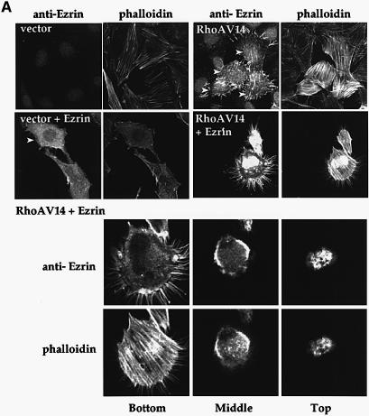
We used transfection assays to test the ability of previously characterized RhoA.V14 effector loop mutants (Sahai et al., 1998) to induce relocalization of ezrin in NIH 3T3 cells. Endogenous ezrin was poorly detectable in serum-deprived cells but, upon co-expression of RhoA.V14, accumulated in actin-containing protrusions from which tubulin was absent (Figure 1A, upper panels and data not shown). Similar results were obtained upon overexpression of ezrin tagged with the VSVG epitope (VSVG-ezrin): upon co-expression of RhoA.V14, VSVG-ezrin relocalized from a diffuse cytoplasmic distribution to actin-containing protrusions and to the dorsal surface of the cells, as assessed by examination of confocal Z-sections (Figure 1A, lower panels and data not shown). Cells expressing RhoA.V14 with or without exogenous ezrin contained substantially increased numbers of actin stress fibres and were markedly decreased in size (Figure 1A, bottom panels). Both endogenous and overexpressed ezrin relocalized similarly in response to RhoA.V14, so we used overexpression of ezrin for further studies.
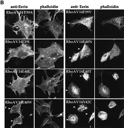
Fig. 1. (A) Activated RhoA relocalizes both endogenous and transiently expressed ezrin into actin-containing dorsal protrusions. NIH 3T3 cells were transfected with 0.3 µg of the RhoA.V14 expression plasmid pEF.RhoA.V14 or its parental vector EFplink (top panels), or with these plasmids together with 0.6 µg of the ezrin expression plasmid pBC6-EzrinWT (lower panels). After 18 h, ezrin and F-actin were detected as described in Materials and methods. The nuclear endogenous ezrin staining is non-specific. RhoA expression was confirmed by 9E10 staining (data not shown). Each panel shows a Z stack of confocal images. (B) Relocalization of ezrin by RhoA effector loop mutants. Cells were transfected with 0.6 µg of pBC6-EzrinWT as above together with 0.3 µg each of expression plasmids encoding the indicated RhoA.V14 effector loop mutants. Transfected cells are indicated by arrowheads. Binding affinities of the effector loop mutants for the Rho-binding domains of ROCK, PKN and citron kinases are as follows (Sahai et al., 1998) (+, binding; –, no binding; +/–, intermediate binding): F39A: PKN–, ROCK–, CitronK–; F39V: PKN–, ROCK+, CitronK–; F39L: PKN+, ROCK+/–, CitronK–; E40L: PKN+, ROCK–, CitronK+; E40W: PKN+, ROCK–, CitronK+; E40N: PKN+, ROCK+, CitronK+; E40T: PKN+, ROCK+, CitronK+; Y42C: PKN–, ROCK+, CitronK+. The proportions of cells displaying dorsally relocalized ezrin were as follows: ezrin alone, 25%; ezrin with RhoA.V14WT, 95%; F39A, 19%; F39L, 24%; E40L, 26%; E40W; 37%; F39V, 60%; E40N, 95%; E40T, 82%; and Y42C, 84%.
We next examined the effects of RhoA.V14 effector loop mutants on ezrin relocalization. RhoA.V14 mutants F39V, E4ON, E40T and Y42C, all of which bind ROCK in yeast two-hybrid and biochemical interaction assays (Sahai et al., 1998), induced ezrin relocalization (Figure 1B, right panels). Of the effector loop mutants that do not bind ROCK effectively in these assays, mutants F39A, E40L and F39L failed to induce ezrin relocalization, while E40W displayed a weak activity (Figure 1B, left panels and legend). The dorsal relocalization of ezrin is unlikely to be the consequence of stress fibre-mediated cell contraction, since it also occurs in cells expressing the RhoA.V14 mutant F39V, which is defective in this contractile response. Binding of the mutants to citron kinase and PKN/PRK1 does not correlate with their ability to relocalize ezrin (see Figure 1 legend). These results strongly suggest that RhoA.V14 must interact with ROCK to induce ezrin relocalization.
Dbl- and Net-induced ezrin relocalization is inhibited by Y27632
We next tested whether activated forms of the RhoGEFs Dbl and Net (NetΔN; Alberts and Treisman, 1998; hereafter referred to as Net) could induce relocalization of exogenously expressed ezrin. To examine involvement of ROCK in this process, we used the ROCK-specific inhibitor Y27632 (Uehata et al., 1997). Our previous studies of RhoGEF-induced transformation suggested that Src-induced signals operate in parallel to or downstream of ROCK (Sahai et al., 1999), so we also tested the effect of v-Src on ezrin relocalization. Both Net and Dbl induced prominent actin stress fibres, with Dbl additionally inducing ruffling of the cell periphery. Dbl induced relocalization of exogenously expressed ezrin into both peripheral and dorsal ruffles, while Net induced relocalization of ezrin into actin-containing dorsal protrusions, although these were not as pronounced as those seen in cells expressing RhoA.V14 (Figure 2A, left panels). Both Net- and Dbl-induced ezrin relocalization were prevented by treatment of the cells with 10 µM Y27632, which also inhibited their contactile response (Figure 2A, compare left and right panels). We think it unlikely that the peripheral ezrin seen in Y27632-treated cells reflects direct association with Dbl (Takahashi et al., 1998) because no general co-localization of Dbl and ezrin was observed, and peripheral ezrin was also observed in cells expressing Net (Figure 2A and data not shown). Although v-Src expression induced an elongated cell shape, it did not cause dorsal relocalization of ezrin (Figure 2A, left panels). These results suggest that the activation of endogenous RhoA by these GEFs induces ezrin relocalization by activation of ROCK. Consistent with this idea, both Y27632 and the kinase-inactive ROCK mutant ROCKΔ3-K105A (Ishizaki et al., 1997) prevented RhoA.V14-induced ezrin relocalization (Figure 2B).
Fig. 2. (A) ROCK activity is required for ezrin relocalization by the oncogenic RhoGEFs Net and Dbl. Cells were transfected with 0.6 µg of pBC6-EzrinWT together with 0.3 µg of EFplink derivatives encoding Net, Dbl or v-Src. Cells were analysed for ezrin and F-actin 18 h later, following a 2 h treatment with Y27632 where indicated, as described in Materials and methods. Panels show a Z stack of confocal images. (B) RhoA.V14-induced ezrin is ROCK dependent. Cells were transfected with 0.6 µg of pBC6-EzrinWT together with pEFplink derivatives encoding either RhoA.V14 (0.3 µg) or the kinase-inactive ROCK mutant ROCKΔ3K105A (0.5 µg), and analysed as above.
ROCK is a candidate for the C-ERMAD kinase
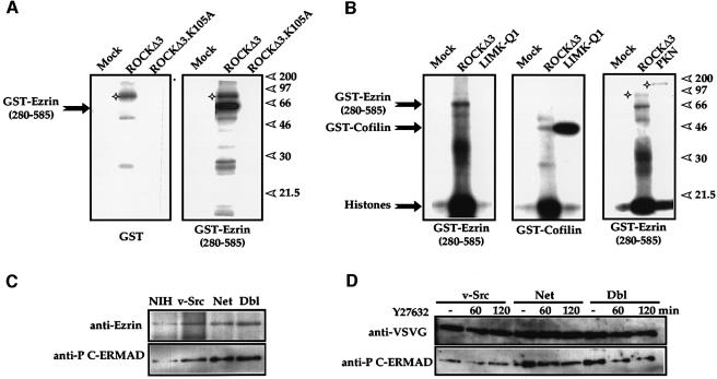
As outlined in the Introduction, the identity of the C-ERMAD kinase has been controversial. We therefore used immune complex kinase assays to reassess the ability of various kinases acting in Rho-dependent pathways to phosphorylate the ezrin C-ERMAD in vitro. 9E10-tagged ROCKΔ3 or its kinase-inactive derivative ROCKΔ3-K105A were expressed in NIH 3T3 cells, and 9E10 immunoprecipitates tested in kinase reactions with recombinant glutathione _S_-transferase (GST) or GST–ezrin(280–585). ROCKΔ3 efficiently immunoprecipitates phosphorylated GST–ezrin(280–585) but not GST or the ezrin N-ERMAD, and this required an intact kinase domain (Figure 3A and data not shown). Similar results were obtained with full-length ROCK (data not shown). We also tested ezrin C-ERMAD phosphorylation by two further RhoA-regulated kinases, PKN/PRK1 and the ROCK effector LIMK-1, both expressed by transfection. In each case, phosphorylation of GST–ezrin(280–585) was not detected under conditions where phosphorylation of their previously characterized histone and cofilin substrates was readily detectable (Figure 3B). Thus, ROCK is both implicated in the promotion of RhoA-induced ezrin relocalization and a candidate ezrin C-ERMAD kinase.
Fig. 3. (A) ROCK can phosphorylate the ezrin C-ERMAD in vitro. NIH 3T3 cells were either mock transfected or transfected with 4 µg of EFplink plasmids encoding 9E10 epitope-tagged ROCKΔ3 or its kinase-inactive derivative ROCKΔ3.K105A. After 18 h, cell extracts were immunoprecipitated with 9E10 antibody and used in immune complex kinases assays with GST or GST–ezrin(280–585) as substrates. Reactions were analysed on a 10% SDS–polyacrylamide gel. Stars indicate presumed autophosphorylation of the kinase. (B) The ROCK effector LIMK-1 and the RhoA effector PKN (PRK1) do not phosphorylate the ezrin C-ERMAD. Cells were transfected with plasmids encoding ROCKΔ3, LIMK-Q1 (the LIMK-1 kinase domain) or intact PKN, and analysed as in (B) with the indicated GST fusion proteins, together with whole calf thymus histones (a kind gift of Dr Robert Nicolas) as substrates. Reactions were analysed on a 15% SDS–polyacrylamide gel. (C) Increased ezrin phosphorylation in transformed cells. Immunoblot analysis of NIH 3T3 cells and derivatives Src-1, Net-e and Dbl-d transformed by the indicated oncogenes using the indicated antibodies. (D) Ezrin phosphorylation is reduced by Y-27632 in RhoGEF-transformed cells. Extracts from derivatives of Src-1-, Net-e- and Dbl-d-transformed cells expressing VSVG-tagged ezrin (Src Ez-1, Net Ez 3-7 and Dbl Ez-2) were analysed using the indicated antibodies after treatment for various times with Y27632.
We next investigated the ability of various activators to induce phosphorylation of ezrin T567 in vivo. For this, we investigated NIH 3T3 cells and their derivatives transformed by Dbl, Net and Src, using the phosphospecific monoclonal antibody 297S to monitor T567 phosphorylation (Matsui et al., 1998). Both Net- and Dbl-transformed cells showed higher levels of ezrin T567 phosphorylation compared with Src-transformed cells, which in turn exhibited higher ezrin T567 phosphorylation than NIH 3T3 cells (Figure 3C). When derivatives of the transformed cell lines overexpressing VSVG-tagged wild-type ezrin were treated with Y27632, we observed a reduction of some 50% in T567 phosphorylation in cells transformed by the RhoGEFs, but not in those transformed by Src (Figure 3D). These results show that ROCK contributes to enhanced levels of ezrin C-ERMAD phosphorylation in vivo.
C-ERMAD phosphorylation is required for relocalization of ezrin
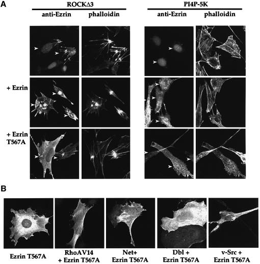
We next investigated whether Rho-induced ezrin localization requires its C-terminal phosphorylation by use of the non-phosphorylatable C-ERMAD mutant T567A (Gautreau et al., 2000). Expression of the activated ROCK mutant ROCKΔ3 induced aggregation of F-actin into striking stellate structures and a strong contractile response, in agreement with previous reports (Leung et al., 1996; Sahai et al., 1998; Figure 4A, left panels). As expected, ROCKΔ3 also induced the relocalization of co-expressed wild-type ezrin and its accumulation in actin-containing protrusions similar to but grosser than those induced by activated RhoA (Figure 4A). We also evaluated the ability of type I PI4P-5K to induce relocalization of ezrin, since this RhoA effector has been proposed to induce ezrin relocalization and T567 phosphorylation independently of ROCK (Matsui et al., 1999). Expression of PI4P-5K induced the formation of F-actin-containing structures reminiscent of the ‘pine-needles’ observed previously (Shibasaki et al., 1997). In contrast to the results obtained with ROCKΔ3, however, PI4P-5K expression relocalized both wild-type ezrin and ezrin T567A into these structures (Figure 4A, right panels and data not shown). Thus, PI4P-5K- and ROCK-induced ezrin relocalization can be distinguished in vivo by their requirement for ezrin T567.
Fig. 4. (A) Ezrin T567 is required for relocalization induced by ROCK but not PI4P-5K. NIH 3T3 cells were transfected with either 0.5 µg of pEF ROCKΔ3 or PI4P-5K either alone or with 0.3 µg of pBC6-EzrinWT or pBC6-EzrinT567A as indicated, and analysed for ezrin localization and F-actin 18 h later. Transfected cells are indicated by arrowheads. (B) Activated RhoA and its GEFs cannot relocalize ezrin T567A. Cells were transfected with plasmids expressing the indicated proteins and analysed as in (A).
We exploited this finding to examine further the involvement of PI4P-5K in RhoA- and RhoGEF-induced ezrin relocalization. Activated RhoA, the RhoGEFs Net and Dbl, and v-Src were tested for their ability to induce relocalization of exogenously expressed ezrin T567A. When expressed alone, ezrin T567A exhibited a diffuse cytoplasmic distribution similar to that of wild-type ezrin (Figure 4B; compare with Figure 1A). Unlike wild-type ezrin, however, ezrin T567A did not relocalize in response to either RhoA.V14, Dbl or Net (Figure 4B; compare with Figure 2A and B) and the cells exhibited a flattened and enlarged morphology; in contrast, ezrin T567A did not prevent the elongation of v-Src-expressing cells. These results are strikingly similar to those obtained upon treatment of cells expressing wild-type ezrin with Y27632 (compare Figures 4B and 2A). Taken together, the results show that ROCK-dependent phosphorylation of ezrin T567 is required for RhoA- and RhoGEF-induced ezrin relocalization.
Ezrin relocalization reflects increased association with the cytoskeleton
Relocalization of ezrin correlates with its increased association with the actin cytoskeleton through its C-ERMAD (Berryman et al., 1995; reviewed by Bretscher, 1999; Mangeat et al., 1999). We tested whether ezrin C-ERMAD phosphorylation is required for this using a Triton X-100 fractionation procedure (Algrain et al., 1993). Expression plasmids encoding VSVG-tagged wild-type ezrin or the ezrin mutant T567A were stably introduced into either NIH 3T3 cells or transformed cell lines stably expressing activated Dbl, Net or Src (Sahai et al., 1999). The cells were left untreated or were treated with Y27632 before being subjected to Triton X-100 fractionation, followed by detection of ezrin by immunoblotting. Wild-type ezrin, but not ezrin T567A, was detectable in the Triton X-100-insoluble fraction from Dbl- and Net-transformed cells, and Y27632 treatment substantially decreased the amount recovered in the Triton X-100-insoluble fraction (Figure 5A). In contrast, in untransformed NIH 3T3 cells and Src-transformed cells expressing wild-type ezrin, no wild-type ezrin was detected in the Triton X-100-insoluble fraction (Figure 5B and data not shown). Taken together, these results strongly suggest that RhoGEF-induced relocalization of ezrin is associated with increased interactions between ezrin and the actin cytoskeleton, and that these interactions require phosphorylation of its C-ERMAD.
Fig. 5. (A) Association of ezrin with the cytoskeleton requires T567. Stable transfectants of Dbl-d- or Net-e-transformed NIH 3T3 cells expressing either VSVG-tagged wild-type ezrin (Dbl Ez-2 and Net Ez-3-7) or ezrin T567A (Dbl EzT567A-10 and Net EzT567A-1) were left untreated or treated for 2 h with Y27632 before fractionation by Triton X-100 extraction. Fractions were analysed for ezrin and actin by immunoblotting. (B) Ezrin is not associated with the cytoskeleton in untransformed or Src-transformed NIH 3T3 cells. Stable transfectants of untransformed Src-transformed NIH 3T3 cell derivatives expressing wild-type VSVG-tagged ezrin (NIH 3T3 Ez-10 and Src-1 Ez-1) were left untreated or treated directly for the indicated times (hours) with Y27632 before fractionation and analysis as above.
C-ERMAD phosphorylation is required for contractility and focal adhesion complex formation in Swiss 3T3 cells
The results described above establish that RhoA and oncogenic RhoGEFs, but not Src, signal through ROCK to relocalize ezrin in a process requiring phosphorylation of its C-ERMAD. The enlarged morphology seen in cells expressing ezrin T567A, however, suggested that expression of this mutant might inhibit the RhoA- and RhoGEF-induced contractile response. Since ERM proteins have also been shown to be required for RhoA-induced focal adhesion formation in permeabilized Swiss 3T3 cells (Mackay et al., 1997), we used Swiss 3T3 cells to examine the requirement for ezrin C-ERMAD phosphorylation in both of these processes. Swiss 3T3 cells were microinjected with plasmids encoding either intact ezrin or ezrin T567A together with RhoA.V14, ROCKΔ3 or Dbl, and maintained in low serum. After 4 h, ezrin expression and focal adhesion complexes were visualized using anti-paxillin antibody. Expression of RhoA.V14 induced the formation of focal adhesion complexes, which coincided with the ends of F-actin stress fibres; formation of both structures was inhibited by Y27632, consistent with the involvement of ROCK (data not shown). Although overexpression of wild-type ezrin resulted in enhanced focal adhesion assembly by RhoA.V14, expression of ezrin T567A inhibited both focal adhesion formation and RhoA-induced contractility (Figure 6). Similar results were observed with both ROCKΔ3 and Dbl (Figure 6). In NIH 3T3 cells, similar but less striking effects were observed owing to a higher basal level of focal adhesions present in these cells (data not shown).
Fig. 6. Ezrin T567A interferes with formation of focal adhesion contacts in Swiss 3T3 cells. Swiss 3T3 cells were microinjected with EFplink derivatives encoding RhoAV14, ROCKΔ3 or Dbl, together with plasmids encoding pBC6-EzrinWT or pBC6-EzrinT567A (50 ng/µl) as indicated. At 4 h post-transfection, cells were fixed, permeabilized and stained for F-actin (green) and paxillin (red). Microinjected cells are indicated by arrowheads.
Ezrin T567A inhibits transformation by RhoGEFs and Ras, but not Src
The data presented above show that ezrin T567A not only fails to relocalize in response to ROCK, but can interfere with ROCK-mediated contractility and focal adhesion assembly. We therefore next tested the effects of ezrin T567A expression on RhoGEF-induced transformation, since our previous studies strongly implicated ROCK in this process (Sahai et al., 1998, 1999). We generated derivatives of both untransformed NIH 3T3 cells and of our previously described Net-, Dbl- and Src-transformed cell lines (Sahai et al., 1999) expressing epitope-tagged forms of either wild-type ezrin or ezrin T567A (see Materials and methods). We were unable to isolate stable cell lines expressing the isolated ezrin N-terminal domain. As judged by immunoblotting, ezrin was expressed at comparable levels in these cell lines, which all grew at somewhat slower rates than the parental cells.
To investigate the effects of ezrin and ezrin T567A contact inhibition, we used a secondary focus formation assay. In this assay, transformed NIH 3T3 cells are seeded amongst untransformed NIH 3T3 cells and their ability to escape contact inhibition is revealed by their ability to form overgrowing foci. Cells transformed by Net, Dbl and v-Src all formed secondary foci in this assay, and focus formation by Net- and Dbl-transformed cells was inhibited by Y27632 (Figure 7A and B; Sahai et al., 1999). Similar results were obtained with the transformed cell derivatives overexpressing wild-type ezrin (Figure 7A and B). In striking contrast, Net- and Dbl-transformed cells expressing ezrin T567A exhibited strongly reduced activity in the secondary focus assays, whereas Src-transformed cells expressing ezrin T567A remained active (Figure 7A and B). Similar results were obtained using other clones of ezrin-expressing cells derived from different Dbl-, Net- and Src-transformed cell lines (data not shown). In some experiments, cells expressing wild-type ezrin showed reduced secondary focus formation. However, differential effects of ezrin T567A and wild-type ezrin expression were only observed in derivatives of the RhoGEF-transformed cells (Figure 7B). When cultured alone, Dbl-, Net- and Src-transformed cells expressing wild-type ezrin overgrew, and bromodeoxyuridine labelling experiments confirmed that they remained in cycle at this point; Dbl- and Net-transformed cells expressing ezrin T567A did not overgrow, in contrast to Src-transformed cells expressing this mutant, which did (data not shown). Neither wild-type ezrin nor ezrin T567A altered the contact inhibition properties of parental NIH 3T3 cells.
Fig. 7. (A) Expression of ezrin T567A but not wild-type ezrin restores contact inhibition in Net- and Dbl-transformed NIH 3T3 cells. Derivatives of both untransformed and transformed NIH 3T3 cells expressing ezrin or ezrin T567A were generated and used in secondary focus formation assays. A total of 5000 test cells were seeded among 30 000 untransformed NIH 3T3 cells and cultured in the presence or absence of 10 µM Y27632 for 10 days, when overgrowing colonies were visualized by crystal violet staining. Upper panel: secondary focus formation by NIH 3T3, v-Src-1, Net-e and Dbl-d cells (Sahai et al., 1999). Middle panel: secondary focus formation by derivatives of these cell lines expressing wild-type ezrin (lines NIH 3T3 Ez-10, v-Src-1 Ez-1, Net-e Ez-1 and Dbl-d Ez-2). Similar results were obtained with a further two cell lines in each case. Bottom panel: secondary focus formation by derivatives of the cell lines shown in the upper panel expressing ezrin T567A (lines NIH 3T3 EzT567A-1, v-Src-1 EzT567A-5, Net-e EzT567A-1 and Dbl-d EzT567A-10). Similar results were obtained with a further ezrin T567A transfectant derived from each of the parental transformed cell lines. In the case of Net and Dbl, two further clones derived from independent Net- and Dbl-transformed cell lines (Net-d and Dbl-e; Sahai et al., 1999) were analysed and a similar result obtained. (B) Summary of secondary focus formation assays. Results from two independent experiments using the cell lines as in (A) are shown. (C) Ezrin T567A inhibits primary focus formation by Net and Ras but not by Src. NIH 3T3 cells were transfected with 0.3 µg of EFplink derivatives encoding v-Src, Net or RasR12 either alone or with increasing doses (0.3 or 0.6 µg) of pBC6-EzrinT567A. DNA inputs were kept constant with pBC6 vector DNA. Cells were maintained for 15 days in 5% donor calf serum before staining with crystal violet to visualize foci, which were then counted and compared with the number of foci induced by the respective oncogenes. Under these conditions, v-Src-transfected cells formed ∼175 foci, RasR12 65 foci and Net 125 foci. The diagram shows the average of three independent experiments.
Finally, we analysed the behaviour of ezrin T567A in primary focus formation assays. We previously showed that ROCK contributes to primary focus forming activity not only by RhoA GEFs but also by activated Ras (Sahai et al., 1999). NIH 3T3 cells were transfected with Net, the activated Ras mutant Ras.R12 or v-Src, with or without ezrin T567A, and transformed foci counted following cultivation for 14 days in 5% serum. In this assay, ezrin T567A interfered with Net-induced transformation and also with transformation by Ras.R12, but only marginally affected v-Src transformation (Figure 7C). Taken together with the secondary focus formation assay results, these data provide strong evidence that phosphorylation of the C-ERMAD is an essential aspect of the ROCK-mediated transforming activity of RhoGEFs.
Discussion
ROCK mediates RhoA- and RhoGEF-induced ezrin relocalization
In this work, we investigated the mechanism by which activated forms of RhoA and its GEFs Net and Dbl can induce relocalization of ezrin, a member of the ERM family of cytoskeletal linker proteins. Several lines of evidence indicate that this process is mediated by the RhoA effector kinase ROCK and requires the phosphorylation of ezrin on the conserved residue T567 in its C-ERMAD. First, the ability of RhoA effector loop mutants to induce ezrin relocalization into dorsal actin-containing structures correlates with their ability to bind ROCK rather than other RhoA effectors. Both Y27632, a specific ROCK inhibitor, and a kinase-defective interfering mutant of ROCK, ROCKΔ3-K105A, inhibited ezrin relocalization induced by both RhoA and its GEFs Dbl and Net. Moreover, expression of a constitutively activated mutant of ROCK, ROCKΔ3, was also sufficient to induce ezrin relocalization. Secondly, ROCK, but not its effector kinase LIMK or the RhoA effector kinase PKN/PRK1, can phosphorylate ezrin T567 in vitro. In line with this, cells transformed by the RhoGEFs Net and Dbl exhibit increased ezrin T567 phosphorylation, which is decreased following Y27632 treatment. Thirdly, a non-phosphorylatable mutant form of ezrin, T567A, fails both to relocalize in response to RhoA and to associate with the actin cytoskeleton. Taken together, these data strongly suggest that phosphorylation of ezrin at T567 is a prerequisite for the rearrangements of the cytoskeleton required for ezrin relocalization.
Our results support previous studies suggesting that direct phosphorylation by ROCK mediates RhoA-induced phosphorylation of the moesin C-ERMAD (Matsui et al., 1998; Oshiro et al., 1998). It is likely that ROCK also contributes to net moesin T558 phosphorylation by inhibiting MLC phosphatase, with which it associates (Fukata et al., 1998). However, PKCθ may phosphorylate the moesin C-ERMAD in leukocytes, suggesting that multiple pathways for ERM regulation exist (Pietromonaco et al., 1998). More recently, expression of another RhoA effector, PI4P-5K, has been reported to induce ERM relocalization and C-ERMAD phosphorylation independently of ROCK, leading to the proposal that these processes are dependent on PI4P-5K rather than ROCK (Matsui et al., 1999). We confirmed that overexpression of PI4P-5K can relocalize ezrin into actin-containing structures; however, this does not require ezrin T567, in contrast to relocalization induced by activated RhoA, the RhoGEFs and ROCK. RhoA- and RhoGEF-induced ezrin relocalization therefore cannot be due solely to activation of PI4P-5K. It remains possible, however, that basal levels of PI4P-5K activity are required for ROCK-induced ezrin relocalization (see below).
Non-phosphorylatable ezrin C-ERMAD mutants interfere with cytoskeletal events
ERM proteins exist in unstimulated cells as soluble cytosolic monomeric or dimeric forms, which translocate to specialized sites in the plasma membrane in response to a variety of signals (Nakamura et al., 1995, 1999; Matsui et al., 1998; Shaw et al., 1998). Such signals, which include phosphorylation of the conserved C-ERMAD threonine residue (Matsui et al., 1998; Huang et al., 1999; Nakamura et al., 1999), disrupt the interaction between the N- and C-ERMADs and allow the ERMs to interact with F-actin and with transmembrane proteins and/or lipids. Our data show that the non-phosphorylatable ezrin T567A mutant is unable to relocalize in response to activated RhoA and RhoGEFs. Surprisingly, however, expression of ezrin T567A also interferes with other ROCK-dependent processes such as contractility, formation of focal adhesions and RhoGEF-induced loss of contact inhibition in fibroblasts. We suggest that unphosphorylated ezrin interferes with cytoskeletal reorganization by interacting with a subset of other molecules involved in ezrin-dependent cytoskeletal reorganization. Recent structural studies of moesin strongly suggest that potential interaction sites within the N-ERMAD are available even when it is complexed with the C-ERMAD (Pearson, 2000), and biochemical studies show that ezrin T567A can interact with other ERM family members (Gautreau et al., 2000). It would appear unlikely that ezrin T567A simply titrates ROCK, because the actin bundles present in NIH 3T3 cell lines expressing ezrin T567A remain sensitive to the ROCK inhibitor Y27632, consistent with basal ROCK activity in these cells (C.Tran Quang, unpublished observation).
A large number of previous studies have implicated the RhoA/ROCK pathway in directing the initial assembly of focal adhesion contacts and in the maturation of the Rac- and Cdc42-induced focal complexes associated with lamellipodia and filopodia into larger complexes (reviewed by Van Aelst and D’Souza-Schorey, 1997). Biochemical studies have shown that ERM proteins are required for RhoA-induced focal adhesion formation in permeabilized Swiss 3T3 cells (Mackay et al., 1997). Although this might reflect the presence of ERM proteins in focal adhesion complexes (discussed by Bretscher et al., 1997), a less controversial alternative view is that the ERMs are required to allow remodelling of the cortical actin cytoskeleton during focal adhesion assembly. In this model, transient agonist-induced phosphorylation of the ezrin C-ERMAD would be required to allow concentration of actin filaments at the specialized membrane sites of contact assembly. In this scenario, ezrin T567A would be unable to undergo signal-induced alterations in its interactions with F-actin and would thereby inhibit focal adhesion formation.
A second proposed mechanism by which ERM protein function is potentially regulated is via the ability of PI4,5P2 to bind the N-ERMAD, which in vitro facilitates ERM interactions with transmembrane partner proteins such as CD44, ICAM1 and ICAM2 (reviewed by Bretscher, 1999; Mangeat et al., 1999). PI4,5P2 binding to moesin also cooperates with phosphorylation of the C-ERMAD site to activate binding to F-actin in vitro (Nakamura et al., 1999). These observations are consistent with the recent finding that overexpression of PI4P-5K, which controls PI4,5P2 synthesis, induces relocalization of ERM proteins in vivo (Matsui et al., 1999). As discussed above, our data show that C-ERMAD phosphorylation is essential for RhoA- and RhoGEF-induced ezrin relocalization, but we cannot exclude the possibility that PI4,5P2 binding is also required. Experiments utilizing dominant-interfering PI4P-5K derivatives or antibodies directed against PI4,5P2 should allow a direct test of the requirement for PI4,5P2 in RhoA-induced ERM protein activation.
Ezrin C-ERMAD phosphorylation and transformation
Our previous studies provided two lines of evidence that the transforming activity of activated RhoA and its GEFs in focus formation assays in NIH 3T3 fibroblasts requires ROCK activity. First, the activity of RhoA effector mutants in focus formation assays correlates with their binding to ROCK in two-hybrid and biochemical assays (Sahai et al., 1998). Secondly, the activity of both activated RhoA and the RhoGEFs Net and Dbl is blocked specifically by Y27632, a small molecule ROCK inhibitor, which does not affect the growth properties of untransformed fibroblasts or transformation by Src (Sahai et al., 1999). In this work, we have shown that overexpressed ezrin T567A, but not wild-type ezrin, specifically inhibited cell transformation by the RhoGEFs Dbl and Net, but did not affect transformation by v-Src, suggesting that ezrin activation is an essential step in transformation induced by these oncogenes. In addition, as observed with Y27632, ezrin T567A also inhibited Ras transformation, consistent with the notion that activation of RhoA-dependent signalling pathways contributes to transformation by Ras (Qiu et al., 1995; Sahai et al., 1999). Our results extend a previous study implicating ezrin expression level in relief of contact inhibition (Kaul et al., 1996).
How might ezrin activation contribute to transformation by RhoA and its GEFs? In our assays, the primary feature of transformation by RhoGEFs appears to be a blockade of contact inhibition and anchorage-dependent growth. Dbl- and Net-transformed cells remain serum dependent and grow at rates similar to untransformed cells, but fail to exit the cell cycle at confluence, overgrow at high density and exhibit reduced anchorage dependence; growth in the presence of Y27632 restores normal growth control (Sahai et al., 1999). These observations suggest that constitutive RhoA activation bypasses contact inhibition by generating a signal, normally associated with adhesion and/or subconfluent growth, which promotes cell cycle progression. The results presented here suggest that constitutive activation of ezrin-mediated cytoskeletal rearrangements is required for generation of this signal. It is tempting to speculate that such rearrangements act to promote the integrity or signalling properties of focal adhesions in confluent cells, maintaining the ‘adhesion’ signal required for continued cell division. Such a possibility would be consistent with a previous study, which concluded that RhoGEFs induce anchorage-independent growth by their ability to mimic signalling by adhesion receptors (Schwartz et al., 1996). In this model, the inability of ezrin T567A to inhibit Src transformation might occur because Src, which is normally associated with focal adhesions, would provide this signal regardless of Rho activity. While this model can rationalize the requirement for ROCK and ezrin phosphorylation in RhoGEF transformation, it does not explain their function in Ras-transformed cells, which contain few actin stress fibres or focal adhesions (Khosravi-Far et al., 1994). Further studies will be necessary to distinguish between this ‘adhesion’ model and others in which ezrin exerts its effects by alterations in the signalling properties of its transmembrane partners such as CD44.
A number of studies have implicated ERM proteins in oncogenesis in vivo. Increased ezrin expression occurs upon transformation of Rat-1 fibroblasts by v-fos (Jooss and Muller, 1995; Lamb et al., 1997), and has been correlated with increased metastatic and invasive potential of adenocarcinoma and endometrial tumour cell lines (Akisawa et al., 1999; Ohtani et al., 1999); ezrin also associates with hamartin, the product of the TSC1 tumour suppressor gene (Lamb et al., 2000). In contrast, expression of the ERM-related protein merlin, the product of the neurofibromatosis type II (NF2) tumour suppressor gene (reviewed by Gusella et al., 1999), can restore contact inhibition and anchorage dependence to Ras-transformed cells (Tikoo et al., 1994). In addition, the RhoA/ROCK pathway has also been implicated in the invasive properties of rat MM1 hepatoma cells (Itoh et al., 1999). Our recent experiments indicate that Y27632 treatment both inhibited growth and delocalized ezrin in five out of seven anchorage-independent colorectal cell lines tested (unpublished observations). It will be interesting to use the ezrin T567A mutant to investigate the role of ezrin C-ERMAD phosphorylation in maintenance of transformation in vivo.
Materials and methods
Cell lines, transfection, microinjections and focus formation assays
Maintenance of NIH 3T3 cells and their transformed derivatives was as previously described; derivatives of NIH 3T3, v-Src1, Net-d, Net-e, Dbl-d and Dbl-e cells (Sahai et al., 1999) expressing VSVG-tagged ezrin or ezrin T567A were generated by transfection with the appropriate pBC6 expression plasmid (Gautreau et al., 2000), followed by selection for growth in 1.2 mg/ml G418. Focus assays and treatment with Y27632 were as described previously (Sahai et al., 1999). Transient transfection using Lipofectamine (Gibco) was according to the manufacturer’s instructions. Cells were then maintained for a further 18 h before analysis. Microinjection of Swiss 3T3 cells was carried out as described (Sahai et al., 1999).
Immunofluorescence analyses
For immunofluorescence, cells grown on glass coverslips were analysed as described previously (Sahai et al., 1999). Additional antibodies were anti-VSVG (1:100; Boehringer Mannheim), affinity-purified rabbit anti-ezrin (1:400, stained at 37°C for 1 h; Algrain et al., 1993) and anti-paxillin (1:100; Transduction Laboratories p13520). Images were obtained using a Zeiss Axiovert 100M confocal microscope; Z stacks are shown in the figures. For analysis of cytoskeleton-associated ezrin, cells were extracted four times for 5 s with MEB (50 mM MES pH 6.4, 3 mM EGTA, 5 mM MgCl2, 0.5% Triton X-100) before processing.
Immune complex kinase assays
NIH 3T3 cells (1 × 10⁶; 90 mm dish) were transfected with 4 µg of pEF.ROCKΔ3 (Ishizaki et al., 1997), pEF.LIMK-Q1 (Sotiropoulos et al., 1999) or pEF.PKN (S.Walker, unpublished). At 48 h post-transfection, cells were washed twice in ice-cold phosphate-buffered saline (PBS), and 200 µl of lysis buffer [1 mM EGTA, 50 mM β-glycerophosphate, 1 mM dithiothreitol (DTT), 1 mM Na3VO4, 1% Triton X-100, 10% glycerol, 50 ng/ml okadaic acid, 10 mM MgCl2, 50 mM Tris–HCl pH 7.5] were added. Following clarification (13 000 r.p.m. at 4°C for 10 min), 1 µg of 9E10 antibody was added, and after 1 h at 4°C proteins were recovered by adsorption to protein G–Sepharose beads (Pharmacia; pre-equilibrated in lysis buffer). After three washes in lysis buffer and three washes with kinase buffer (50 mM Tris–HCl pH 7.5, 1 mM EDTA, 10 mM MgCl2, 50 mM NaCl, 0.03% Brij35, 1 mM DTT), the beads were resuspended in 30 µl of kinase buffer containing 10 µM ATP (Pharmacia) and 4 µCi of [γ-32P]ATP (Amersham; 5000 Ci/mmol). Reactions were carried out at 30°C for 30 min, and analysed by SDS–PAGE and autoradiography. Substrates (1.5 µg) were GST, GST–ezrin(1–310), GST–ezrin(280–585) or GST–cofilin, purified by standard methods, or calf thymus histones (gift from Dr Robert Nicolas).
Cell fractionation, immunoprecipitation and immunoblotting
Triton X-100 fractionation was carried out as described (Algrain et al., 1993). For immunoprecipitation, cells were lysed in 150 mM NaCl, 1 mM EDTA, 1% Triton X-100, 25 mM Tris–HCl pH 7.4 and centrifuged at 100 000 g for 30 min. Lysates were incubated with 0.8 µg of anti-VSVG antibody for 1 h at 4°C, 40 µl of protein G–Sepharose added, and incubated for a further hour. Beads were recovered by centrifugation and washed three times with lysis buffer. Proteins were resolved by 10% SDS–PAGE, transferred to nitrocellulose, and ezrin was detected with anti-VSVG monoclonal antibody (1:1000; Boehringer) and anti-phospho-C-ERMAD 297S (Matsui et al., 1998; from S.Tsukita) with horseradish peroxidase-conjugated anti-rat or anti-mouse goat antibodies, respectively (1:100; Dako), with visualization by ECL (Amersham).
Acknowledgments
Acknowledgements
We thank Peter Jordan of the ICRF microscopy department for assistance with confocal imaging, Dr Laura Machesky for the gift of PI4P-5K cDNA, Dr Shuh Narumiya for the gift of Y27632, and Dr Sachiko Tsukita for the generous gift of the 297S antibody. We thank Dr Jacques Ghysdael and Dr Erik Sahai for helpful comments and advice on the manuscript, as well as members of the laboratory for entertaining discussions. C.T.Q. was supported by a postdoctoral fellowship from the Human Frontier Science Program.
References
- Akisawa N., Nishimori,I., Iwamura,T., Onishi,S. and Hollingsworth,M.A. (1999) High levels of ezrin expressed by human pancreatic adenocarcinoma cell lines with high metastatic potential. Biochem. Biophys. Res. Commun., 258, 395–400. [DOI] [PubMed] [Google Scholar]
- Alberts A.S. and Treisman,R. (1998) Activation of RhoA and SAPK/JNK signalling pathways by the RhoA-specific exchange factor mNET1. EMBO J., 17, 4075–4085. [DOI] [PMC free article] [PubMed] [Google Scholar]
- Algrain M., Turunen,O., Vaheri,A., Louvard,D. and Arpin,M. (1993) Ezrin contains cytoskeleton and membrane binding domains accounting for its proposed role as a membrane–cytoskeletal linker. J. Cell Biol., 120, 129–139. [DOI] [PMC free article] [PubMed] [Google Scholar]
- Amano M., Chihara,K., Kimura,K., Fukata,Y., Nakamura,N., Matsuura,Y. and Kaibuchi,K. (1997) Formation of actin stress fibers and focal adhesions enhanced by Rho-kinase. Science, 275, 1308–1311. [DOI] [PubMed] [Google Scholar]
- Berryman M., Gary,R. and Bretscher,A. (1995) Ezrin oligomers are major cytoskeletal components of placental microvilli: a proposal for their involvement in cortical morphogenesis. J. Cell Biol., 131, 1231–1242. [DOI] [PMC free article] [PubMed] [Google Scholar]
- Bretscher A. (1999) Regulation of cortical structure by the ezrin–radixin–moesin protein family. Curr. Opin. Cell Biol., 11, 109–116. [DOI] [PubMed] [Google Scholar]
- Bretscher A., Gary,R. and Berryman,M. (1995) Soluble ezrin purified from placenta exists as stable monomers and elongated dimers with masked C-terminal ezrin–radixin–moesin association domains. Biochemistry, 34, 16830–16837. [DOI] [PubMed] [Google Scholar]
- Bretscher A., Reczek,D. and Berryman,M. (1997) Ezrin: a protein requiring conformational activation to link microfilaments to the plasma membrane in the assembly of cell surface structures. J. Cell Sci., 110, 3011–3018. [DOI] [PubMed] [Google Scholar]
- Fukata Y., Kimura,K., Oshiro,N., Saya,H., Matsuura,Y. and Kaibuchi,K. (1998) Association of the myosin-binding subunit of myosin phosphatase and moesin: dual regulation of moesin phosphorylation by Rho-associated kinase and myosin phosphatase. J. Cell Biol., 141, 409–418. [DOI] [PMC free article] [PubMed] [Google Scholar]
- Gary R. and Bretscher,A. (1995) Ezrin self-association involves binding of an N-terminal domain to a normally masked C-terminal domain that includes the F-actin binding site. Mol. Biol. Cell, 6, 1061–1075. [DOI] [PMC free article] [PubMed] [Google Scholar]
- Gautreau A., Louvard,D. and Arpin,M. (2000) Morphogenic effects of ezrin require a phosphorylation-induced transition from oligomers to monomers at the plasma membrane. J. Cell Biol., 150, 193–204. [DOI] [PMC free article] [PubMed] [Google Scholar]
- Gusella J.F., Ramesh,V., MacCollin,M. and Jacoby,L.B. (1999) Merlin: the neurofibromatosis 2 tumor suppressor. Biochim. Biophys. Acta, 1423, M29–M36. [DOI] [PubMed] [Google Scholar]
- Huang L., Wong,T.Y., Lin,R.C. and Furthmayr,H. (1999) Replacement of threonine 558, a critical site of phosphorylation of moesin in vivo, with aspartate activates F-actin binding of moesin. Regulation by conformational change. J. Biol. Chem., 274, 12803–12810. [DOI] [PubMed] [Google Scholar]
- Ishizaki T., Naito,M., Fujisawa,K., Maekawa,M., Watanabe,N., Saito,Y. and Narumiya,S. (1997) p160ROCK, a Rho-associated coiled-coil forming protein kinase, works downstream of Rho and induces focal adhesions. FEBS Lett., 404, 118–124. [DOI] [PubMed] [Google Scholar]
- Itoh K., Yoshioka,K., Akedo,H., Uehata,M., Ishizaki,T. and Narumiya,S. (1999) An essential part for Rho-associated kinase in the transcellular invasion of tumor cells. Nature Med., 5, 221–225. [DOI] [PubMed] [Google Scholar]
- Jooss K.U. and Muller,R. (1995) Deregulation of genes encoding microfilament-associated proteins during Fos-induced morphological transformation. Oncogene, 10, 603–608. [PubMed] [Google Scholar]
- Kaul S.C., Mitsui,Y., Komatsu,Y., Reddel,R.R. and Wadhwa,R. (1996) A highly expressed 81 kDa protein in immortalized mouse fibroblast: its proliferative function and identity with ezrin. Oncogene, 13, 1231–1237. [PubMed] [Google Scholar]
- Khosravi-Far R., Chrzanowska-Wodnicka,M., Solski,P., Eva,A., Burridge,K. and Der,C. (1994) Dbl and Vav mediate transformation via mitogen-activated protein kinase pathways that are distinct from those activated by oncogenic Ras. Mol. Cell. Biol., 14, 6848–6857. [DOI] [PMC free article] [PubMed] [Google Scholar]
- Khosravi-Far R., Solski,P.A., Clark,G.J., Kinch,M.S. and Der,C.J. (1995) Activation of Rac1, RhoA and mitogen-activated protein kinases is required for Ras transformation. Mol. Cell. Biol., 15, 6443–6453. [DOI] [PMC free article] [PubMed] [Google Scholar]
- Kimura K. et al. (1996) Regulation of myosin phosphatase by Rho and Rho-associated kinase (Rho-kinase). Science, 273, 245–248. [DOI] [PubMed] [Google Scholar]
- Lamb R.F., Ozanne,B.W., Roy,C., McGarry,L., Stipp,C., Mangeat,P. and Jay,D.G. (1997) Essential functions of ezrin in maintenance of cell shape and lamellipodial extension in normal and transformed fibroblasts. Curr. Biol., 7, 682–688. [DOI] [PubMed] [Google Scholar]
- Lamb R.F., Roy,C., Diefenbach,T.J., Vinters,H.V., Johnson,M.W., Jay,D.G. and Hall,A. (2000) The TSC1 tumour suppressor hamartin regulates cell adhesion through ERM proteins and the GTPase Rho. Nature Cell Biol., 2, 281–287. [DOI] [PubMed] [Google Scholar]
- Leung T., Chen,X.Q., Manser,E. and Lim,L. (1996) The p160 RhoA-binding kinase ROK α is a member of a kinase family and is involved in the reorganization of the cytoskeleton. Mol. Cell. Biol., 16, 5313–5327. [DOI] [PMC free article] [PubMed] [Google Scholar]
- Mackay D.J., Esch,F., Furthmayr,H. and Hall,A. (1997) Rho- and rac-dependent assembly of focal adhesion complexes and actin filaments in permeabilized fibroblasts: an essential role for ezrin/radixin/moesin proteins. J. Cell Biol., 138, 927–938. [DOI] [PMC free article] [PubMed] [Google Scholar]
- Maekawa M. et al. (1999) Signaling from Rho to the actin cytoskeleton through protein kinases ROCK and LIM-kinase. Science, 285, 895–898. [DOI] [PubMed] [Google Scholar]
- Mangeat P., Roy,C. and Martin,M. (1999) ERM proteins in cell adhesion and membrane dynamics. Trends Cell Biol., 9, 187–192. [DOI] [PubMed] [Google Scholar]
- Matsui T., Maeda,M., Doi,Y., Yonemura,S., Amano,M., Kaibuchi,K. and Tsukita,S. (1998) Rho-kinase phosphorylates COOH-terminal threonines of ezrin/radixin/moesin (ERM) proteins and regulates their head-to-tail association. J. Cell Biol., 140, 647–657. [DOI] [PMC free article] [PubMed] [Google Scholar]
- Matsui T., Yonemura,S. and Tsukita,S. (1999) Activation of ERM proteins in vivo by Rho involves phosphatidyl-inositol 4-phosphate 5-kinase and not ROCK kinases. Curr. Biol., 9, 1259–1262. [DOI] [PubMed] [Google Scholar]
- Nakamura F., Amieva,M.R. and Furthmayr,H. (1995) Phosphorylation of threonine 558 in the carboxyl-terminal actin-binding domain of moesin by thrombin activation of human platelets. J. Biol. Chem., 270, 31377–31385. [DOI] [PubMed] [Google Scholar]
- Nakamura F., Huang,L., Pestonjamasp,K., Luna,E.J. and Furthmayr,H. (1999) Regulation of F-actin binding to platelet moesin in vitro by both phosphorylation of threonine 558 and polyphosphatidylinositides. Mol. Biol. Cell, 10, 2669–2685. [DOI] [PMC free article] [PubMed] [Google Scholar]
- Ohtani K., Sakamoto,H., Rutherford,T., Chen,Z., Satoh,K. and Naftolin,F. (1999) Ezrin, a membrane–cytoskeletal linking protein, is involved in the process of invasion of endometrial cancer cells. Cancer Lett., 147, 31–38. [DOI] [PubMed] [Google Scholar]
- Olson M.F., Ashworth,A. and Hall,A. (1995) An essential role for Rho, Rac and Cdc42 GTPases in cell cycle progression through G1. Science, 269, 1270–1272. [DOI] [PubMed] [Google Scholar]
- Olson M., Paterson,H. and Marshall,C. (1998) Signals from Ras and Rho GTPases interact to regulate expression of p21Waf1/Cip1. Nature, 394, 295–259. [DOI] [PubMed] [Google Scholar]
- Oshiro N., Fukata,Y. and Kaibuchi,K. (1998) Phosphorylation of moesin by rho-associated kinase (Rho-kinase) plays a crucial role in the formation of microvilli-like structures. J. Biol. Chem., 273, 34663–34666. [DOI] [PubMed] [Google Scholar]
- Oude-Weernink P.A. et al. (2000) Stimulation of phosphatidylinositol-4-phosphate 5-kinase by Rho-kinase. J. Biol. Chem., 275, 10168–10174. [DOI] [PubMed] [Google Scholar]
- Pearson M.A., Reczek,D., Bretscher,A. and Karplus,A.P. (2000) Structure of the ERM protein moesin reveals the FERM domain fold masked by an extended actin binding tail domain. Cell, 101, 259–270. [DOI] [PubMed] [Google Scholar]
- Pietromonaco S.F., Simons,P.C., Altman,A. and Elias,L. (1998) Protein kinase C-θ phosphorylation of moesin in the actin-binding sequence. J. Biol. Chem., 273, 7594–7603. [DOI] [PubMed] [Google Scholar]
- Qiu R., Chen,J., McCormick,F. and Symons,M. (1995) A role for Rho in Ras transformation. Proc. Natl Acad. Sci. USA, 92, 11781–11785. [DOI] [PMC free article] [PubMed] [Google Scholar]
- Sahai E., Alberts,A.S. and Treisman,R. (1998) RhoA effector mutants reveal distinct effector pathways for cytoskeletal reorganization, SRF activation and transformation. EMBO J., 17, 1350–1361. [DOI] [PMC free article] [PubMed] [Google Scholar]
- Sahai E., Ishizaki,T., Narumiya,S. and Treisman,R. (1999) Transformation mediated by RhoA requires activity of ROCK kinases. Curr. Biol., 9, 136–145. [DOI] [PubMed] [Google Scholar]
- Schwartz M.A., Toksoz,D. and Khosravi Far,R. (1996) Transformation by Rho exchange factor oncogenes is mediated by activation of an integrin-dependent pathway. EMBO J., 15, 6525–6530. [PMC free article] [PubMed] [Google Scholar]
- Shaw R.J., Henry,M., Solomon,F. and Jacks,T. (1998) RhoA-dependent phosphorylation and relocalization of ERM proteins into apical membrane/actin protrusions in fibroblasts. Mol. Biol. Cell, 9, 403–419. [DOI] [PMC free article] [PubMed] [Google Scholar]
- Shibasaki Y., Ishihara,H., Kizuki,N., Asano,T., Oka,Y. and Yazaki,Y. (1997) Massive actin polymerization induced by phosphatidylinositol-4-phosphate 5-kinase in vivo. J. Biol. Chem., 272, 7578–7581. [DOI] [PubMed] [Google Scholar]
- Sotiropoulos A., Gineitis,D., Copeland,J. and Treisman,R. (1999) Signal-regulated activation of serum response factor is mediated by changes in actin dynamics. Cell, 98, 159–169. [DOI] [PubMed] [Google Scholar]
- Takahashi K., Sasaki,T., Mammoto,A., Takaishi,K., Kameyama,T., Tsukita,S. and Takai,Y. (1997) Direct interaction of the Rho GDP dissociation inhibitor with ezrin/radixin/moesin initiates the activation of the Rho small G protein. J. Biol. Chem., 272, 23371–23375. [DOI] [PubMed] [Google Scholar]
- Takahashi K., Sasaki,T., Mammoto,A., Hotta,I., Takaishi,K., Imamura,H., Nakano,K., Kodama,A. and Takai,Y. (1998) Interaction of radixin with Rho small G protein GDP/GTP exchange protein Dbl. Oncogene, 16, 3279–3284. [DOI] [PubMed] [Google Scholar]
- Tikoo A., Varga,M., Ramesh,V., Gusella,J. and Maruta,H. (1994) An anti-Ras function of neurofibromatosis type 2 gene product (NF2/Merlin). J. Biol. Chem., 269, 23387–23390. [PubMed] [Google Scholar]
- Tsukita S., Oishi,K., Sato,N., Sagara,J., Kawai,A. and Tsukita,S. (1994) ERM family members as molecular linkers between the cell surface glycoprotein CD44 and actin-based cytoskeletons. J. Cell Biol., 126, 391–401. [DOI] [PMC free article] [PubMed] [Google Scholar]
- Uehata M. et al. (1997) A key role for p160ROCK-mediated Ca++ sensitization of smooth muscle in hypertension. Nature, 389, 990–994. [DOI] [PubMed] [Google Scholar]
- Van Aelst L. and D’Souza-Schorey,C. (1997) Rho GTPases and signaling networks. Genes Dev., 11, 2295–2322. [DOI] [PubMed] [Google Scholar]
- Watanabe N., Kato,T., Fujita,A., Ishizaki,T. and Narumiya,S. (1999) Cooperation between mDia1 and ROCK in Rho-induced actin reorganization. Nature Cell Biol., 136, 136–143. [DOI] [PubMed] [Google Scholar]
- Weber J., Hu,W., Jefcoat,S.J., Raben,D. and Baldassare,J. (1997) Ras-stimulated extracellular signal-related kinase 1 and RhoA activities coordinate platelet-derived growth factor-induced G1 progression through the independent regulation of cyclin D1and p27. J. Biol. Chem., 272, 32966–32971. [DOI] [PubMed] [Google Scholar]
- Whitehead I.P., Campbell,S., Rossman,K.L. and Der,C.J. (1997) Dbl family proteins. Biochim. Biophys. Acta, 1332, F1–F23. [DOI] [PubMed] [Google Scholar]


