TNF-induced necroptosis in L929 cells is tightly regulated by multiple TNFR1 complex I and II members (original) (raw)
Abstract
TNF receptor 1 signaling induces NF-_κ_B activation and necroptosis in L929 cells. We previously reported that cellular inhibitor of apoptosis protein-mediated receptor-interacting protein 1 (RIP1) ubiquitination acts as a cytoprotective mechanism, whereas knockdown of cylindromatosis, a RIP1-deubiquitinating enzyme, protects against tumor necrosis factor (TNF)-induced necroptosis. We report here that RIP1 is a crucial mediator of canonical NF-_κ_B activation in L929 cells, therefore questioning the relative cytoprotective contribution of RIP1 ubiquitination versus canonical NF-_κ_B activation. We found that attenuated NF-_κ_B activation has no impact on TNF-induced necroptosis. However, we identified A20 and linear ubiquitin chain assembly complex as negative regulators of necroptosis. Unexpectedly, and in contrast to RIP3, we also found that knockdown of RIP1 did not block TNF cytotoxicity. Cell death typing revealed that RIP1-depleted cells switch from necroptotic to apoptotic death, indicating that RIP1 can also suppress apoptosis in L929 cells. Inversely, we observed that Fas-associated protein via a death domain, cellular FLICE inhibitory protein and caspase-8, which are all involved in the initiation of apoptosis, counteract necroptosis induction. Finally, we also report RIP1-independent but RIP3-mediated necroptosis in the context of TNF signaling in particular conditions.
Keywords: TNF-induced necroptosis, RIP1, A20, LUBAC
Tumor necrosis factor (TNF) is a pleiotropic cytokine that binds and activates TNF receptor 1 and 2 (TNFR1 and TNFR2). In most cell types, TNFR1 signaling promotes cell survival by triggering expression of pro-survival genes through activation of the canonical NF-_κ_B pathway. However, when protein synthesis is inhibited, TNFR1 activation turns into a death signal.1 Upon activation, TNFR1 trimers undergo a conformational change that allows the cytosolic part of the receptor to recruit TNFR-associated death domain (TRADD), receptor-interacting protein 1 (RIP1) and TNFR-associated factor 2 (TRAF2).2, 3 Subsequently, TRAF2 binds to cellular inhibitor of apoptosis proteins 1 and 2 (cIAP1/2), which allow recruitment of the linear ubiquitin chain assembly complex (LUBAC) to finally generate a membrane-proximal supramolecular structure called the TNFR1 complex I (TNFR1-CI).4 Within this complex, non-degradative polyubiquitin chains are conjugated to RIP1 by cIAP1/2 (Bertrand et al.5) and those chains were reported to serve as scaffolds for the recruitment and activation of the TAB–transforming growth factor-_β_-activated kinase 1 (TAK1) complex and the inhibitor of NF-_κ_B kinase (IKK)_α_–IKK_β_–NF-_κ_B essential modulator (NEMO) complex, resulting in NF-_κ_B activation and transcriptional upregulation of pro-survival genes.6, 7 A recent study using RIP1-deficient cells, however, challenged the common notion that RIP1 is absolutely required for TNF-induced NF-_κ_B activation, suggesting that other ubiquitinated proteins within complex I can recruit and activate the TAB-TAK1 and the IKK_α_–IKK_β_–NEMO complexes.8
Upon internalization of ligand-bound TNFR1, the molecular composition of the TNFR1-CI changes and initiates the formation of a cytosolic death-inducing signaling complex, better known as TNFR1 complex II (TNFR1-CII).3 RIP1 polyubiquitination not only affects the NF-_κ_B activation, but also influences the transition from TNFR1-CI to TNFR1-CII.5, 9, 10 Upon deubiquitination of RIP1 by cylindromatosis (CYLD),11 RIP1 is recruited to supramolecular complexes that include at least TRADD, Fas-associated protein with a death domain (FADD), RIP1, RIP3 and caspase-8.3, 12 It remains unclear whether other RIP1-deubiquitinating enzymes such as A20 also stimulate the lethal functions of RIP1. In TNFR1-CII, caspase-8 inactivates RIP1 and RIP3 by proteolytic cleavage and initiates the pro-apoptotic caspase activation cascade.12, 13 The activation and release of caspase-8 is controlled by cellular FLICE inhibitory protein (c-FLIP).14
By contrast, when caspase-8 is deleted, depleted or inhibited by CrmA or pharmacological agents, TNFR1-CII cannot enter the ‘apoptotic mode' and TNFR1 ligation results (at least in some cell types) in necroptosis.15, 16, 17 These findings are underscored by the observation that the embryonic lethality of caspase-8-deficient mice is rescued by RIP3 deletion.17, 18 Whether FADD or TRADD is strictly required to assemble the necroptosis signaling complex, or ‘necrosome', is still controversial.15 In favor of an anti-necrotic function for FADD is the observation that FADD-deficient mice display massive necrosis, contain elevated expression levels of RIP1, are embryonic lethal and could be rescued by RIP1 deletion.19 Both RIP1 and RIP3 kinase activities are crucial for TNF-induced necroptosis,20 but the molecular mechanisms regulating this type of cell death are still enigmatic. Until recently, necrosis was defined, in contrast to apoptosis, as an accidental and uncontrolled type of cell death. The term ‘necroptosis' was originally introduced to indicate regulated necrosis,21 but it was later on found to be RIP1 dependent.22 However, RIP3-dependent necrosis can also proceed without the need for RIP1. Indeed, RIP3 overexpression or viral infection has been reported to induce necrosis independently of RIP1.23, 24 Therefore, the Nomenclature Committee on Cell Death proposed that ‘necroptosis' can be used to indicate RIP1- and/or RIP3-dependent regulated necrosis.25 Understanding the regulation of necroptosis is a key priority, especially in the light of recent in vivo studies reporting the importance of necroptosis in several pathological conditions.20 The murine fibrosarcoma L929 cell line is a well-established cellular model to study necroptosis. In these cells, TNFR1 signaling induces NF-_κ_B activation and necroptosis in the absence of caspase or protein synthesis inhibitors.16 Using this model, we previously reported a cytoprotective role for cIAP1 and TAK1 in TNF-induced necroptosis.26 Our results indicated that cIAP1-mediated RIP1 ubiquitination acts as a major protective mechanism against necrosome formation. Consistently, we and others found that repressing CYLD, a RIP1-deubiquitinating enzyme, protects L929 cells against TNF-induced necroptosis.26, 27
In this study, we found that RIP1 is a crucial mediator of canonical NF-_κ_B activation in L929 cells, therefore questioning the relative contribution of RIP1 ubiquitination versus canonical NF-_κ_B activation as cytoprotective events against TNF-induced necroptosis. To answer this question, we investigated the role of other TNFR1-CI components in our necroptotic cellular model. Interestingly, we found that attenuation of NF-_κ_B activation has no impact on TNF-induced necroptosis, which led us to the identification of an unexpected regulatory role of A20, another RIP1-deubiquitinating enzyme, and for LUBAC in necroptosis induction. In the light of the still controversial contribution of the TNFR1-CII members to the cell death outcome, we investigated their role in the cell death modality decision by depleting them. This approach revealed that FADD, c-FLIPL and caspase-8, which are all involved in the initiation of apoptosis, counteract necroptosis induction. In addition, we found that RIP1 but not RIP3 repression induces a switch from TNF-induced necroptosis to TNF-mediated apoptosis, indicating that RIP1 suppresses apoptosis in this cellular system. Finally, we also report RIP1-independent but RIP3-dependent necroptosis in the context of TNF signaling in L929 cells.
Results
Knockdown of heme-oxidized iron regulatory protein 2 ubiquitin ligase-1 (HOIL-1), HOIL-1-interacting protein (HOIP) or A20 sensitizes cells to TNF-induced necroptosis, whereas depletion of TRADD or NEMO does not affect cell viability
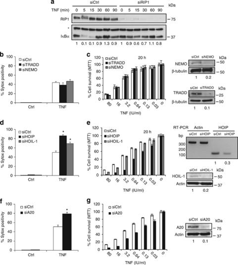
In contrast to the situation observed in most cell types, TNFR1 signaling in the mouse L929 fibrosarcoma cell line activates NF-κ_B and also induces cell death (necroptosis), in the absence of synthetic caspase or protein synthesis inhibitors. 16 Using this cellular system, we previously reported that depletion of cIAPs resulted in necrosome complex formation and strongly sensitized the cells to TNF-induced necroptosis.26 Interestingly, we now provide data showing that repression of RIP1 levels by RNAi almost completely inhibits TNF-mediated I_κ_B_α degradation in L929 cells (Figure 1a), indicating that RIP1 is a major regulator of TNF-induced NF-_κ_B activation in these cells. Within TNFR1-CI, cIAP-mediated RIP1 ubiquitination is important to prevent RIP1 from integrating into death-inducing TNFR1-CII.5, 9 In addition, the conjugation of non-degradative ubiquitin chains to RIP1 is important for NF-_κ_B activation, another cytoprotective event. To explore the contribution of each process to cell death prevention, we analyzed the effect of depleting proteins reported to be involved in the regulation of each cytoprotective process on TNF-induced necroptosis. TRADD,28, 29 NEMO30 and LUBAC (HOIL-1 and HOIP)4, 31 were all reported to be important positive regulators of TNF-mediated NF-κ_B activation and we found that repressing each of these proteins by RNAi led to an approximately similar inhibition of I_κ_B_α degradation upon TNF stimulation in L929 cells (Supplementary Figure 1a). Interestingly, silencing of NEMO and TRADD had no impact on TNF-mediated necroptosis (Figures 1b and c). In contrast, repression of the LUBAC component HOIL-1 or HOIP enhanced TNF-induced cell death (Figure 1d), indicating that the sensitization to death upon LUBAC knockdown is independent of NF-_κ_B activation. However, it should be noted that knockdown of HOIP is more potent in promoting TNF-induced cell death than silencing of HOIL-1 (Figure 1e). To determine the cell death type in these sensitized conditions upon knockdown of HOIL-1 or HOIP, we stably transfected a Förster Resonance Energy Transfer (FRET)-based apoptosis sensor construct into L929 cells to detect caspase-3 activity, hereafter referred to as L929pCasper3-BG cells. In these cells, silencing of HOIL-1 or HOIP increased the amount of cell death (Supplementary Figure 2a) without any caspase-3 activity (Supplementary Figure 2b), indicating that the enhanced cell death type was not due to a shift to apoptosis. CYLD is a RIP1-deubiquitinating enzyme,11 and we and others previously showed that CYLD repression protects L929 cells from TNF-induced necroptosis,26, 27 consistent with the cytoprotective role of non-degradative ubiquitination of RIP1. A20 is another RIP1-deubiquitinating enzyme reported to negatively regulate TNF-induced NF-_κ_B activation.32 Repression of A20 was therefore expected to protect cells from TNF-induced necroptosis. Instead, we were surprised to find that A20 depletion had an opposite effect and greatly sensitized the cells to death (Figures 1f and g). Performing this experiment in L929pCasper3-BG cells demonstrated that A20 silencing enhances cell death (Supplementary Figure 2c) in the absence of any caspase-3 activity upon TNF stimulation (Supplementary Figure 2d). It is important to note that this sensitization is also NF-_κ_B independent, because A20 depletion did not inhibit early NF-_κ_B activation (Supplementary Figure 1b). Together, these results indicate that NF-_κ_B activation does not have a major protective role against TNF-induced necroptosis in L929 cells. In addition, we identified the LUBAC components HOIL-1 and HOIP, as well as the deubiquitinase A20, as negative regulators of TNF-mediated necroptosis.
Figure 1.
Knockdown of HOIL-1, HOIP or A20 sensitizes to TNF-induced necroptosis, whereas depletion of TRADD or NEMO does not affect the cell viability. (a) Upon knockdown of RIP1, L929 cells were stimulated with TNF for the indicated times. I_κ_B_α_ degradation and RIP1 knockdown efficiency was analyzed using western blotting. Asterisk indicates an nonspecific band. Flow cytometric analysis of the percentage of Sytox-positive cells after TNF stimulation for 4 h (reaching around 50% cell death in control setup) of L929 cells in which TRADD or NEMO (b), HOIL-1 or HOIP (d), or A20 was knocked down (f). Error bars indicate S.D.s of triplicates of three independent experiments. Cell survival analysis using a MTT assay after 20 h of TNF stimulation of L929 cells in which TRADD or NEMO (c), HOIL-1 or HOIP (e), or A20 was knocked down (g). Error bars indicate S.Ds of triplicates. Knockdown efficiency of the indicated proteins was determined by western blotting or RT-PCR. *P<0.05 (Mann–Whitney _U_-test)
Knockdown of FADD, caspase-8 or c-FLIP sensitizes to TNF-induced necroptosis
Upon TNF stimulation, deubiquitination of RIP1 allows formation of the cytosolic death-inducing TNFR1-CII.3.This complex, which consists of at least TRADD, FADD, caspase-8, RIP1 and RIP3, allows the induction of apoptosis or necroptosis, depending on the cellular context.20 To test the requirement for these proteins in TNF-induced necroptosis in L929 cells, we targeted them by RNAi. Knockdown of FADD or caspase-8 strongly enhanced TNF-induced cell death as a function of time (Figures 2a and b), and sensitized TNF-induced cell death by 30-fold based on IC50 values (Figures 2a and c). In L929pCasper3-BG cells, knockdown of caspase-8 enhanced the amount of cell death (Supplementary Figure 3a) upon TNF stimulation without showing any caspase-3 activity (Supplementary Figure 3b). In addition, the increased amount of cell death following caspase-8 knockdown and TNF treatment was strongly reduced in the presence of the RIP1 kinase inhibitor necrostatin-1 (Nec-1) (Supplementary Figure 3a). Together, these data demonstrate that silencing of caspase-8 sensitizes L929 cells to TNF-induced necroptosis and are in agreement with other studies reporting the negative regulatory role of caspase-8 in necroptosis in Jurkat and primary T cells33 and in benzyloxycarbonyl-Val-Ala-Asp(OMe)-fluoromethylketone (zVAD)-fmk-induced RIP3-dependent cell death in L929 cells.17 Interestingly, downregulation of c-FLIP, a regulator of caspase-8 activation,17 also strongly enhanced (Figure 2b) and sensitized cell death by 10-fold on TNF stimulation (Figure 2c). We reasoned that c-FLIP downregulation would result in increased formation of active caspase-8 dimers within TNFR1-CII, and would induce a switch to apoptotic cell death. However, the use of the FRET-based apoptosis sensor revealed that the L929p Casper3-BG cells depleted of c-FLIP still died in a caspase-independent way when exposed to TNF (Supplementary Figures 4a and b), thereby confirming the study of Oberst et al.17 showing that in L929 cells, c-FLIP silencing promotes zVAD-fmk-induced cell death that is RIP3-dependent. Live-cell imaging of TNF-treated cells confirmed the necrotic morphology (Supplementary Figure 4c) of these sensitized cells. In contrast, our control, consisting of Fas receptor-mediated apoptosis, showed increased apoptosis upon knockdown of c-FLIP (Supplementary Figures 4a and b). Together with the previous results, these data indicate that the adaptor proteins TRADD and FADD apparently have different roles in TNFR1-induced necroptosis. Whereas TRADD is dispensable for TNF-induced necroptosis, the key molecules of the apoptotic axis, FADD, caspase-8 and c-FLIP, counteract TNF-induced necroptosis.
Figure 2.
FADD, caspase-8 or c-FLIP knockdown sensitizes to TNF-induced necroptosis. (a) Flow cytometric analysis of the percentage of Sytox-positive cells after 3 h of TNF stimulation (reaching around 25% cell death in control setup) and cell survival analysis using a MTT assay after 20 h of TNF stimulation of L929 cells in which FADD was knocked down. (b) Flow cytometric analysis of the percentage of Sytox-positive cells after 3 h of TNF stimulation of L929 cells in which caspase-8 or c-FLIP was knocked down. Error bars indicate S.D.s of triplicates of at least two independent experiments. (c) Cell survival analysis using a MTT assay after 20 h of TNF stimulation of L929 cells in which caspase-8 or c-FLIP were knocked down. Error bars indicate S.Ds of triplicates and data are representative of at least three independent experiments. Knockdown efficiency of the indicated proteins was determined by western blotting or RT-PCR
Knockdown of RIP1 but not of RIP3 switches TNF-induced TRADD-independent necroptosis to TRADD-dependent apoptosis
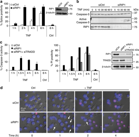
RIP1 and RIP3 kinases have a crucial role in TNF-induced necroptosis.12, 15, 23, 34 Previous reports showed that silencing RIP3 expression rescued L929 cells from TNF-induced necroptosis.12, 23, 26, 34 Surprisingly, RIP1 knockdown did not protect L929 cells from TNF-induced cell death (Figure 3a). Because RIP1 depletion does block TNF-induced necroptosis in the presence of the pan-caspase inhibitor zVAD-fmk in L929 cells,12, 35 we reasoned that RIP1 depletion may have resulted in a shift to apoptosis. This hypothesis is underscored by our previous finding that chemical inhibition of heat shock protein 90, which results in degradation of its client proteins, such as RIP1,36 resulted in a shift from TNF-induced necroptosis to apoptosis.37 Use of the FRET-based apoptosis sensor demonstrated that lowering RIP1 levels by RNAi indeed resulted in rapid induction of apoptosis in response to TNF stimulation (Figure 3c), as also revealed by proteolytic activation of caspase-3 (Figure 3b) and apoptotic blebbing (Figure 3d). Because TRADD and RIP1 compete for TNFR1 binding,38 we reasoned that TRADD contributes to this switch to apoptosis upon RIP1 loss. Indeed, knockdown of TRADD blocked TNF-induced caspase activity in the absence of RIP1 and strongly reduced cell death (Figure 3c). In summary, these data show that RIP1 depletion in L929 cells results in a shift from TRADD-independent necroptosis (Figure 1b) to TRADD-dependent apoptosis in response to TNF.
Figure 3.
RIP1 knockdown shifts TNF-induced TRADD-independent necroptosis to TRADD-dependent apoptosis. (a) Flow cytometric analysis of the percentage of Sytox-positive cells at the indicated time points after TNF stimulation of L929 cells in which RIP1 is knocked down. Knockdown efficiency was determined by western blotting. (b) Western blot analysis of caspase-3 processing at the indicated time points after TNF stimulation of L929 cells in which RIP1 is knocked down. (c) Flow cytometric analysis of the percentage of caspase-3 activity and PI-positive cells at the indicated time points after TNF stimulation of L929pCasper3-BG cells in which RIP1 or RIP1+TRADD are knocked down. Knockdown efficiency of the indicated proteins was determined by western blotting. (d) Cells were stained with Hoechst 33242 (blue) and PI (red) for live-cell imaging and monitored for 8 h. Overlay snapshots of the fluorescent and transmitted light images at the indicated time points after TNF stimulation of L929 cells in which RIP1 is knocked down. Error bars indicate S.Ds of triplicates and data are representative of at least three independent experiments.
RIP3-dependent necroptosis upon knockdown of RIP1 and caspase-8
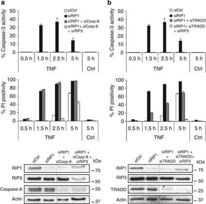
Using a similar approach as above, we found that RIP1 and caspase-8 double knockdown completely inhibits caspase-mediated apoptosis on TNF stimulation without affecting the extent of cell death (Figure 4a), indicating a switch to a RIP1-independent type of necrosis. A triple knockdown revealed that silencing RIP1, caspase-8 and RIP3 resulted in complete protection, demonstrating that the absence of RIP1 and caspase-8 leads to a strong bias toward RIP3-dependent necrosis (Figure 4a). Similarly, TNF-induced necrosis of cells in which both RIP1 and TRADD were knocked down was completely rescued when RIP3 was also silenced (Figure 4b). We conclude that knockdown of caspase-8 in RIP1-deficient conditions clearly prevents TNF-induced apoptosis but still allows a regulated form of necrosis, which depends solely on RIP3. These results indicate that under particular conditions, TNF-mediated RIP3-dependent necrosis can occur in the absence of RIP1.
Figure 4.
RIP3-dependent necroptosis when RIP1 and caspase-8 or TRADD are knocked down. (a) Flow cytometric analysis of the percentage of caspase-3 activity and PI-positive cells at the indicated time points after TNF stimulation of L929pCasper3-BG (FRET-based apoptosis sensor) cells in which RIP1, RIP1+caspase-8, or RIP1+caspase-8+RIP3 are knocked down. (b) Flow cytometric analysis of the percentage of caspase-3 activity and PI-positive cells at the indicated time points after TNF stimulation of L929pCasper3-BG cells in which RIP1, RIP1+TRADD and RIP1+TRADD+RIP3 are knocked down. Error bars indicate S.Ds of triplicates and data are representative of at least two independent experiments. Knockdown efficiency of the indicated proteins was determined by western blotting
Discussion
TNF-induced necroptosis is a regulated form of cell death involving the kinase activity of RIP1 and RIP3 in its initiation phase.20 However, still little is known about the regulatory mechanisms that modulate sensitivity to TNF-induced necroptosis. In this study, we examined the impact of repressing proteins known to be recruited to TNFR1-CI and/or TNFR1-CII on the decision of L929 cells to die in a necroptotic way.
Within TNFR1-CI, cIAP1/2 conjugates RIP1 with non-degradative ubiquitin chains,5 and this event has been reported to prevent RIP1 from integrating in complex II.5, 9 Our initial observation that cIAP1 protects L929 cells from TNF-induced necroptosis suggested that RIP1 ubiquitination also inhibits necrosome formation.26 The finding that RIP1 is a crucial mediator of TNF-induced NF-_κ_B activation in L929 cells then questioned the relative contribution of NF-κ_B activation to the cytoprotective mechanism mediated by ubiquitinated RIP1. Silencing of NEMO or TRADD did not sensitize to TNF-induced necroptosis, whereas knockdown of the LUBAC component HOIP and to lesser extent HOIL-1 enhanced TNF-induced necroptosis, although all repressions resulted in approximately similar reductions of I_κ_B_α degradation in response to TNF. Together, these results indicate that NF-_κ_B activation does not have a major cytoprotective role in this cellular system and revealed a negative regulatory function of LUBAC on TNF-induced necroptosis. Recruitment of LUBAC to TNFR1-CI has been reported to stabilize the complex.4 It is therefore conceivable that repressing LUBAC components destabilizes TNFR1-CI and facilitates necrosome formation.
The observation that CYLD knockdown rescues L929 cells from TNF-induced necroptosis26, 27 suggests that RIP1 deubiquitination is required for the initiation of necroptosis. Beside CYLD,11 RIP1 is also deubiquitinated by A20 in the TNF signaling pathway.32 However, in contrast to the suppression of necroptosis by CYLD knockdown, A20 silencing elevated the sensitivity to TNF-induced necroptotic cell death. Thus, although both CYLD and A20 are RIP1 lysine 63-deubiquitinating enzymes, they seem to act differently during TNF-induced necroptosis. The sensitization to necroptosis by A20 depletion agrees with the observation that A20 overexpression inhibits TNF-induced necroptosis in L929 cells,39 as well as the TNF-induced caspase-independent cell death of macrophages.40 In contrast to CYLD, A20 has been reported to possess E3 ubiquitin ligase activity in addition to its deubiquitinase properties.32 It is therefore possible that either the deubiquitinase activity or the E3 ubiquitin ligase activity of A20 inhibits or activates the pro-death or pro-survival potential, respectively, of components of TNFR1-CI, but also of TNFR1-CII. It is also important to note that the cytotoxic role of CYLD in necroptosis induction might not be at the level of RIP1 or not only at the level of RIP1. Indeed, we found that CYLD repression also protects L929 cells from hydrogen peroxide-induced necrosis (Supplementary Figure 5), which occurs independently of RIP1 and RIP3.12, 35
TNFR1-CII is formed downstream of TNFR1-CI. This cytosolic death-inducing complex is composed of RIP1, RIP3, FADD, TRADD and caspase-8. Whether TRADD is strictly required in TNF-induced necroptosis signaling is controversial. In Jurkat T cells, TRADD depletion had no effect on TNF-induced necroptosis,38 whereas TRADD-deficient mouse embryonic fibroblast (MEF) cells, but not macrophages, are protected against necroptosis induced by TNF in the presence of CHX and zVAD-fmk.28 In line with the Jurkat data, which do not need CHX for necroptosis induction, we found that TRADD depletion in L929 cells did not affect TNF-induced necroptosis. However, in RIP1-knockdown conditions, we observed that TRADD is important for TNF-mediated apoptosis, suggesting that the role of TRADD in cell death induction depends on the cellular condition.
Several independent reports have shown that RIP1 depletion blocks TNF-induced necroptosis in the presence of the caspase inhibitor zVAD-fmk,12, 35 but fails to do so in the absence of zVAD-fmk.27 We found that in L929 cells knockdown of RIP1, but not RIP3, even potentiates TNF-induced cell death and shifts it from necroptosis to apoptosis (Figure 5), which most probably explains why RIP1 depletion in the absence of zVAD-fmk does not block cell death in L929 cells.27 RIP1 knockout mice die shortly after birth due to massive apoptosis, which was initially explained by the absence of NF-_κ_B activation and concomitant reduced survival signaling.41 The exclusive link between RIP1 and NF-_κ_B activation has now been challenged because several cell types from RIP1-deficient embryos still activated NF-_κ_B upon TNF stimulation.8 We observed in L929 cells a switch to apoptosis even when RIP1 knockdown was less efficient and did not affect NF-_κ_B activation (data not shown), indicating that RIP1 may fulfill an anti-apoptotic role independently of its NF-_κ_B-modulating activity. RIP1-mediated suppression of apoptosis is not due to phosphorylation of the pro-apoptotic proteins FADD and caspase-8 because inhibition of RIP1 kinase activity by Nec-1 did not induce apoptosis upon TNF stimulation.26, 35 RIP1 and TRADD compete for TNFR1 binding.2, 38 Therefore, it is conceivable that loss of RIP1 enhances the recruitment of TRADD, which has been reported to favor the induction of apoptosis.38 Additionally, RIP1 deficiency was shown to result in depletion of c-FLIP,42 which is also an apoptosis sensitizing condition. Taken together, both enhanced TRADD recruitment and decreased levels of c-FLIP may account for the shift from necroptosis to apoptosis in response to TNF.
Figure 5.
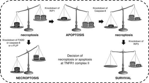
Summary of the RNAi study of TNF-induced necroptosis in L929 cells. At TNFR1 complex II, the apoptotic proteins FADD, caspase-8 and c-FLIP suppress TNF-induced necroptosis. RIP1 knockdown shifts TNF-induced TRADD-independent necroptosis to TRADD-dependent apoptosis. The combined knockdown of RIP1 and caspase-8 blocks apoptosis but does not affect the extent of cell death. This RIP1-independent necroptotic cell death can be blocked by deleting RIP3 as well.
We observed that combined knockdown of RIP1 and TRADD completely blocks TNF-induced apoptosis, but allows, to some extent, RIP3-dependent induction of necrotic cell death in L929 cells. Simultaneous knockdown of RIP1 and caspase-8 also rescues from apoptotic cell death, but still allows full blown induction of cell death, which can be inhibited by RIP3 silencing. This indicates that when both RIP1 and caspase-8 are absent, TRADD probably recruits FADD/RIP3 because these proteins interact constitutively.12 Although it was shown that the TNF-induced assembly of the necrosome requires RIP1 kinase activity,12, 34 our results provide a model in which necrosome formation and subsequent necroptosis occur in the absence of RIP1. This implies that certain cellular conditions may bypass the need for RIP1 in necrosome assembly and RIP3 activation. Indeed, a similar RIP1-independent but RIP3-dependent necroptosis upon ectopic expression of RIP3 has been described in RIP1-deficient MEF cells23 and more recently during viral infection.24
This RNAi study in L929 cells demonstrates how knockdown of several TNF-signaling proteins involved in TNFR1-CI and II strongly modulate TNF-induced necroptosis (Table 1). Considering the similarity between the observations in L929 cells and other cellular systems related to TNF-induced signaling,12, 15, 22, 23, 34 and the confirmation thereof in an in vivo context,17, 18, 23 we consider L929 cells as a good in vitro cellular system to study molecular cell death signaling in response to TNF. These results provide a lead for testing the role of these proteins in experimental in vivo models of diseases in which necroptosis is presumed to have a role. This is the first report (i) illustrating a cytoprotective role for A20 and LUBAC in TNF-induced necroptosis, (ii) explaining why RIP1 depletion in the absence of zVAD-fmk does not block cell death, due to a switch toward apoptosis and (iii) showing RIP3-dependent necroptosis induction in the absence of RIP1 in the context of TNF signaling. Finally, our results could have important therapeutic implications for strategies aiming to sensitize necroptosis to bypass the resistance of tumor cells to apoptosis or to induce a more immunogenic form of cell death. Desensitizing necroptosis could be therapeutically useful in degenerative diseases such as ischemia-reperfusion damage during heart failure, stroke, diabetes type II and organ transplantation.20
Table 1. Overview of knockdowns for TNF-induced necroptosis in L929 cells.
| RNAi target | Role in TNF-induced necroptosis |
|---|---|
| Complex I | |
| cIAP1 | Anti-necroptotic |
| HOIL-1 HOIP | |
| TAK1 NEMO | No apparent role or redundancy |
| I and II | |
| TRADD | No apparent role or redundancy |
| RIP1 | Pro-necroptotic and anti-apoptotic |
| Complex II | |
| FADD caspase-8 c-FLIP | Anti-necroptotic |
| RIP3 | Pro-necroptotic |
| DUB | |
| A20 | Anti-necroptotic |
| CYLD | Pro-necroptotic |
Materials and Methods
Materials
We used TNF and Fas antibody to stimulate cell death. Recombinant human TNF was produced and purified in our laboratory to at least 99% homogeneity, and its specific biological activity was 3 × 10⁷ IU/mg. We used human TNF because it is a specific agonist for murine TNFR1 and does not act on TNFR2. Anti-human Fas antibody clone 2R2 was from Cell Diagnostica, Munster, Germany. H2O2 30% and 3-(4,5-dimethylthiazol-2-yl)-2,5-diphenyltetrazolium bromide (MTT) (Sigma Aldrich, St. Louis, MO, USA) was used at 2 mM and 500 _μ_g/ml, respectively. Loss of plasma membrane permeability was monitored using 5 nM Sytox Red Dead Cell Stain (Molecular Probes–Invitrogen, Eugene, OR, USA) and 3 _μ_M propidium iodide (PI, Sigma Aldrich). Nuclei were stained using Hoechst 33242 (Molecular Probes–Invitrogen). The following antibodies were used for western blot analysis: anti-HOIL-1 antibody (gift from H Walczak); anti-_β_-tubulin (HRP) (Ab21058, Abcam, Cambridge, UK); anti-β_-actin (Clone C4, MP Biomedicals Europe NV, Illkirch, France); anti-caspase-8 (1G12, Alexis, Enzo Life Sciences, Zandhoven, Belgium); anti-RIP1 (610459, BD Biosciences, Franklin Lakes, NJ, USA); anti-RIP3 (R4277, Sigma Aldrich); rabbit polyclonal antibody against full-length murine caspase-3 (made in-house); cleaved caspase-3 antibody (9661; Cell Signaling Technology, Danvers, MA, USA). Antibodies to A20 (A-12), FADD (M-19), I_κ_B_α (C-21), NEMO (FL-419) and TRADD (H-278) were purchased from Santa-Cruz Biotechnology (Santa Cruz, CA, USA).
Cell lines
L929sAhFas cells were produced by expressing the human Fas gene in L929sA cells, a TNF-sensitive derivative of the murine fibrosarcoma cell line L929.16.These cells are referred to as L929 cells and were cultured in Dulbecco's modified Eagle's medium supplemented with 10% fetal calf serum, penicillin (100 IU/ml), streptomycin (0.1 mg/ml), and -glutamine (0.03%). L929pCasper3-BG cells were made by transfecting the pCasper3-BG vector (FP970; Evrogen, Moscow, Russia) into L929sAhFas cells by the calcium phosphate method. After transfection, cells were sorted four times using the Beckman Coulter Altra flow cytometer (Beckman Coulter, Brea, CA, USA) to obtain L929pCasper3-BG cells with strong expression of pCasper3-BG.
Analysis of cell survival, cell death and caspase-3 activity
L929 or L929pCasper3-BG cells were pretreated with Nec-1 for 1 h and then stimulated with TNF (10 000 IU/ml) or anti-Fas (125 ng/ml) for the indicated periods. Death of L929 cells was analyzed by flow cytometry on a dual-laser FACSCalibur (488 nm, 635 nm) with Cellquest software (BD Biosciences). Simultaneous analysis of cell death and caspase-3 activity in L929pCasper3-BG cells was performed using a triple-laser LSR-II (405, 488 and 635 nm) with FACSDiva software (BD Biosciences). Cell death or loss of plasma membrane integrity was determined by measuring Sytox Red (Molecular Probes–Invitrogen, Eugene, OR, USA) or propidium iodide (PI)-emitted fluorescence. Caspase-3 activity was determined by Casper3-BG, a FRET-based apoptosis sensor consisting of the blue fluorescent protein TagBFP connected by a DEVD sequence with a green fluorescent protein, TagGFP2. During apoptosis, cleavage of DEVD by caspase-3 eliminates FRET and increases the TagBFP:TagGFP2 ratio. Only viable cells (PI negative) were gated for analysis of caspase-3 activity. All experiments were performed at least twice in triplicate. Cell survival was determined by a MTT assay. After 20 h of TNF stimulation, MTT was added at 500 _μ_g/ml for 4 h, followed by lysis in 10% SDS/ 0.1 M HCl. Absorbance was measured using a standard microplate reader with a 595-nm filter and a 655-nm reference filter.
RNAi-mediated knockdown
L929 and L929pCasper3-BG cells were transfected in six-well plates according to the manufacturer's protocol using 20 nM siRNA targeting A20, caspase-8, c-FLIP, HOIL-1, HOIP, NEMO, RIP1, RIP3 and TRADD (mixture of four siRNAs, ON-TARGET_plus_ SMART pool siRNA, Dharmacon, Thermo Fisher Scientific, Waltham, MA, USA). As a negative control, we used siCONTROL non-targeting siRNA (ON-TARGET_plus_ SMART pool siRNA, Dharmacon). INTERFERin (Polyplus-transfection SA, Illkirch, France) was used as a transfection reagent. Knockdown of FADD was performed by retroviral infection with a pRetroSuper construct (DNAengine Inc., Seattle, WA, USA) containing the sense-strand sequence 5′-CAGACTTTCAGACTCTCCCTT-3′. A scrambled pRetroSuper shRNA was used as a negative control. First, shRNAs were introduced in QNX-A cells (Nolan Lab, Stanford, CA, USA) by calcium phosphate transfection. After 24 and 48 h, viral supernatant of QNX-A cells was harvested, polybrene (Sigma Aldrich) was added (16 _μ_g/ml) and viral supernatant was used to infect L929 cells by centrifugation at 1500 r.p.m. at 32 °C for 90 min. After 72 or 96 h of knockdown by siRNA or shRNA, cells were stimulated with TNF and cell death was determined as described above. Knockdown efficiency was checked by western blot or RT-PCR.
RT-PCR
RNA was prepared from L929 cells using the RNeasy Plus Mini Kit (Qiagen, Hilden, Germany). Starting with 2 _μ_g RNA, cDNA was synthesized using the SuperScript Reverse Transcriptase III kit (Invitrogen, Carlsbad, CA, USA). PCR was performed using GoTaq Green Mastermix (Promega, Madison, WI, USA). PCR products were separated on 2% agarose gel and visualized by SYBR Safe DNA gel stain (Molecular Probes–Invitrogen). The following primers were used: c-FLIPL: Fwd 5′- CCTCCAGCTCATCCTCTGTG-3′ and Rev 5′-TTTGTCCATGAGTTCAACGTG-3′ c-FLIPR: Fwd 5′- GCTGCTGTGGTTCTGAACATG-3′ and Rev 5′-GGTACTCCATACACTGGCTCC-3′ HOIP: Fwd 5′- GCTTCATCTATGAACGCGAAC-3′ and Rev 5′- ATTGGTGCGTTTCCAGTTCT-3′.
Microscopy
Time-lapse microscopy of L929 cells grown in eight-well Lab-Tek chambers (Nunc, VWR international, Heverlee, Belgium) was carried out using a Leica AS MDW live cell imaging system (Leica Microsystems, Mannheim, Germany). This system includes a DM IRE2 microscope (Leica Microsystems, Mannheim, Germany) equipped with an HCX PL APO 63 × /1.3 glycerin corrected 37 °C objective and a 12-bit Coolsnap HQCamera. The microscope is surrounded by an incubation chamber, which maintains the cells at 37 °C in a 5% CO2 environment. Cell morphology was observed by using differential interference contrast (DIC). Cells were monitored for 8 h, and multiple image z-stacks were captured every 5 min. Within the z-stack, different focal planes were set at 1-_μ_m intervals to prevent loss of focus during the process of cell death. Hoechst 33342 was excited at 380 nm and emission was detected using a BP340/80/FT400/LP425 filter cube. PI was excited at 540 nm and the emission was detected using a BP515-560/FT580/LP590 filter. From each image stack, maximum intensity projections (for Hoechst and PI) and autofocus images (for DIC) were made for each time point by using a script developed in-house for Fiji public domain imaging software (http://fiji.sc/wiki/index.php/Fiji).
Acknowledgments
We apologize for not referring to all relevant publications because of reference limit. We thank Dr. A Bredan for editing, Dr. C Guérin, E Parthoens and S Noppen of the DMBR Microscopy Core Facility for imaging and L Duprez for critical feedback. We are grateful to Professor Dr. H Walczak for providing the HOIL-1 antibody. TVB and MJMB hold a postdoctoral fellowship from the FWO, PB is paid by VIB, and NV obtained a predoctoral fellowship from the BOF, Ghent University, and has been paid by the Methusalem grant. Research in the Vandenabeele group is funded by the European grants (FP6 ApopTrain, MRTN-CT-035624; FP7 EC RTD Integrated Project, Apo-Sys, FP7-200767; Euregional PACT II), the Belgian grants (Interuniversity Attraction Poles, IAP 6/18), the Flemish grants (Research Foundation Flanders, FWO G.0875.11 and FWO G.0973.11), the Ghent University grants (MRP, GROUP-ID consortium) and grants from the Flanders Institute for Biotechnology (VIB). PV holds a Methusalem grant (BOF09/01M00709) from the Flemish Government. MB has a tenure track position with the Multidisciplinary Research Program from the Ghent University (GROUP-ID).
Glossary
c-FLIP
cellular FLICE inhibitory protein
CHX
cycloheximide
cIAP1
cellular inhibitor of apoptosis protein
CYLD
cylindromatosis
FADD
Fas-associated protein via a death domain
FRET
Förster Resonance Energy Transfer
HOIP
HOIL-1-interacting protein
HOIL-1
heme-oxidized iron regulatory protein 2 ubiquitin ligase-1
IKK
inhibitor of NF-_κ_B kinase
I_κ_B
inhibitor of NF-_κ_B
LUBAC
linear ubiquitin chain assembly complex
MEF
mouse embryonic fibroblast
Nec-1
necrostatin-1
NEMO
NF-_κ_B essential modulator
PI
propidium iodide
RIP
receptor-interacting protein
TAK1
transforming growth factor-_β_-activated kinase 1
TNF
tumor necrosis factor
TNFR
tumor necrosis factor receptor
TRADD
TNF receptor-associated death domain
TRAF2
TNF receptor-associated factor 2
zVAD-fmk
benzyloxycarbonyl-Val-Ala-Asp(OMe)-fluoromethylketone
The authors declare no conflict of interest.
Footnotes
Supplementary Information accompanies the paper on Cell Death and Disease website (http://www.nature.com/cddis)
Edited by P Salomoni
Supplementary Material
Supplementary Figure 1
Supplementary Figure 2
Supplementary Figure 3
Supplementary Figure 4
Supplementary Figure 5
Supplementary Figure Legends
References
- Van Antwerp DJ, Martin SJ, Kafri T, Green DR, Verma IM. Suppression of TNF-alpha-induced apoptosis by NF-kappaB. Science. 1996;274:787–789. doi: 10.1126/science.274.5288.787. [DOI] [PubMed] [Google Scholar]
- Harper N, Hughes M, MacFarlane M, Cohen GM. Fas-associated death domain protein and caspase-8 are not recruited to the tumor necrosis factor receptor 1 signaling complex during tumor necrosis factor-induced apoptosis. J Biol Chem. 2003;278:25534–25541. doi: 10.1074/jbc.M303399200. [DOI] [PubMed] [Google Scholar]
- Micheau O, Tschopp J. Induction of TNF receptor I-mediated apoptosis via two sequential signaling complexes. Cell. 2003;114:181–190. doi: 10.1016/s0092-8674(03)00521-x. [DOI] [PubMed] [Google Scholar]
- Haas TL, Emmerich CH, Gerlach B, Schmukle AC, Cordier SM, Rieser E, et al. Recruitment of the linear ubiquitin chain assembly complex stabilizes the TNF-R1 signaling complex and is required for TNF-mediated gene induction. Mol Cell. 2009;36:831–844. doi: 10.1016/j.molcel.2009.10.013. [DOI] [PubMed] [Google Scholar]
- Bertrand MJ, Milutinovic S, Dickson KM, Ho WC, Boudreault A, Durkin J, et al. cIAP1 and cIAP2 facilitate cancer cell survival by functioning as E3 ligases that promote RIP1 ubiquitination. Mol Cell. 2008;30:689–700. doi: 10.1016/j.molcel.2008.05.014. [DOI] [PubMed] [Google Scholar]
- Ea CK, Deng L, Xia ZP, Pineda G, Chen ZJ. Activation of IKK by TNFalpha requires site-specific ubiquitination of RIP1 and polyubiquitin binding by NEMO. Mol Cell. 2006;22:245–257. doi: 10.1016/j.molcel.2006.03.026. [DOI] [PubMed] [Google Scholar]
- Wu CJ, Conze DB, Li T, Srinivasula SM, Ashwell JD. Sensing of Lys 63-linked polyubiquitination by NEMO is a key event in NF-kappaB activation [corrected] Nat Cell Biol. 2006;8:398–406. doi: 10.1038/ncb1384. [DOI] [PubMed] [Google Scholar]
- Wong WW, Gentle IE, Nachbur U, Anderton H, Vaux DL, Silke J. RIPK1 is not essential for TNFR1-induced activation of NF-kappaB. Cell Death Differ. 2010;17:482–487. doi: 10.1038/cdd.2009.178. [DOI] [PubMed] [Google Scholar]
- O′Donnell MA, Legarda-Addison D, Skountzos P, Yeh WC, Ting AT. Ubiquitination of RIP1 regulates an NF-kappaB-independent cell-death switch in TNF signaling. Curr Biol. 2007;17:418–424. doi: 10.1016/j.cub.2007.01.027. [DOI] [PMC free article] [PubMed] [Google Scholar]
- Wang L, Du F, Wang X. TNF-alpha induces two distinct caspase-8 activation pathways. Cell. 2008;133:693–703. doi: 10.1016/j.cell.2008.03.036. [DOI] [PubMed] [Google Scholar]
- Wright A, Reiley WW, Chang M, Jin W, Lee AJ, Zhang M, et al. Regulation of early wave of germ cell apoptosis and spermatogenesis by deubiquitinating enzyme CYLD. Dev Cell. 2007;13:705–716. doi: 10.1016/j.devcel.2007.09.007. [DOI] [PubMed] [Google Scholar]
- Cho YS, Challa S, Moquin D, Genga R, Ray TD, Guildford M, et al. Phosphorylation-driven assembly of the RIP1-RIP3 complex regulates programmed necrosis and virus-induced inflammation. Cell. 2009;137:1112–1123. doi: 10.1016/j.cell.2009.05.037. [DOI] [PMC free article] [PubMed] [Google Scholar]
- Feng S, Yang Y, Mei Y, Ma L, Zhu DE, Hoti N, et al. Cleavage of RIP3 inactivates its caspase-independent apoptosis pathway by removal of kinase domain. Cell Signal. 2007;19:2056–2067. doi: 10.1016/j.cellsig.2007.05.016. [DOI] [PubMed] [Google Scholar]
- Irmler M, Thome M, Hahne M, Schneider P, Hofmann K, Steiner V, et al. Inhibition of death receptor signals by cellular FLIP. Nature. 1997;388:190–195. doi: 10.1038/40657. [DOI] [PubMed] [Google Scholar]
- Holler N, Zaru R, Micheau O, Thome M, Attinger A, Valitutti S, et al. Fas triggers an alternative, caspase-8-independent cell death pathway using the kinase RIP as effector molecule. Nat Immunol. 2000;1:489–495. doi: 10.1038/82732. [DOI] [PubMed] [Google Scholar]
- Vercammen D, Beyaert R, Denecker G, Goossens V, Van Loo G, Declercq W, et al. Inhibition of caspases increases the sensitivity of L929 cells to necrosis mediated by tumor necrosis factor. J Exp Med. 1998;187:1477–1485. doi: 10.1084/jem.187.9.1477. [DOI] [PMC free article] [PubMed] [Google Scholar]
- Oberst A, Dillon CP, Weinlich R, McCormick LL, Fitzgerald P, Pop C, et al. Catalytic activity of the caspase-8-FLIP(L) complex inhibits RIPK3-dependent necrosis. Nature. 2011;471:363–367. doi: 10.1038/nature09852. [DOI] [PMC free article] [PubMed] [Google Scholar]
- Kaiser WJ, Upton JW, Long AB, Livingston-Rosanoff D, Daley-Bauer LP, Hakem R, et al. RIP3 mediates the embryonic lethality of caspase-8-deficient mice. Nature. 2011;471:368–372. doi: 10.1038/nature09857. [DOI] [PMC free article] [PubMed] [Google Scholar]
- Zhang H, Zhou X, McQuade T, Li J, Chan FK, Zhang J. Functional complementation between FADD and RIP1 in embryos and lymphocytes. Nature. 2011;471:373–376. doi: 10.1038/nature09878. [DOI] [PMC free article] [PubMed] [Google Scholar]
- Vandenabeele P, Galluzzi L, Vanden Berghe T, Kroemer G. Molecular mechanisms of necroptosis: an ordered cellular explosion. Nat Rev Mol Cell Biol. 2010;11:700–714. doi: 10.1038/nrm2970. [DOI] [PubMed] [Google Scholar]
- Degterev A, Huang Z, Boyce M, Li Y, Jagtap P, Mizushima N, et al. Chemical inhibitor of nonapoptotic cell death with therapeutic potential for ischemic brain injury. Nat Chem Biol. 2005;1:112–119. doi: 10.1038/nchembio711. [DOI] [PubMed] [Google Scholar]
- Degterev A, Hitomi J, Germscheid M, Ch′en IL, Korkina O, Teng X, et al. Identification of RIP1 kinase as a specific cellular target of necrostatins. Nat Chem Biol. 2008;4:313–321. doi: 10.1038/nchembio.83. [DOI] [PMC free article] [PubMed] [Google Scholar]
- Zhang DW, Shao J, Lin J, Zhang N, Lu BJ, Lin SC, et al. RIP3, an energy metabolism regulator that switches TNF-induced cell death from apoptosis to necrosis. Science. 2009;325:332–336. doi: 10.1126/science.1172308. [DOI] [PubMed] [Google Scholar]
- Upton JW, Kaiser WJ, Mocarski ES. Virus inhibition of RIP3-dependent necrosis. Cell Host Microbe. 2010;7:302–313. doi: 10.1016/j.chom.2010.03.006. [DOI] [PMC free article] [PubMed] [Google Scholar]
- Galluzzi L, Vitale I, Abrams JM, Alnemri ES, Baehrecke EH, Blagosklonny MV, et al. Molecular definitions of cell death subroutines: recommendations of the Nomenclature Committee on Cell Death 2012 Cell Death Differ 2011. e-pub ahead of print 15 July 2011. [DOI] [PMC free article] [PubMed]
- Vanlangenakker N, Vanden Berghe T, Bogaert P, Laukens B, Zobel K, Deshayes K, et al. cIAP1 and TAK1 protect cells from TNF-induced necrosis by preventing RIP1/RIP3-dependent reactive oxygen species production. Cell Death Differ. 2011;18:656–665. doi: 10.1038/cdd.2010.138. [DOI] [PMC free article] [PubMed] [Google Scholar]
- Hitomi J, Christofferson DE, Ng A, Yao J, Degterev A, Xavier RJ, et al. Identification of a molecular signaling network that regulates a cellular necrotic cell death pathway. Cell. 2008;135:1311–1323. doi: 10.1016/j.cell.2008.10.044. [DOI] [PMC free article] [PubMed] [Google Scholar]
- Pobezinskaya YL, Kim YS, Choksi S, Morgan MJ, Li T, Liu C, et al. The function of TRADD in signaling through tumor necrosis factor receptor 1 and TRIF-dependent Toll-like receptors. Nat Immunol. 2008;9:1047–1054. doi: 10.1038/ni.1639. [DOI] [PMC free article] [PubMed] [Google Scholar]
- Ermolaeva MA, Michallet MC, Papadopoulou N, Utermohlen O, Kranidioti K, Kollias G, et al. Function of TRADD in tumor necrosis factor receptor 1 signaling and in TRIF-dependent inflammatory responses. Nat Immunol. 2008;9:1037–1046. doi: 10.1038/ni.1638. [DOI] [PubMed] [Google Scholar]
- Yamaoka S, Courtois G, Bessia C, Whiteside ST, Weil R, Agou F, et al. Complementation cloning of NEMO, a component of the IkappaB kinase complex essential for NF-kappaB activation. Cell. 1998;93:1231–1240. doi: 10.1016/s0092-8674(00)81466-x. [DOI] [PubMed] [Google Scholar]
- Tokunaga F, Sakata S, Saeki Y, Satomi Y, Kirisako T, Kamei K, et al. Involvement of linear polyubiquitylation of NEMO in NF-kappaB activation. Nat Cell Biol. 2009;11:123–132. doi: 10.1038/ncb1821. [DOI] [PubMed] [Google Scholar]
- Wertz IE, O′Rourke KM, Zhou H, Eby M, Aravind L, Seshagiri S, et al. De-ubiquitination and ubiquitin ligase domains of A20 downregulate NF-kappaB signalling. Nature. 2004;430:694–699. doi: 10.1038/nature02794. [DOI] [PubMed] [Google Scholar]
- Bell BD, Leverrier S, Weist BM, Newton RH, Arechiga AF, Luhrs KA, et al. FADD and caspase-8 control the outcome of autophagic signaling in proliferating T cells. Proc Natl Acad Sci USA. 2008;105:16677–16682. doi: 10.1073/pnas.0808597105. [DOI] [PMC free article] [PubMed] [Google Scholar]
- He S, Wang L, Miao L, Wang T, Du F, Zhao L, et al. Receptor interacting protein kinase-3 determines cellular necrotic response to TNF-alpha. Cell. 2009;137:1100–1111. doi: 10.1016/j.cell.2009.05.021. [DOI] [PubMed] [Google Scholar]
- Vanden Berghe T, Vanlangenakker N, Parthoens E, Deckers W, Devos M, Festjens N, et al. Necroptosis, necrosis and secondary necrosis converge on similar cellular disintegration features. Cell Death Differ. 2010;17:922–930. doi: 10.1038/cdd.2009.184. [DOI] [PubMed] [Google Scholar]
- Lewis J, Devin A, Miller A, Lin Y, Rodriguez Y, Neckers L, et al. Disruption of hsp90 function results in degradation of the death domain kinase, receptor-interacting protein (RIP), and blockage of tumor necrosis factor-induced nuclear factor-kappaB activation. J Biol Chem. 2000;275:10519–10526. doi: 10.1074/jbc.275.14.10519. [DOI] [PubMed] [Google Scholar]
- Vanden Berghe T, Kalai M, van Loo G, Declercq W, Vandenabeele P. Disruption of HSP90 function reverts tumor necrosis factor-induced necrosis to apoptosis. J Biol Chem. 2003;278:5622–5629. doi: 10.1074/jbc.M208925200. [DOI] [PubMed] [Google Scholar]
- Zheng L, Bidere N, Staudt D, Cubre A, Orenstein J, Chan FK, et al. Competitive control of independent programs of tumor necrosis factor receptor-induced cell death by TRADD and RIP1. Mol Cell Biol. 2006;26:3505–3513. doi: 10.1128/MCB.26.9.3505-3513.2006. [DOI] [PMC free article] [PubMed] [Google Scholar]
- Heyninck K, Denecker G, De Valck D, Fiers W, Beyaert R. Inhibition of tumor necrosis factor-induced necrotic cell death by the zinc finger protein A20. Anticancer Res. 1999;19 (4B:2863–2868. [PubMed] [Google Scholar]
- Tran TM, Temkin V, Shi B, Pagliari L, Daniel S, Ferran C, et al. TNFalpha-induced macrophage death via caspase-dependent and independent pathways. Apoptosis. 2009;14:320–332. doi: 10.1007/s10495-009-0311-4. [DOI] [PMC free article] [PubMed] [Google Scholar]
- Kelliher MA, Grimm S, Ishida Y, Kuo F, Stanger BZ, Leder P. The death domain kinase RIP mediates the TNF-induced NF-kappaB signal. Immunity. 1998;8:297–303. doi: 10.1016/s1074-7613(00)80535-x. [DOI] [PubMed] [Google Scholar]
- Gentle IE, Wong WW, Evans JM, Bankovacki A, Cook WD, Khan NR, et al. In TNF-stimulated cells, RIPK1 promotes cell survival by stabilizing TRAF2 and cIAP1, which limits induction of non-canonical NF-{kappa}B and activation of caspase-8. J Biol Chem. 2011;286:13282–13291. doi: 10.1074/jbc.M110.216226. [DOI] [PMC free article] [PubMed] [Google Scholar]
Associated Data
This section collects any data citations, data availability statements, or supplementary materials included in this article.
Supplementary Materials
Supplementary Figure 1
Supplementary Figure 2
Supplementary Figure 3
Supplementary Figure 4
Supplementary Figure 5
Supplementary Figure Legends


