Epithelial mouse mammary cell line exhibiting normal morphogenesis in vivo and functional differentiation in vitro (original) (raw)
Abstract
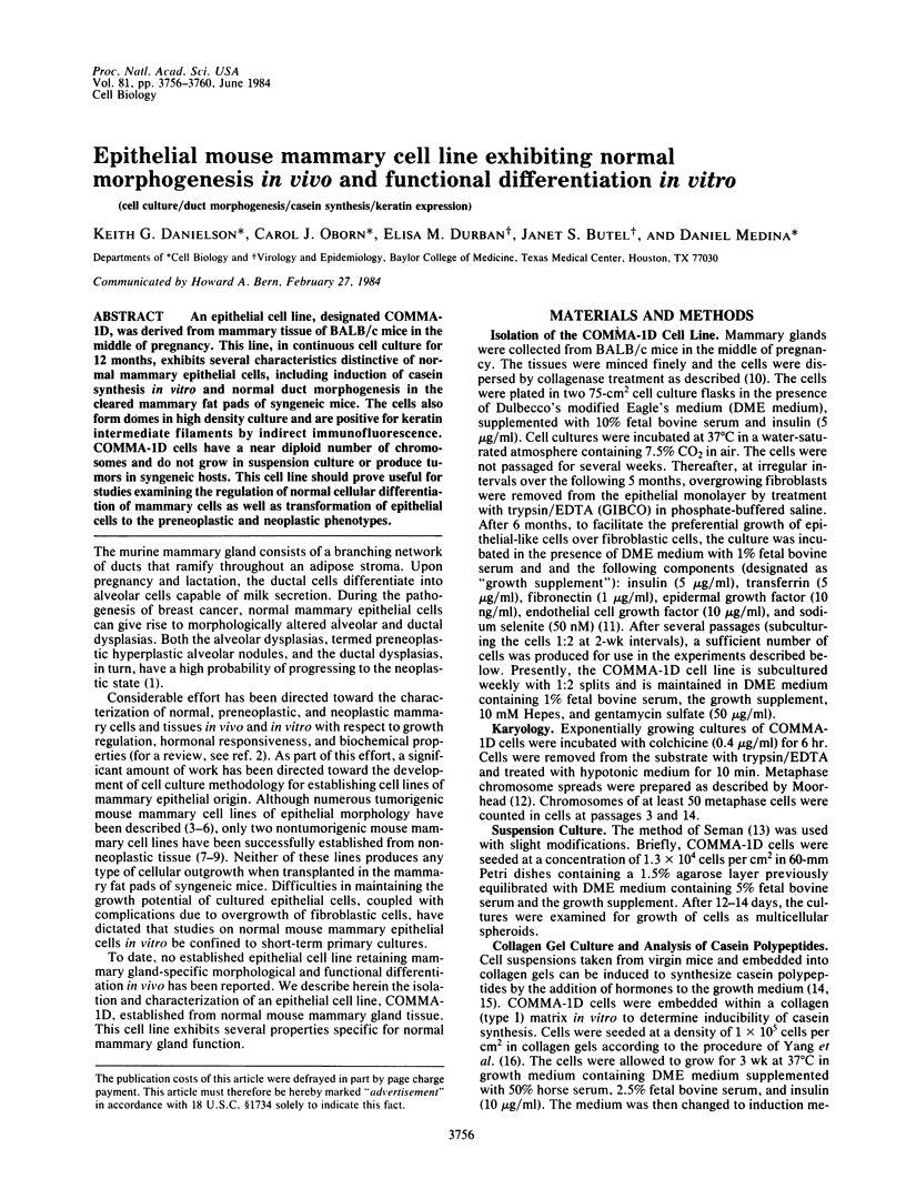
An epithelial cell line, designated COMMA-1D, was derived from mammary tissue of BALB/c mice in the middle of pregnancy. This line, in continuous cell culture for 12 months, exhibits several characteristics distinctive of normal mammary epithelial cells, including induction of casein synthesis in vitro and normal duct morphogenesis in the cleared mammary fat pads of syngeneic mice. The cells also form domes in high density culture and are positive for keratin intermediate filaments by indirect immunofluorescence. COMMA-1D cells have a near diploid number of chromosomes and do not grow in suspension culture or produce tumors in syngeneic hosts. This cell line should prove useful for studies examining the regulation of normal cellular differentiation of mammary cells as well as transformation of epithelial cells to the preneoplastic and neoplastic phenotypes.

Images in this article
Selected References
These references are in PubMed. This may not be the complete list of references from this article.
- Anderson L. W., Danielson K. G., Hosick H. L. New cell line. Epithelial cell line and subline established from premalignant mouse mammary tissue. In Vitro. 1979 Nov;15(11):841–843. doi: 10.1007/BF02618037. [DOI] [PubMed] [Google Scholar]
- Asch B. B., Burstein N. A., Vidrich A., Sun T. T. Identification of mouse mammary epithelial cells by immunofluorescence with rabbit and guinea pig antikeratin antisera. Proc Natl Acad Sci U S A. 1981 Sep;78(9):5643–5647. doi: 10.1073/pnas.78.9.5643. [DOI] [PMC free article] [PubMed] [Google Scholar]
- Asch B. B., Medina D. Concanavalin A-induced agglutinability of normal, preneoplastic, and neoplastic mouse mammary cells. J Natl Cancer Inst. 1978 Dec;61(6):1423–1430. [PubMed] [Google Scholar]
- Burnette W. N. "Western blotting": electrophoretic transfer of proteins from sodium dodecyl sulfate--polyacrylamide gels to unmodified nitrocellulose and radiographic detection with antibody and radioiodinated protein A. Anal Biochem. 1981 Apr;112(2):195–203. doi: 10.1016/0003-2697(81)90281-5. [DOI] [PubMed] [Google Scholar]
- Chen T. R. In situ detection of mycoplasma contamination in cell cultures by fluorescent Hoechst 33258 stain. Exp Cell Res. 1977 Feb;104(2):255–262. doi: 10.1016/0014-4827(77)90089-1. [DOI] [PubMed] [Google Scholar]
- Danielson K. G., Anderson L. W., Hosick H. L. Selection and characterization in culture of mammary tumor cells with distinctive growth properties in vivo. Cancer Res. 1980 Jun;40(6):1812–1819. [PubMed] [Google Scholar]
- Dexter D. L., Kowalski H. M., Blazar B. A., Fligiel Z., Vogel R., Heppner G. H. Heterogeneity of tumor cells from a single mouse mammary tumor. Cancer Res. 1978 Oct;38(10):3174–3181. [PubMed] [Google Scholar]
- Howard D. K., Schlom J., Fisher P. B. Chemical carcinogen-mouse mammary tumor virus interactions in cell transformation. In Vitro. 1983 Jan;19(1):58–66. doi: 10.1007/BF02617995. [DOI] [PubMed] [Google Scholar]
- Medina D., Oborn C. J. Growth of preneoplastic mammary epithelial cells in serum-free medium. Cancer Res. 1980 Nov;40(11):3982–3987. [PubMed] [Google Scholar]
- Owens R. B., Smith H. S., Hackett A. J. Epithelial cell cultures from normal glandular tissue of mice. J Natl Cancer Inst. 1974 Jul;53(1):261–269. doi: 10.1093/jnci/53.1.261. [DOI] [PubMed] [Google Scholar]
- Stampfer M., Hallowes R. C., Hackett A. J. Growth of normal human mammary cells in culture. In Vitro. 1980 May;16(5):415–425. doi: 10.1007/BF02618365. [DOI] [PubMed] [Google Scholar]
- Tonelli Q. J., Sorof S. Expression of a phenotype of normal differentiation in cultured mammary glands is promoted by epidermal growth factor and blocked by cyclic adenine nucleotide and prostaglandins. Differentiation. 1981;20(3):253–259. doi: 10.1111/j.1432-0436.1981.tb01180.x. [DOI] [PubMed] [Google Scholar]
- Towbin H., Staehelin T., Gordon J. Electrophoretic transfer of proteins from polyacrylamide gels to nitrocellulose sheets: procedure and some applications. Proc Natl Acad Sci U S A. 1979 Sep;76(9):4350–4354. doi: 10.1073/pnas.76.9.4350. [DOI] [PMC free article] [PubMed] [Google Scholar]
- Vaidya A. B., Lasfargues E. Y., Sheffield J. B., Coutinho W. G. Murine mammary tumor virus (MuMTV) infection of an epithelial cell line established from C57BL/6 mouse mammary glands. Virology. 1978 Oct 1;90(1):12–22. doi: 10.1016/0042-6822(78)90328-8. [DOI] [PubMed] [Google Scholar]
- White M. T., Hu A. S., Hamamoto S. T., Nandi S. In vitro analysis of proliferating epithelial cell populations from the mouse mammary gland: fibroblast-free growth and serial passage. In Vitro. 1978 Mar;14(3):271–281. doi: 10.1007/BF02616036. [DOI] [PubMed] [Google Scholar]
- Yagi M. J. Cultivation and characterization of BALB-cfC3H mammary tumor cell lines. J Natl Cancer Inst. 1973 Dec;51(6):1849–1860. doi: 10.1093/jnci/51.6.1849. [DOI] [PubMed] [Google Scholar]
- Yang J., Richards J., Bowman P., Guzman R., Enami J., McCormick K., Hamamoto S., Pitelka D., Nandi S. Sustained growth and three-dimensional organization of primary mammary tumor epithelial cells embedded in collagen gels. Proc Natl Acad Sci U S A. 1979 Jul;76(7):3401–3405. doi: 10.1073/pnas.76.7.3401. [DOI] [PMC free article] [PubMed] [Google Scholar]