Jarid2 is a PRC2 component in embryonic stem cells required for multi-lineage differentiation and recruitment of PRC1 and RNA Polymerase II to developmental regulators (original) (raw)
. Author manuscript; available in PMC: 2013 Feb 14.
Published in final edited form as: Nat Cell Biol. 2010 May 16;12(6):618–624. doi: 10.1038/ncb2065
Abstract
Polycomb Repressor Complexes (PRCs) are important regulators of embryogenesis. In embryonic stem (ES) cells many genes that regulate subsequent stages in development are enriched at their promoters for PRC1, PRC2 and Ser 5-phosphorylated RNA Polymerase II (RNAP), and contain domains of ‘bivalent’ chromatin (enriched for H3K4me3; histone H3 di- or trimethylated at Lys 4 and H3K27me3; histone H3 trimethylated at Lys 27). Loss of individual PRC components in ES cells can lead to gene de-repression and to unscheduled differentiation. Here we show that Jarid2 is a novel subunit of PRC2 that is required for the co-recruitment of PRC1 and RNAP to genes that regulate development in ES cells. Jarid2-deficient ES cells showed reduced H3K4me2/me3 and H3K27me3 marking and PRC1/PRC2 recruitment, and did not efficiently establish Ser 5-phosporylated RNAP at target genes. ES cells lacking Jarid2, in contrast to previously characterized PRC1 and PRC2 mutants, did not inappropriately express PRC2 target genes. Instead, they show a severely compromised capacity for successful differentiation towards neural or mesodermal fates and failed to correctly initiate lineage-specific gene expression in vitro. Collectively, these data indicate that transcriptional priming of bivalent genes in pluripotent ES cells is Jarid2-dependent, and suggests that priming is critical for subsequent multi-lineage differentiation.
In undifferentiated ES cells, many genes that encode transcription factors that are required for subsequent stages of development are enriched with histones modified simultaneously for active transcription (H3K4me2/me3) and PRC2-mediated repression (H3K27me3)1-4. It has been suggested that this chromatin profile—often referred to as bivalent—serves to prime undifferentiated cells to respond rapidly to lineage induction5-7. Although there is a paucity of experimental data to validate or refute this idea, reports have shown evidence of transcriptional priming in ES cells, as well as other cell types8-10. Importantly, RNAP is enriched at many PRC1/PRC2 target genes in mouse ES cells in a Ser 5-phosphorylated form that is ‘poised’ but inhibited (at least in part) by PRC1-mediated H2Aub1, and fails to efficiently elongate or produce mature transcripts8. Experimental removal of PRC1 or PRC2 in ES cells leads to gene derepression in vitro2-4,8,11-13, and early embryonic lethality in vivo14-17. Although these data show that bivalent genes are poised for expression in ES cells, they do not address whether transcriptional priming is central to the core biological properties of ES cells to self-renew and execute multi-lineage differentiation.
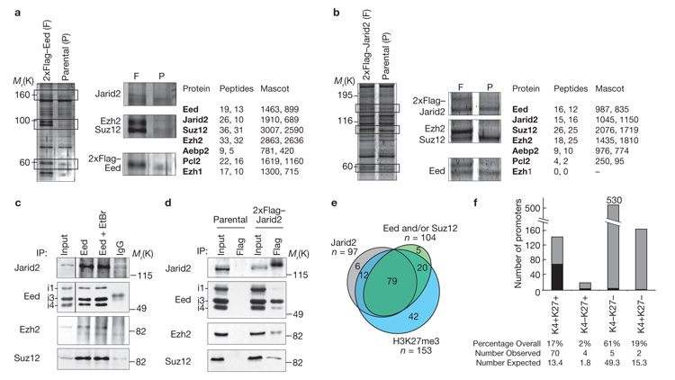
To identify PRC2-interacting proteins in mouse ES cells we used mass spectroscopy, and generated an ES cell line that stably expressed amino-terminal 2×Flag-tagged Eed isoform 3 as bait. Western blot analysis confirmed that expression of 2×Flag–Eed in these cells was moderate, and did not substantially elevate overall Eed levels (Supplementary Information, Fig. S1a). Flag antibodies immunoprecipitated proteins with the expected sizes of PRC2 core components from 2×Flag–Eed-transfected, but not parental, ES cells (Fig. 1a). Mass spectrometry of the immunoprecipitated material confirmed the presence of PRC2 core components, and in addition, showed that Jarid2 and Pcl2 were present in abundance (Fig. 1a).
Figure 1.
Jarid2 selectively associates with PRC2 proteins in mouse ES cells and localizes to PRC2-bound promoters. (a, b) Mass spectrometric analysis of Flag-immunoprecipitates from 2×Flag–Eed- or 2×Flag–Jarid2-expressing ES cells. Silver staining of 2×Flag–Eed immunoprecipitation (a) and Coomassie staining of 2×Flag–Jarid2 immunoprecipitation (b) is shown. Insets show bands present only in the Flag-transfected samples that correspond to PRC2 subunits, assigned according to molecular size. Unique peptides detected in two independent mass spectrometric experiments and their mascot scores are indicated. (c) Western blot analysis of nuclear extracts of E14 ES cells immunoprecipitated with a mouse anti-Eed (in the presence or absence of ethidium bromide; EtBr) and blotted with antiserum to Jarid2, Eed, Ezh2 and Suz12. Mouse immunoglobulin-G (IgG) was used as a negative control. Anti-Eed detected Eed isoforms 1, 3 and 4 as indicated (i1, i3 and i4). (d) Western blot analysis using antibodies described in (c) of anti-Flag immunoprecipitated material from parental (E14) and 2×Flag–Jarid2-transfected ES cells. (e) Overlay of data from 2×Flag–Jarid2 ChIP-chip genomic tiling array (grey, two replicates; as designed in ref. 19) with published ChIP-chip promoter array analysis of polycomb group proteins (green) and H3K27 trimethylation (blue)2. (f) Comparison of 2×Flag–Jarid2 (black) with ChIP-chip data for H3K4me2 (K4+) and H3K27me3 (K27+; ref. 20) shows significant (P = 2.31×10−62, chi-squared test) preference of Jarid2 for bivalent (K4+K27+) promoters.
To verify that Jarid2 associates with the PRC2 complex in vivo, we generated 2×Flag–Jarid2 ES cells. Immunoprecipitation with Flag antibodies from 2×Flag–Jarid2 ES cells showed bands, not seen in samples from parental ES cells, with the expected sizes of the PRC2 core members Eed, Ezh2 and Suz12, in addition to 2×Flag–Jarid2 (Fig. 1b). Mass spectrometry of the immunoprecipitated material confirmed the presence of Suz12, Ezh2 and Eed, as well as Aebp2 (Fig. 1b); a zinc-finger protein that has been reported to enhance H3K27 methylation activity by PRC2 (ref. 18). Ezh1 and Pcl2 were readily co-immunoprecipitated with Eed (Fig. 1a), but not with Jarid2 (Fig. 1b), suggesting that Jarid2 and Ezh1/Pcl2 reside in distinct PCR2 sub-complexes. To confirm that Jarid2 interacts with Eed in vivo, we immunoprecipitated endogenous Eed from wild-type ES cells. Western blotting demonstrated that Eed co-immunoprecipitated Jarid2 as well as Ezh2 and Suz12 (Fig. 1c). Interaction between Jarid2 and PRC2 components persisted in the presence of ethidium bromide, suggesting that it was direct and not mediated by DNA. The reverse immunoprecipitation experiment in 2×Flag–Jarid2 ES cells confirmed that 2×Flag–Jarid2 interacts with Ezh2, Suz12 and Eed (particularly isoform 3) (Fig. 1d). These data indicate that Jarid2 is a component of PRC2 in ES cells.
The genomic targets of Jarid2 in mouse ES cells were identified by chromatin immunoprecipitation followed by genomic tiling arrays (ChIP-chip). These contained regions of 200 kilobases (kb) to 2 megabases (Mb) surrounding 120 selected loci, and a contiguous section of mouse chromosome 17 (approximately 3% of the unique sequence content of the mouse genome covering 1,098 annotated genes19). Flag-ChIP showed localization of Jarid2 to 626 sites (false discovery rate 5%), of which 109 mapped within 1 kb upstream of transcription start sites (TSS) at promoter regions (< 1 kb upstream of TSS). This represents a 12-fold preference of Jarid2 for 5′ upstream sequences, compared with other regions of the genome (P = 2.2 × 10⁻¹⁶, chi-squared test). More than 90% of promoters bound by Jarid2 were also associated with PRC2 components (Eed/Suz12) and/or enriched in H3K27me3, based on comparison with published tiling array data2 covering 97 of these promoters (Fig. 1e). Binding of histone H3K27me3 or H3K4me2 had been reported for 81 of our Jarid2 targets20, and 70 of these were marked by both H3K27me3 and H3K4me2. This represents a striking preference of Jarid2 for bivalent promoters (P = 2.31 × 10−62, chi-squared test; Fig. 1f). Jarid2 also showed a significant preference for promoters with high CpG density and was underrepresented at promoters with low CpG content (P = 6.15 × 10⁻⁶, chi-squared test; data not shown). Flag-ChIP of 2×Flag-Jarid2 followed by quantitative PCR (qPCR) confirmed binding of Jarid2 to each of seven bivalent promoters tested (Supplementary Information, Fig. S1b). For three bivalent loci (Dach1, Sox7 and Nkx2-2) we profiled binding over a larger region around the TSS and found that Jarid2 recapitulated the profile of H3K4me2 enrichment (Supplementary Information, Fig. S1c). Thus, genome-scale analysis of Jarid2 targets shows a highly significant overlap between Jarid2 and PRC2 at the promoters of bivalent genes in mouse ES cells.
To probe the effect of Jarid2 on histone methylation levels, we transfected HEK293T cells with Jarid2 (Supplementary Information, Fig. S1d) and found H3K4me2/3 levels unchanged (Supplementary Information, Fig. S1e). As a control, overexpression of the H3K4-specific demethylase JARID1A (RBP2; Supplementary Information, Fig. S1d) resulted in significantly reduced methylation levels (Supplementary Information, Fig. S1e). H3K9 methylation was unaffected by both Jarid2 and RBP2 overexpression and Jarid2 did not measurably affect H3K27me3, H3K36me2 and H4K20me2/me3 levels (Supplementary Information, Fig. S1e), suggesting that Jarid2 is not catalytically active in these assays.
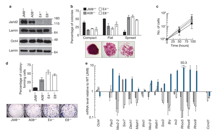
Jarid2 heterozygous ES cells were obtained through the knockout mouse project (KOMP), and we generated homozygous mutant Jarid2 cells by targeting the second allele (Supplementary Information, Fig. S2a). Long-range PCR confirmed that heterozygous (A08) and homozygous _Jarid2_-knockout lines (E4 and E8) contained one- or two-targeted Jarid2 alleles, respectively (Supplementary Information, Figures S2a, S2b). Western blotting showed that Jarid2 protein was reduced in Jarid2+/− and undetectable in _Jarid2_−/− samples relative to wild-type (Fig. 2a). _Jarid2_−/− cells retained normal levels of Oct4 protein (Fig. 2a), Oct4, Sox2 and Nanog transcripts, and expressed similar levels of PRC2 components (Supplementary Information, Fig. S2c, d). _Jarid2_−/− ES cell colonies showed similar levels of alkaline phosphatase activity as controls (Supplementary Information, Fig. S2e), although Jarid2-deficient and -null lines had a more dispersed appearance (Fig. 2b). Jarid2-deficient ES cells grew faster (Fig. 2c), and when plated at low density generated more colonies than wild-type (Fig. 2d). Assessment of cell-cycle phases using propidium iodide labelling (Supplementary Information, Fig. S2f) showed that _Jarid2_−/−, Jarid2+/− and wild-type cells all had broadly similar profiles; immunofluorescence labelling confirmed that most cells within these cultures remained undifferentiated and expressed Oct4 (> 95%, Supplementary Information, Fig. S2g) and SSEA1 (> 99%, data not shown). These results show that Jarid2-deficient ES cells retain pluripotency markers and have enhanced growth and cloning efficiencies without marked disturbance in cell-cycle progression.
Figure 2.
_Jarid2_-null ES cells express pluripotency-associated factors and do not show global de-repression of PRC2-target genes. (a) Western blot, using anti-Jarid2 and anti-Oct4 antibodies, of whole cell extracts of wild-type (JM8+/+) ES cells, a Jarid2 heterozygous (A08+/−) line and two Jarid2 knockout cell lines (E4−/− and E8−/−). Lamin was used as a loading control. Uncropped versions of the western blots are shown in Supplementary Information, Figure S7. (b) Percentage of alkaline phosphatase-positive colonies showing a compact, flat or spread morphology (mean ± s.d.). (c) Growth curve of JM8 (black line), A08 (grey line), E4 (dashed line) and E8 (dashed line) ES cells over 4 days. (d) Percentage of methylene blue-stained colonies (mean ± s.d.) detected 8 days after plating 100 cells per well; representative images of methylene blue-stained colonies plated at 500 cells per well. (e) Expression of PRC2-regulated genes in Jarid2-deficient cells (A08; grey, E4 and E8; white) relative to wild-type control (JM8). Analysis of an Eed deficient ES cell line (clone B1.3), in which PRC2 genes are de-repressed (blue), was included as a control. Mean ± s.d. of three experiments is shown.
ES cells lacking individual PRC2 components inappropriately express PRC2 target genes and are prone to unscheduled differentiation2-4,11,13,21. To investigate whether Jarid2 has a similar repressive function we examined transcript levels in wild-type, Jarid2+/− and _Jarid2_−/− ES cells, using reverse transcription quantitative PCR (RT-qPCR**)** and _Eed_−/− ES cells as controls. In contrast to _Eed_−/− ES cells (Fig. 2e; blue), Jarid2-deficiency did not result in derepression of most of the PRC2 target genes tested (Fig. 2e), including those bound by Jarid2 (Supplementary Information, Fig. S1b). Jarid2-deficient cells tended to express even lower levels of most of these genes than parental cells. These results suggest that Jarid2, unlike the PRC2 components Eed, Suz12 and Ezh2, is not essential for PRC2-mediated gene repression in ES cells.
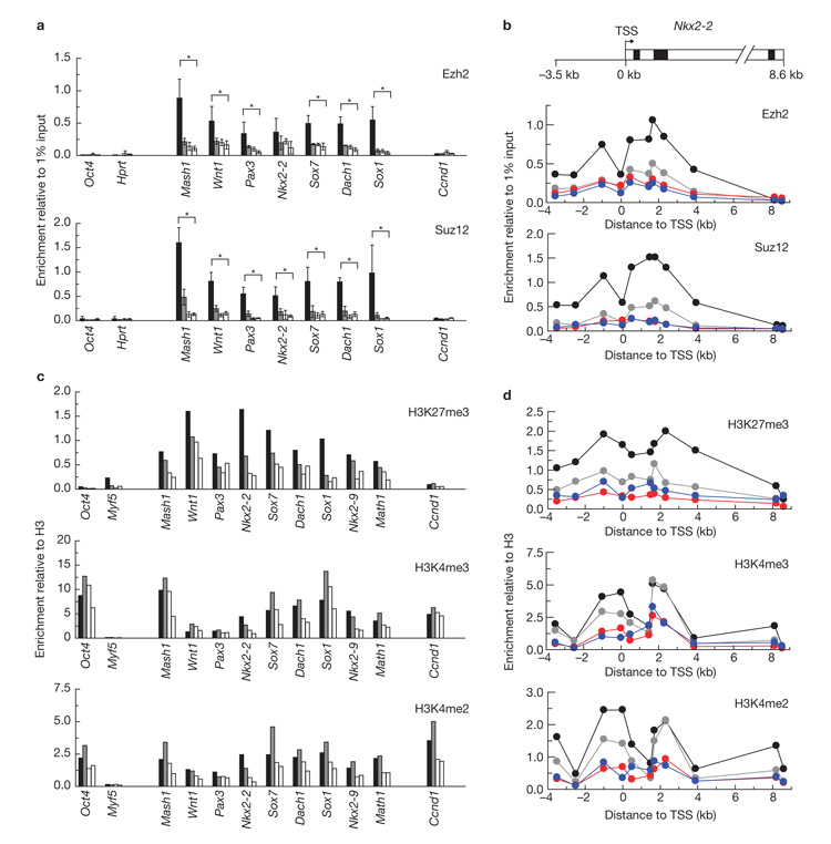
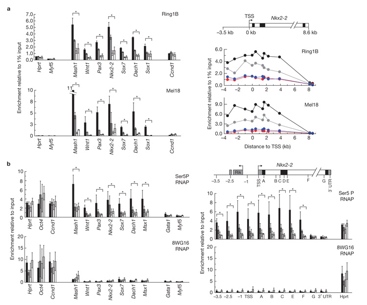
To determine whether Jarid2 is important for the recruitment of PRC1 and PRC2 components to target genes in ES cells we performed ChIP analysis. Jarid2-deficient ES cells showed reduced binding of Ezh2 and Suz12 to the promoters of each of seven PRC2 target genes tested (Mash1, Wnt1, Pax3, Nkx2-2, Sox7, Dach1 and Sox1) (Fig. 3a), and at the Nkx2-2 locus (Fig. 3b; Supplementary Information, Fig. S3); an extensively characterized bivalent domain8. H3K27me3, H3K4me2 and H3K4me3 levels at each of these loci were diminished in Jarid2-deficient ES cells, compared with wild-type (Fig. 3c, d; Supplementary Information, Fig. S3), although levels remained above the background signal. The binding of PRC1 components Ring1B and Mel18 to target promoters was also reduced in _Jarid2_−/− ES cells (Fig. 4a, left) as well as across the Nkx2-2 locus (Fig. 4a right; Supplementary Information, Fig. S3). Hence, PRC1 is not efficiently recruited to PRC2 target genes in the absence of Jarid2.
Figure 3.
Jarid2 is required to recruit PRC2 to target genes in ES cells. (a, b) Binding of Ezh2 and Suz12 to the promoter regions of genes, or across the Nkx2-2 locus, was assessed by ChIP. Results show mean ± s.d. of three experiments where values are expressed relative to input. Asterisks indicate statistical significant differences between JM8 wild-type and Jarid2 knockout cells (p < 0.05; Student’s _t_-test). (c, d) The abundance of modified histones was assessed by ChIP using specific antibodies. Results show mean of two experiments and values are expressed relative to H3 abundance. In (b) and (d) the abundance (mean) along Nkx2-2, using primer pairs detailed in Supplementary Information, Figure S3, is plotted as a function of distance to TSS (error bars are shown in Supplementary Information, Fig. S3). Experiments were performed using wild-type (JM8, black), heterozygous (A08, grey) and _Jarid2_-knockout ES cells (E4, E8, white bars or E4; red and E8; blue lines). A scheme of the Nkx2-2 locus displaying TSS (arrow) and three exons (black boxes) is shown in b. Ccnd1, a putative Jarid2 target gene in cardiac myocytes29, and Oct4, Hprt and Myf5 were included as controls.
Figure 4.
Efficient recruitment of PRC1 and Ser5-phosphorylated RNAP to target genes in ES cells requires Jarid2. (a) Ring1B and Mel18 binding at gene promoter regions (left panel), or across the Nkx2-2 locus (right panel), was assessed by ChIP. Values and error bars are shown in Supplementary Information, Figure S3, and the distance from the Nkx2-2 TSS is indicated. (b) RNAP binding (Ser 5 phosphorylated or detected by 8WG16) was assessed by ChIP. Right panel shows a schematic representation of the Nkx2-2 locus where the TSS, a conserved region at −2.5 kb (light grey box), the coding region (8.7 kb containing three exons, black boxes), untranslated regions (light grey boxes) and the position of primer pairs are indicated. Results show mean ± s.d. of three experiments, where values are expressed relative to input chromatin. Experiments were performed using wild-type (JM8, black), heterozygous (A08, grey) and _Jarid2_-knockout ES cells (E4, E8, white bars or E4; red and E8; blue lines). Hprt, Oct4 and Ccnd1 (active genes), and Gata1 and Myf5 (silent genes) provide controls. Asterisks indicate statistical significant differences between JM8 wild-type and Jarid2 knockout cells (P < 0.05; Student’s _t_-test).
As PRC1 is thought to mediate the silencing of many PRC2 target genes, it was surprising that PRC2 targets are not de-repressed in ES cells lacking Jarid2. Lack of de-repression could not be ascribed to increased DNA methylation, as bisulphite sequencing of five PRC2 target genes (Sox30, Pax5, Hoxa10, Irx3 and Math1) showed no appreciable differences between _Jarid2_−/− and wild-type ES cells (Supplementary Information, Fig. S4). We therefore examined alternative mechanisms to explain the continued transcriptional inactivity of PRC2 target genes in Jarid2-deficient ES cells. H2Aub1, the histone modification catalysed by the PRC1 complex was reduced in _Jarid2_−/− cells (Supplementary Information, Fig. S5a), although this was less striking than the decrease of Ring1B and Mel18.
In wild-type ES cells RNAP is engaged at PRC2 target gene promoters8,9, but shows an unusual set of post-translational modifications22 in which high levels of phosphorylated Ser 5 are present, but detection of phosphorylated Ser 2 and reactivity with 8WG16 (an antibody that preferentially recognizes non-phosphorylated Ser2 residues of RNAP) is low. Consistent with this, phosphorylated Ser 5 RNAP was abundant at the promoters of actively expressed genes (such as Hprt, Oct4 and Ccnd1) and PRC2-target genes in wild-type ES cells (Fig. 4b, black bars, upper panel). In striking contrast, only low levels of phosphorylated Ser 5 RNAP were detected in _Jarid2_−/− ES cells at six of seven PRC2 target genes and across the Nkx2-2 locus (Fig. 4b; Supplementary Information, Fig. S5a). As expected, hypophosphorylated RNAP was detected at active genes (Hprt, Oct and Ccnd1) in both wild-type and Jarid2-deficient ES cells (Fig. 4b, lower panels), but was not detected at silent (Gata1 and Myf5) or bivalent loci (Mash1, Wnt1, Pax3, Nkx2-2, Sox7, Dach1 and Msx1), regardless of Jarid2 status. TATA-binding protein (TBP), a transcription factor known to bind polycomb-repressed promoters in Drosophila cells23, was detected at the promoters of expressed and bivalent genes in _Jarid2_-deficient and wild-type ES cells (Supplementary Information, Fig. S5b). These data reveal a defect in recruiting phosphorylated Ser 5 RNAP to PRC2 target genes in Jarid2-deficient ES cells, and provide evidence that Jarid2 is critical for the transcriptional priming of these genes in ES cells.
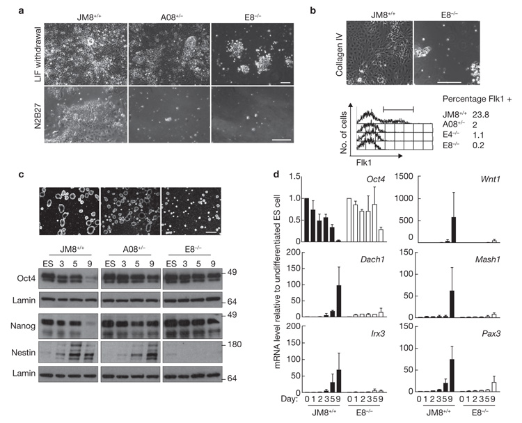
Jarid2 expression is dependent on the triad of transcription factors Oct4, Sox2 and Nanog, and is downregulated upon ES cell differentiation24-26. Consistently with this, Jarid2 was downregulated when wild-type ES cells were induced to differentiate into neuronal cells or mesoderm (Supplementary Information, Fig. S6). In contrast to wild-type ES cells, _Jarid2_−/− ES cells were unable to differentiate efficiently in response to withdrawal of leukaemia inhibitory factor (LIF), neural induction (Fig. 5a) or mesoderm induction (Fig. 5b). Differentiation of _Jarid2_−/− ES cells towards Flk1-expressing hemangioblasts27 was also severely compromised (Fig. 5b, lower panel). Jarid2-deficient cells formed only rudimentary embryoid bodies, which, in contrast to wild-type embryoid bodies, continued to express Oct4 and Nanog protein and failed to express Nestin (Fig. 5c). Jarid2+/− cells showed an intermediate phenotype, with a partial downregulation of Oct4 and Nanog and delayed induction of Nestin (Fig. 5c). Similarly, Dach1, Irx3, Wnt1, Mash1 and Pax3 transcripts were upregulated on neural induction of wild-type (Fig. 5d, black bars), but not in _Jarid2_−/− ES cells (Fig. 5d, white bars). These data show that Jarid2 is important for the successful execution of multiple differentiation pathways.
Figure 5.
Jarid2 is required for successful ES cell differentiation. Brightfield images of parental (JM8+/+), heterozygous (A08+/−) and _Jarid2_-null (E8−/−) ES cells induced by (a) withdrawal of LIF (upper panel, day 9), culture in neural-inducing N2B27 media34 (lower panel, day 5) and (b) culture on Collagen IV-coated plates under mesoderm-inducing conditions35 (upper panel, day 4). Flk1-expressing hemangioblasts were induced from embryoid bodies using standard approaches27 and quantified by FACS (lower panel, day 3.75) where bar shows gates used to identify Flk1+ cells. (c) Upper panel shows brightfield images where the absence of Jarid2 results in fewer and smaller embryoid bodies induced upon LIF withdrawal, compared with wild-type (day 5). Time course analysis of Oct4, Nanog and Nestin expression in embryoid bodies by western blot (lower panel). (d) Kinetics of gene expression during embryoid body formation (relative to undifferentiated ES cell controls). Values for parental (black) and Jarid2-null (E8, white) ES cells are shown. Scale bars, 100 μm.
Jarid2 is essential for mouse development28,29, and is a recognized component of pluripotency networks24-26. Recently, Jarid2 was shown to associate with the PRC2 complex in ES cells and to occupy many polycomb targets genome-wide30-33. These reports have suggested that Jarid2 modulates H3K27me3 levels in ES cells through PRC2 recruitment, but have offered contradictory views on whether Jarid2 inhibits30,31 or stimulates32,33 PRC2 histone methyltransferase activity. Our data show that in the absence of Jarid2, PRC2-target genes in ES cells show modest reductions in both H3K27me3 and H3K4me2/3 levels, a corresponding reduction in PRC1 recruitment and rather mild effects on downstream H2Aub1. Perhaps the most surprising observation is that ES cells lacking Jarid2 failed to efficiently establish ‘initiating’ Ser 5 RNAP at PRC2 target genes. In wild-type ES cells, where PRC1 and Ser 5 RNAP are bound at many PRC2 targets2,8, withdrawal of Eed, Suz12, Ezh2 or Ring1A/B results in the de-repression of target genes2-4,8,11-13. In Jarid2-deficient ES cells a functionally ‘poised’ or ‘primed’ state is clearly not established, a point that is reinforced by the finding that expression of PRC2 target genes in Jarid2-depleted ES cells is actually lower than the basal transcript levels detected in wild-type ES cells. In addition, we have shown that Jarid2-deficient ES cells are unable to efficiently differentiate to mesoderm or neural lineages in vitro but exhibit enhanced proliferation when maintained in conditions that support the undifferentiated state. The compromised differentiation of Jarid2-deficient ES cells described here is similar to recently reported deficits in ‘cell fate transition’ and lineage induction reported for Jarid2 knockout31 or Jarid2-shRNA-treated (knockdown) ES cells33. Our data, showing that in the absence of Jarid2 important development regulator genes in ES cells are not efficiently primed, may provide an explanation for the failure of these cells to respond in a timely and appropriate way to developmental cues. The requirement for Jarid2 during ES cell differentiation is particularly intriguing as ES cells normally downregulate Jarid2 expression soon after the onset of differentiation. This suggests that Jarid2 has a critical role in undifferentiated ES cells either in preserving their multi-lineage potential status or in mediating the successful switching of ES cells from a proliferative to a differentiating state. The fact that Jarid2 is also required to efficiently establish a poised RNAP configuration at PRC2 targets in ES cells provides further evidence that transcriptional priming of key regulators is important for pluripotent function.
METHODS
Generation of Flag-fusion-expressing cell lines
Jarid2 and Eed isoform 3 cDNA were cloned from OS25 ES cell mRNA using oligonucleotides: 5′-GGATCCATGTCCGAGAGGGAAGTGTCG-3′ 5′-GAATTCTTATCGAAGTCGATCCCATCGC-3′ for Eed and 5′-TGATCAATGAGCAAGGAAAGACCCAAG-3′ and 5′-TCTAGATCATGAGGATGGGAGCCG-3′ for Jarid2. cDNA was cloned into 2×FlagpCDNA3 through _Bam_HI/_Eco_RI (Eed) and _Bam_HI/_Xba_I (Jarid2) restriction sites. cDNA from 2×Flag-Eed and 2×Flag-Jarid2 cells was excised and cloned into the chicken β-actin promoter-driven expression vector pCBA36 and used to stably transfect E14 (2× Flag-Jarid2) and Ezfl (2×Flag-Eed) murine ES cell lines using Lipofectamine 2000 (Invitrogen) and G418 selection (300 μg ml−1 for 6 days). ES cells were cultured on gelatin coated dishes in the presence of LIF at 37 °C and 5% CO2. Eed expression by 2×Flag-Eed ES cells is shown in Supplementary Information, Figure S1a. Jarid2 expression by 2×Flag-Jarid2 ES cells was modestly increased over endogenous levels (~20%, data not shown).
Immunoprecipitation and Mass Spectrometric analysis
Immunoprecipitation, silver staining and mass spectrometric analysis were carried out as described37 with the following antibodies: mouse anti-Eed (A. Otte, University of Amsterdam, NL), mouse anti-Flag (Sigma) and mouse IgG (Santa Cruz Biotechnology).
Jarid2 and RBP2 expression in HEK293T cells
HEK293T cells were transiently transfected with pCDNA3 (Invitrogen), pCDNA3 containing 2×Flag-Jarid2 (as above) or human RBP2 cDNA, using calcium phosphate.
Western blot analysis
Western blots were carried out using the following antibodies: rabbit antisera to Jarid2 (1:1,000) (Abcam or Y. Lee, University Wisconsin, USA), histone H3 (1:30,000; Abcam), H3K4me3 (1:1,000; Abcam), H3K4me2 (1:1,000; Millipore), H3K36me3 (1:1,000; Abcam), H3K9me3 (1:1,000; Millipore), H3K9me2 (1:1,000; Diagenode), H3K36me2 (1:1,000; Millipore), H3K27me3 (1:1,000; Millipore), H4K20me3 (1:1,000; Abcam), H4K20me2 (1:1,000; Millipore), Suz12 (1:1,000; Diagenode), Ezh2 (1:1,000; Millipore) and Nanog (1:200; Cosmo Bio Co.); mouse antibodies to Eed (1:2,000; A. Otte), Nestin (1:500; BD Biosciences) and Flag (1:1000; Sigma) and goat antibodies to LaminB (1:30,000; Santa Cruz Biotechnology) and Oct4 (1:5,000; Santa Cruz Biotechnology). Secondary species-specific antibodies were anti-goat-HRP (Santa Cruz), anti-mouse-HRP (GE Healthcare) and anti-rabbit-HRP (GE Healthcare). For detection Amersham ECL Plus (GE Healthcare) was used.
2×Flag-Jarid2 ChIP-on-chip analysis
Cells were fixed in PBS in 1% paraformaldehyde (15 min, 25 °C), quenched with 125mM glycine and lysed in a solution containing 5mM PIPES at pH 8.0, 85mM KCl and 0.5% NP-40 for 20 min at 4 °C. After spinning, nuclei were resuspended in a solution containing 1% SDS, 10mM EDTA at pH 8.0 and 50mM Tris at pH 8.1, diluted 1:1 in radio immunoprecipitation assay (RIPA) buffer, and sonicated until DNA fragment size was 0.2-2 kb. Flag M2 antibody (25 μg; Sigma) and Protein-G-Dynabeads (Invitrogen) were used. Elution was performed with 250 μl of RIPA buffer containing 0.05 mg of 3×Flag peptide (Sigma). Reverse crosslinked DNA was purified using QIAquick spin columns and eluted in 60μl Buffer EB (Qiagen). ChIP samples were amplified according to manufacturer’s instructions (Nimblegen) for hybridization to custom tiling arrays with 380,000 features at 100 bp resolution, and results were analysed using Tilemap software. The comparison with the Mohn et al. data set20 was based on chromosomal coordinates available at (http://www.fmi.ch/groups/schubeler.d/web/supplements/Mohn_2008_promoters.txt) after conversion from mm8 (NCBI build 36) to mm5 (NCBI build 33) by using the UCSC LiftOver tool.
Histone modifications, polycomb proteins and RNAP ChIP analysis
ChIP experiments for methylated histones were performed as described above with the following modifications. For each immunoprecipitation, 100 μg of chromatin was incubated with 10 μg of one of the following antibodies: anti-H3 (Abcam), anti-H3K4me2 (Millipore), anti-H3K4me3 (Abcam), anti-H3K27me3 (Millipore) and Protein A-Dynabeads (Invitrogen). ChIP experiments of Ezh2 and Suz12 were performed as above, but the cells were fixed in medium with 1% paraformaldehyde for 10 min and using 10 μg of anti-Ezh2 (Diagenode) and anti-Suz12 (Cell signaling), respectively. ChIP experiments for H2Aub1, Mel18, Ring1B and Tbp were performed using conditions previously described8 and anti-ubiquityl-Histone H2A (Millipore), anti-Mel18 (Santa Cruz), anti-Ring1B (MBL) or anti-TBP (Santa Cruz), respectively, and ProteinA/G-Dynabeads (Invitrogen). ChIP experiments for phosphorylated Ser 5 and 8WG16 RNAP were performed using anti-RNAP CTD4H8 (Covance) or Ms 8WG16 (Covance), respectively, as described previously8 with the following modifications. Magnetic beads (Active Motif) were incubated with rabbit anti-mouse antibodies (Jackson Immunoresearch; 10 μg per 50 μl of beads) for 1 h at 4 °C and washed with sonication buffer. Chromatin (700 μg, determined by measuring absorbance of alkaline-lysed, crosslinked chromatin) was immunoprecipitated (overnight at 4 °C) with 10 μl antibody and 50 μl of beads (with bridging antibody). Immunoprecipitated DNA of the different ChIP experiments was analysed using real-time PCR4. Primer sequences are available upon request.
Gene expression analysis by RT-qPCR
RNA was isolated using the RNeasy Kit (Qiagen), reverse transcribed using SuperScriptIII as recommended by the manufacturer (Invitrogen) and analysed by real-time PCR using Quantitect SYBR green PCR kit (Qiagen), as described previously4. Primer sequences are available upon request.
Jarid2 KO ES targeting, genotyping and cell culture
Feeder-free adapted parental JM8 (wild-type) mouse ES cells are derived from the C57BL6/N mouse strain38. Jarid2 mutant mouse embryonic stem (mES) cell lines were obtained using targeted genetrapping approaches39. JM8 and Jarid2 heterozygous mutant mES cells were generated by the Wellcome Trust Sanger Institute’s knockout mouse project (http://www.komp.org) production center (W.C. Skarnes, et al., manuscript in preparation). The Jarid2 heterozygous mutant ‘KO first’ allele contains a standard β-geo trapping cassette targeted by replacement mutagenesis into the intron 1-2. Jarid2 homozygous mutant mES cell lines were generated using an insertion type vector containing a hygromycin resistance gene, an enhanced GFP marker, and a poly(A) sequence in a gene trapping cassette that targets intron 2-3. Further details on the second allele targeting strategy will be published elsewhere (C. Fisher and W.C. Skarnes, in preparation). Correct targeting of the first allele was reconfirmed by genomic long-range PCR (LRPCR) using the gene-specific primer: 5′-AGGTATTTTGTTCGCCGTGTGTTTC-3′ and the vector-specific primer: 5′-GCCTCTTCGCTATTACGCCAGCTG-3′. Correct targeting of the second allele was reconfirmed by LRPCR using the gene-specific primer: 5′-TCTTTCTGTGCCTTGTTCTGACCTG-3′ and the vector-specific primer: 5′-GCAGCTATTTACCCGCAGGA-3′. PCR reactions were performed using the ‘Expand Long Template’ PCR system (Roche).
Alkaline phosphatase activity was determined using alkaline phosphatase kit (Sigma). Cell-cycle profile was analysed by staining ice-cold ethanol fixed cells with propidium iodide followed by analysis with FACSCalibur (Beckton Dickinson). Oct4 immunofluorescence analysis was carried out using Ms anti-Oct3/4 (BD bioscience) antibody.
ES cell culture and differentiation
Bry201 ES cells (V. Kouskoff, University of Manchester, UK) were maintained and differentiated as described elsewhere27. Differentiation of JM8 and Jarid2-deficient ES cells was induced by LIF withdrawal in monolayer cultures (5 × 10³ cells per cm2, plated on gelatinized flasks in ES medium). Monolayer differentiation towards neural lineage in serum-free neural differentiation medium N2B27 (Stem Cell Sciences) was carried out as described previously elsewhere34. Monolayer differentiation to mesoderm was carried out as described35. For embryoid body differentiation, 7 × 10⁶ cells were plated in non-adherent plates in ES cell medium without LIF. To obtain mesoderm-enriched embryoid bodies, cells were differentiated as described elsewhere27. At 3.75 days of differentiation EBs were stained for FACS analysis using PE-Flk1 antibody (BD Bioscience,).
Bisulphite genomic sequencing
Bisulphite genomic sequencing was carried out as described previously40 with oligonucleotides detailed in Supplementary Information, Table S1.
Statistical analysis
Statistical significance of PCR and ChIP-chip experiments was determined by applying a one-tailed Student’s _t_-test to raw enrichment values and Chi-squared test, respectively.
Supplementary Material
Supplementary Material
ACKNOWLEDGMENTS
We are grateful to our colleagues Y. Lee, V. Kouskoff and A. Otte for supplying reagents and advice. We thank N. Ryan, A. Terry and the MRC CSC flow cytometry facility for technical assistance. This work was funded by the Medical Research Council (MRC) and the EU Epigenome Network of Excellence.
Footnotes
Note: Supplementary Information is available on the Nature Cell Biology website
AUTHOR CONTRIBUTIONS: D.L., S.S., M.M. and A.G.F. participated in the design of the study and wrote the manuscript. D.L., S.S., M.D., L.M., H.F.J., C.F.P., M.L., F.M.P., M.S., E.B., C.F., T.S., K.B., J.D., M.C. and L.T. performed experiments. R.P., A.P., W.C.S., R.J.K., N.B., M.M. and A.G.F. provided conceptual advice on study design and the interpretation of results.
ACCESSION NUMBER: Flag-Jarid2 genomic tiling array experiment is available in ArrayExpress, accession number E-TABM-963.
COMPETING FINANCIAL INTERESTS: The authors declare no competing financial interests.
References
- 1.Bernstein BE, et al. A bivalent chromatin structure marks key developmental genes in embryonic stem cells. Cell. 2006;125:315–326. doi: 10.1016/j.cell.2006.02.041. [DOI] [PubMed] [Google Scholar]
- 2.Boyer LA, et al. Polycomb complexes repress developmental regulators in murine embryonic stem cells. Nature. 2006;441:349–353. doi: 10.1038/nature04733. [DOI] [PubMed] [Google Scholar]
- 3.Lee TI, et al. Control of developmental regulators by Polycomb in human embryonic stem cells. Cell. 2006;125:301–313. doi: 10.1016/j.cell.2006.02.043. [DOI] [PMC free article] [PubMed] [Google Scholar]
- 4.Azuara V, et al. Chromatin signatures of pluripotent cell lines. Nat. Cell Biol. 2006;8:532–538. doi: 10.1038/ncb1403. [DOI] [PubMed] [Google Scholar]
- 5.Jaenisch R, Young R. Stem cells, the molecular circuitry of pluripotency and nuclear reprogramming. Cell. 2008;132:567–582. doi: 10.1016/j.cell.2008.01.015. [DOI] [PMC free article] [PubMed] [Google Scholar]
- 6.Jorgensen HF, et al. Stem cells primed for action: polycomb repressive complexes restrain the expression of lineage-specific regulators in embryonic stem cells. Cell Cycle. 2006;5:1411–1414. doi: 10.4161/cc.5.13.2927. [DOI] [PubMed] [Google Scholar]
- 7.Buszczak M, Spradling AC. Searching chromatin for stem cell identity. Cell. 2006;125:233–236. doi: 10.1016/j.cell.2006.04.004. [DOI] [PubMed] [Google Scholar]
- 8.Stock JK, et al. Ring1-mediated ubiquitination of H2A restrains poised RNA polymerase II at bivalent genes in mouse ES cells. Nat. Cell Biol. 2007;9:1428–1435. doi: 10.1038/ncb1663. [DOI] [PubMed] [Google Scholar]
- 9.Guenther MG, Levine SS, Boyer LA, Jaenisch R, Young RA. A chromatin landmark and transcription initiation at most promoters in human cells. Cell. 2007;130:77–88. doi: 10.1016/j.cell.2007.05.042. [DOI] [PMC free article] [PubMed] [Google Scholar]
- 10.Dellino GI, et al. Polycomb silencing blocks transcription initiation. Mol. Cell. 2004;13:887–893. doi: 10.1016/s1097-2765(04)00128-5. [DOI] [PubMed] [Google Scholar]
- 11.Pasini D, Bracken AP, Hansen JB, Capillo M, Helin K. The polycomb group protein Suz12 is required for embryonic stem cell differentiation. Mol. Cell Biol. 2007;27:3769–3779. doi: 10.1128/MCB.01432-06. [DOI] [PMC free article] [PubMed] [Google Scholar]
- 12.Endoh M, et al. Polycomb group proteins Ring1A/B are functionally linked to the core transcriptional regulatory circuitry to maintain ES cell identity. Development. 2008;135:1513–1524. doi: 10.1242/dev.014340. [DOI] [PubMed] [Google Scholar]
- 13.Shen X, et al. EZH1 mediates methylation on histone H3 lysine 27 and complements EZH2 in maintaining stem cell identity and executing pluripotency. Mol. Cell. 2008;32:491–502. doi: 10.1016/j.molcel.2008.10.016. [DOI] [PMC free article] [PubMed] [Google Scholar]
- 14.Faust C, Schumacher A, Holdener B, Magnuson T. The eed mutation disrupts anterior mesoderm production in mice. Development. 1995;121:273–285. doi: 10.1242/dev.121.2.273. [DOI] [PubMed] [Google Scholar]
- 15.O’Carroll D, et al. The polycomb-group gene Ezh2 is required for early mouse development. Mol. Cell Biol. 2001;21:4330–4336. doi: 10.1128/MCB.21.13.4330-4336.2001. [DOI] [PMC free article] [PubMed] [Google Scholar]
- 16.Voncken JW, et al. Rnf2 (Ring1b) deficiency causes gastrulation arrest and cell cycle inhibition. Proc. Natl Acad. Sci. USA. 2003;100:2468–2473. doi: 10.1073/pnas.0434312100. [DOI] [PMC free article] [PubMed] [Google Scholar]
- 17.Pasini D, Bracken AP, Jensen MR, Lazzerini Denchi E, Helin K. Suz12 is essential for mouse development and for EZH2 histone methyltransferase activity. EMBO J. 2004;23:4061–4071. doi: 10.1038/sj.emboj.7600402. [DOI] [PMC free article] [PubMed] [Google Scholar]
- 18.Cao R, Zhang Y. SUZ12 is required for both the histone methyltransferase activity and the silencing function of the EED-EZH2 complex. Mol. Cell. 2004;15:57–67. doi: 10.1016/j.molcel.2004.06.020. [DOI] [PubMed] [Google Scholar]
- 19.Parelho V, et al. Cohesins functionally associate with CTCF on mammalian chromosome arms. Cell. 2008;132:422–433. doi: 10.1016/j.cell.2008.01.011. [DOI] [PubMed] [Google Scholar]
- 20.Mohn F, et al. Lineage-specific polycomb targets and de novo DNA methylation define restriction and potential of neuronal progenitors. Mol. Cell. 2008;30:755–766. doi: 10.1016/j.molcel.2008.05.007. [DOI] [PubMed] [Google Scholar]
- 21.Chamberlain SJ, Yee D, Magnuson T. Polycomb Repressive Complex 2 is Dispensable for Maintenance of Embryonic Stem Cell Pluripotency. Stem Cells. 2008;26:1496–1505. doi: 10.1634/stemcells.2008-0102. [DOI] [PMC free article] [PubMed] [Google Scholar]
- 22.Brookes E, Pombo A. Modifications of RNA polymerase II are pivotal in regulating gene expression. EMBO Rep. 2009;10:1213–1219. doi: 10.1038/embor.2009.221. [DOI] [PMC free article] [PubMed] [Google Scholar]
- 23.Breiling A, Turner BM, Bianchi ME, Orlando V. General transcription factors bind promoters repressed by Polycomb group proteins. Nature. 2001;412:651–655. doi: 10.1038/35088090. [DOI] [PubMed] [Google Scholar]
- 24.Boyer LA, et al. Core transcriptional regulatory circuitry in human embryonic stem cells. Cell. 2005;122:947–956. doi: 10.1016/j.cell.2005.08.020. [DOI] [PMC free article] [PubMed] [Google Scholar]
- 25.Kim J, Chu J, Shen X, Wang J, Orkin SH. An extended transcriptional network for pluripotency of embryonic stem cells. Cell. 2008;132:1049–1061. doi: 10.1016/j.cell.2008.02.039. [DOI] [PMC free article] [PubMed] [Google Scholar]
- 26.Loh YH, et al. The Oct4 and Nanog transcription network regulates pluripotency in mouse embryonic stem cells. Nat. Genet. 2006;38:431–440. doi: 10.1038/ng1760. [DOI] [PubMed] [Google Scholar]
- 27.Fehling HJ, et al. Tracking mesoderm induction and its specification to the hemangioblast during embryonic stem cell differentiation. Development. 2003;130:4217–4227. doi: 10.1242/dev.00589. [DOI] [PubMed] [Google Scholar]
- 28.Jung J, Mysliwiec MR, Lee Y. Roles of JUMONJI in mouse embryonic development. Dev. Dyn. 2005;232:21–32. doi: 10.1002/dvdy.20204. [DOI] [PubMed] [Google Scholar]
- 29.Takeuchi T, Watanabe Y, Takano-Shimizu T, Kondo S. Roles of jumonji and jumonji family genes in chromatin regulation and development. Dev. Dyn. 2006;235:2449–2459. doi: 10.1002/dvdy.20851. [DOI] [PubMed] [Google Scholar]
- 30.Peng JC, et al. Jarid2/Jumonji coordinates control of PRC2 enzymatic activity and target gene occupancy in pluripotent cells. Cell. 2009;139:1290–1302. doi: 10.1016/j.cell.2009.12.002. [DOI] [PMC free article] [PubMed] [Google Scholar]
- 31.Shen X, et al. Jumonji modulates polycomb activity and self-renewal versus differentiation of stem cells. Cell. 2009;139:1303–1314. doi: 10.1016/j.cell.2009.12.003. [DOI] [PMC free article] [PubMed] [Google Scholar]
- 32.Li G, et al. Jarid2 and PRC2, partners in regulating gene expression. Genes Dev. 2010;24:368–380. doi: 10.1101/gad.1886410. [DOI] [PMC free article] [PubMed] [Google Scholar]
- 33.Pasini D, et al. JARID2 regulates binding of the Polycomb repressive complex 2 to target genes in ES cells. Nature. 2010;464:306–310. doi: 10.1038/nature08788. [DOI] [PubMed] [Google Scholar]
- 34.Ying QL, Smith AG. Defined conditions for neural commitment and differentiation. Methods Enzymol. 2003;365:327–341. doi: 10.1016/s0076-6879(03)65023-8. [DOI] [PubMed] [Google Scholar]
- 35.Fraser ST, et al. In vitro differentiation of mouse embryonic stem cells: hematopoietic and vascular cell types. Methods Enzymol. 2003;365:59–72. doi: 10.1016/s0076-6879(03)65004-4. [DOI] [PubMed] [Google Scholar]
- 36.Gontan C, et al. Exportin 4 mediates a novel nuclear import pathway for Sox family transcription factors. J Cell Biol. 2009;185:27–34. doi: 10.1083/jcb.200810106. [DOI] [PMC free article] [PubMed] [Google Scholar]
- 37.van den Berg DL, et al. An Oct4-centered protein interaction network in embryonic stem cells. Cell Stem Cell. 2010;6:369–381. doi: 10.1016/j.stem.2010.02.014. [DOI] [PMC free article] [PubMed] [Google Scholar]
- 38.Pettitt SJ, et al. Agouti C57BL/6N embryonic stem cells for mouse genetic resources. Nat. Methods. 2009;6:493–495. doi: 10.1038/nmeth.1342. [DOI] [PMC free article] [PubMed] [Google Scholar]
- 39.Friedel RH, et al. Gene targeting using a promoterless gene trap vector (“targeted trapping”) is an efficient method to mutate a large fraction of genes. Proc. Natl Acad. Sci. USA. 2005;102:13188–13193. doi: 10.1073/pnas.0505474102. [DOI] [PMC free article] [PubMed] [Google Scholar]
- 40.Pereira CF, et al. Heterokaryon-based reprogramming of human B lymphocytes for pluripotency requires Oct4 but not Sox2. PLoS Genet. 2008;4:e1000170. doi: 10.1371/journal.pgen.1000170. [DOI] [PMC free article] [PubMed] [Google Scholar]
Associated Data
This section collects any data citations, data availability statements, or supplementary materials included in this article.
Supplementary Materials
Supplementary Material
