Shaping organisms with apoptosis (original) (raw)
Abstract
Programmed cell death is an important process during development that serves to remove superfluous cells and tissues, such as larval organs during metamorphosis, supernumerary cells during nervous system development, muscle patterning and cardiac morphogenesis. Different kinds of cell death have been observed and were originally classified based on distinct morphological features: (1) type I programmed cell death (PCD) or apoptosis is recognized by cell rounding, DNA fragmentation, externalization of phosphatidyl serine, caspase activation and the absence of inflammatory reaction, (2) type II PCD or autophagy is characterized by the presence of large vacuoles and the fact that cells can recover until very late in the process and (3) necrosis is associated with an uncontrolled release of the intracellular content after cell swelling and rupture of the membrane, which commonly induces an inflammatory response. In this review, we will focus exclusively on developmental cell death by apoptosis and its role in tissue remodeling.
Keywords: apoptosis, development, sculpting, morphogenesis, Drosophila
Facts
- Apoptosis has a crucial role in a variety of morphogenetic events.
- Apoptosis can either shape an organ by the simple elimination of cells that are no longer required, without inducing tissue remodeling (e.g. digit individualization), or participate in morphogenesis by inducing cellular reorganization in the surrounding tissue (e.g. dorsal closure or genitalia rotation).
- In normal conditions, apoptotic cells induce the formation of an actomyosin ring in their neighbors for their extrusion.
- In stress conditions, apoptotic cells can also produce mitotic signals to induce compensatory proliferation.
- Depending on the context, apoptosis can either generate a pulling force or act as a biological scissor to release the neighboring tissue.
Open Questions
- How is the communication between apoptotic cells and their surroundings established?
- What kind of signal is sent by the apoptotic cells?
- What is the participation of dying cell elimination in tissue remodeling?
- Is the communication between apoptotic cells and their neighbors dependent on the type of morphogenetic event?
The term ‘apoptosis' was introduced by Kerr et al.1 to describe naturally occurring cell death in mammalian tissues. Since then, work in various model organisms has been instrumental in gaining insights into the mechanism and regulation of apoptosis, and in identifying the diverse biological roles of this process. The apoptotic pathway was first characterized in C. elegans where somatic cell death occurs in an invariant, cell lineage-directed pattern. In this organism, programmed cell death allows a single-cell lineage program to operate in each sex or in different parts of the animal, with only minor modifications (for review, see2). In other organisms, including insects and mammals, apoptosis is epigenetically regulated and is also important for the removal of potentially harmful cells. In addition, apoptosis can function in a variety of morphogenetic events, such as, digits individualization.3
Cells undergoing apoptosis show a series of well-characterized physical changes such as plasma membrane blebbing, permeabilization of the mitochondrial outer membrane, DNA fragmentation, nucleus disintegration and eventually cell disintegration into apoptotic bodies that are then engulfed and degraded by phagocytes. These steps involve a conserved molecular program that leads to the activation of caspases, a family of cysteine proteases that proteolyses numerous substrates in dying cells to facilitate cell clearance.4, 5, 6, 7, 8, 9, 10
The rearrangement of cytoskeletal elements during apoptosis is well documented. Actin is first reorganized into a peripheral ring that contracts with the collaboration of myosin II to form dynamic membrane protrusions or blebs. Subsequently, blebbing stops, actin depolymerizes and apoptotic bodies are formed. Intermediate filaments, such as lamins, are also modified during apoptosis. Finally, microtubules, first depolymerized in early stage apoptosis, reform during later stages and appear to assist fragmentation at least in some cell types.11
Interestingly, caspases are key mediators of cell degradation during apoptosis, but are also involved in non-apoptotic cellular remodeling, such as spermatid individualization, macrophage differentiation, cornification, skeletal differentiation, erythropoiesis and lens cells differentiation. This highlights an important role of caspases in controlling cell-shape modification in apoptotic or non-apoptotic processes.12
At the cellular level, considerable progress has been made in understanding the key biochemical steps during apoptosis. However, much remains to be learned about how the striking cellular reorganization and loss of cells affect the surrounding tissue. Here, we review advances in understanding how apoptosis helps in shaping organs and body structure during animal morphogenesis.
Sculpting with Apoptosis
Digit individualization is the oldest model of programmed cell death in vertebrates and is considered the classical morphogenetic example for how programmed cell death sculpts an organ (recently reviewed in Hernandez-Martinez et al.13 The very restricted cell death pattern in the developing limb suggests that the degeneration of a preformed interdigital tissue is required to free the digits. In agreement with this model, species with webbed limbs, such as the duck14 and the bat,15 show scarce cell death in the interdigit region. Initial data came from the observation of digit malformations linked to an aberrant cell death pattern in the interdigital webs. In these mutants, the lack of cell death in specific web regions leads to the fusion of the corresponding digits or syndactyly. Interestingly, the defect in cell death pattern has no impact on skeleton formation and can be rescued by retinoic acid-induced cell death.3 These studies have lead to ‘the sculpting model' in which free digit formation results from the elimination of the interdigital tissue by apoptosis. In this process, the role of apoptosis can be compared with the work of a stone sculptor who shapes stone by progressively chipping off small fragments of material from a crude block, eventually creating a form (Figure 1). Here, the major purpose of apoptosis is to eliminate excessive cells, to reveal a new shape in the tissue. It seems that during digits individualization, apoptosis has little effect on the surrounding tissue. However, the expression of matrix metalloproteinases (MMP11 and stromelysin), enzymes involved in extracellular matrix degradation, coincides with the appearance of apoptotic cells in interdigital regions, and could be involved in the final remodeling of the digits.16, 17 Consistently, in the context of other developmental phenomena, there is evidence that apoptosis can more actively induce the remodeling of the surrounding cells to shape the tissue. For example, it is becoming increasingly clear that the elimination of excess and abnormal neuronal connections during vertebrate nervous system development has direct implications on the organization and maintenance of optimal brain function.18 Likewise, apoptosis seems to have a more active role during amphibian metamorphosis, including larval to adult remodeling of organs such as the brain19, 20 and the intestine.21 Apoptosis occurs at the onset of intestinal remodeling and coincides with extracellular matrix reorganization in this tissue, suggesting that it may also have a role in shaping the adult intestine. More direct evidence for the influence of apoptotic cells on their microenvironment has come from Cuervo and Covarrubias.22 who have shown that during vertebrate palate formation, the fusion of both palate shelves requires the degradation of the extracellular matrix in response to local apoptosis. Interestingly, extracellular matrix remodeling also coincides with abundant cell death in other developmental models like amphibian intestine remodeling or digits individualization, suggesting that extracellular matrix redistribution through apoptosis could be a general mechanism involved in morphogenesis.13, 21 Finally, a direct implication of apoptosis in tissue remodeling has come from the segmentation of Drosophila embryo23 and the morphogenetic model of leg joint formation in Drosophila,24 in which, localized programmed cell death in a monolayer epithelium is required to create a fold in the epithelium. In both cases, in the absence of apoptosis, fold formation is impaired. In digit formation, the elimination of cells from the whole interdigital zone is sufficient to explain digit sculpting: part of the tissue is destroyed without affecting the living cells forming the digits, thus revealing a new shape. However, in the case of embryo segmentation or leg joint formation, the cellular mechanism must be different. Here, a few cells are dying in a monolayer epithelium and the epithelial barrier must be maintained while the dying cells are eliminated. Epithelial integrity is maintained by the reorganization of the neighboring cells, filling the gap that would be created by the removal of dead cells and restoring a flat epithelium. Thus, the fold cannot be the direct consequence of dead cell elimination creating a ‘hole' in the tissue. It may rely on cell shape remodeling of the non-dying cells that will create the fold in response to a signal coming from the dying cells.
Figure 1.
Separation of the digits: an example of morphogenetic apoptosis acting as a stone sculptor. A schematic representation of a developing limb is shown with apoptosis indicated in red (up). When cells from the interdigital zones are removed, a new shape is revealed (down)
Collectively, these studies have revealed that apoptotic cells profoundly influence their environment to promote morphogenetic changes. However, the precise nature of the underlying signals and their mechanisms remain to be discovered.
Communication Between Dying Cells and their Surroundings
A number of studies have focused on the communication between dying cells and their neighbors. Original efforts were concentrated on identifying ‘find-me' signals that apoptotic cells use to attract phagocytes which then migrate towards apoptotic cells. These cells will then communicate an ‘eat-me' signal in order to be removed by phagocytes. The fact that phagocytes are often at a certain distance from the place where cells are dying indicates that dying cells are able to send a long range signal to their surroundings.25
Another interesting set of data focused on the direct interaction of dying cells and their neighbors. Initially, Rosenblatt et al.26 have shown that apoptotic cells communicate with their direct surrounding for their proper extrusion from an epithelium. Cell extrusion relies on the formation of an actomyosin ring both in the apoptotic cell and in its direct neighbors. The contraction of the ring squeezes the dying cell out of the epithelium, either apically or basally, depending on the localization of the actomyosin ring.27 This extrusion depends on a pathway involving the production of a lipid, sphingosine 1 phosphate (S1P) by the dying cell, binding of S1P to its receptor S1P2 on neighboring cells and the subsequent activation of Rho by 115RhoGEF that induces myosin reorganization and ring formation.28 Altogether, these data demonstrate the direct influence of apoptotic cells on the cytoskeleton of neighboring cells.
Further insights into the ability of dying cells to communicate came from studies of stress-induced apoptosis in Drosophila. These studies revealed that cells stimulated to undergo apoptosis in response to stress or injury can produce mitogenic signals to promote the proliferation of surrounding living cells.29, 30, 31, 32, 33 Under these circumstances, apoptotic cells can transcriptionally activate signaling genes, including wingless (wg) and decapentaplegic (dpp), which have a known role in stimulating cell proliferation (reviewed in Bermann A et al34). If cells are stimulated to undergo apoptosis but kept alive with an effector caspase inhibitor, these ‘undead cells' secrete excessive amounts of mitogens that cause hyperplastic disc growth, and this overgrowth requires wg and dpp.32 Both the JNK pathway and p53 have a critical role in this response.34, 35
The ability of apoptotic cells to secrete mitogenic factors has many interesting potential implications for wound healing, tissue regeneration and tumor development. Direct support for the idea that apoptotic cells release mitogenic signals to stimulate tissue regeneration has come from diverse models, including hydra, xenopus, planaria, newts and mice (reviewed in.12 For example, in hydra, apoptotic cells stimulate the formation of a new head after amputation in a Wnt3-dependent manner, and factors secreted by apoptotic cells can stimulate stem cell proliferation, tissue repair and tumor growth in mammals.36, 37, 38, 39 Interestingly, in Drosophila, p53 is involved not only in apoptosis-induced proliferation that is provoked by stress, but also in the regulation of primordial germ cells number during normal development.40 Therefore, it is possible that apoptosis-induced proliferation is not restricted to stress-induced paradigms and that similar mechanisms may operate to generate proliferative signals during normal development.
Altogether, these examples illustrate the existence of extensive communication networks between apoptotic cells and their surviving neighbors. However, much remains to be learned about how apoptosis acts during normal development to actively induce tissue modification, and to what extent signals involved in injury and stress paradigms also contribute to normal development and morphogenesis.
Functions of Apoptosis as a Force Generator and Brake-releaser
Apoptosis can generate a pulling force to regulate the speed of cell migration
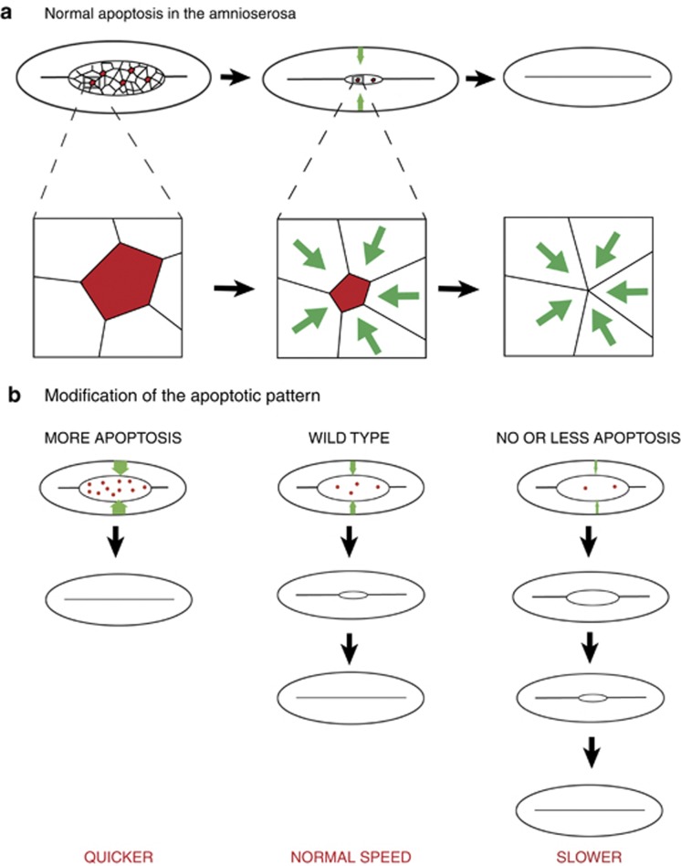
A few years ago, an active mechanical function of apoptosis was revealed during Drosophila dorsal closure, a powerful model system to study wound healing in vivo.41, 42 Dorsal closure is a morphogenetic process taking place at the end of embryogenesis. Before dorsal closure, only the ventral and lateral surfaces of the embryo are covered by epidermal cells, whereas the dorsal side is covered by the amnioserosa, a transient tissue that is eventually eliminated. During dorsal closure, the lateral epidermis moves dorsalward to cover the amnioserosa. This morphogenetic process is accompanied by cellular elongation and formation of an acting cable in the leading edge, the most dorsal row of lateral epidermis.43, 44 Synchronized cellular forces from both the contracting amnioserosa and the migrating dorsal epithelium, which tightly contacts the amnioserosa throughout the process, contribute to the movement. Interestingly, a small fraction of cells from the amniosera (around 10%) were shown to constrict their apical surface and drop out of the amnioserosa surface plane.45 More recently, Toyama et al.42 showed that these cells present all the hallmarks of apoptosis, including blebbing and fragmentation, and that they induce cell-shape modification of their neighbors. Furthermore, apoptosis has an active role in regulating the speed of dorsal closure; if apoptosis is inhibited in the amniosera, closure is delayed and the force produced by the amnioserosa on the dorsal epithelium is reduced. In contrast, ectopic induction of apoptosis can accelerate the movement.41, 42 Therefore, in this system, apoptosis has an active role in promoting tissue dynamics, though it is not strictly essential for this process. Although the precise underlying molecular mechanisms remain to be elucidated, one can speculate that the modification of the actomyosin network in the neighboring cells through, for instance, the formation of actomyosin extrusion rings, creates a pulling force that is propagated throughout the amnioserosa to the epidermis, thus enhancing the movement (Figure 2).
Figure 2.
Apoptosis acting as a pulling force. (a; up) Schematic representation of a dorsal view of a Drosophila embryo during dorsal closure. Apoptotic cells are indicated in red (in the amnioserosa), the force generated by apoptosis in the lateral epidermis is represented by the green arrows. Successive stages of dorsal closure are represented. (a; down) Representation of an apoptotic cells and its closest neighbors at higher magnification. The progressive stretching of the neighboring cells is proposed to be the origin of the pulling force generated by apoptosis on the surrounding tissue.(b) Dorsal closure defects when apoptosis pattern is modified. Dorsal closure can be either accelerated when apoptosis is promoted (left), or slowed down when apoptosis is inhibited (right)
Interestingly, a recent study suggests that apoptosis may have a similar function during neural tube closure in mouse. Large amounts of apoptotic cells were originally observed during neural tube closure.46 More recently, the dynamic pattern of apoptosis has been determined in this tissue, revealing extensive apoptosis in the dorsal ridge of the neural plates and in the boundary domain between neural plates and surface ectoderm. Similar to what is observed during dorsal closure in Drosophila, apoptosis does not seem to be essential for neural tube closure, but is required for accelerating the process and ensure zipping.47
Role of apoptosis in tissue-brake release
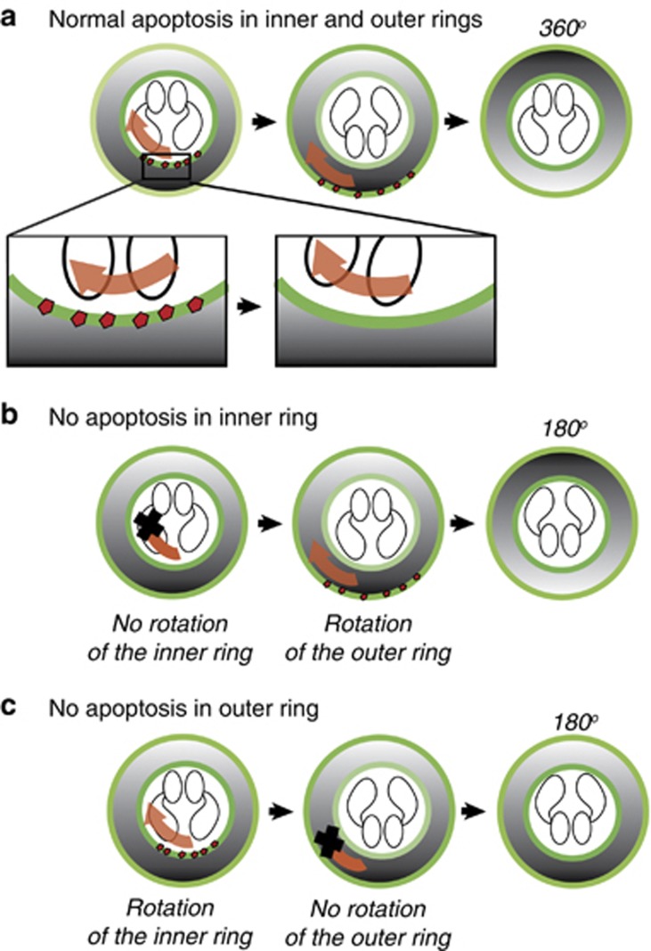
More recently, apoptosis has been shown to act as a ‘brake-release' signal during genitalia rotation in Drosophila, a developmental process that involves dramatic tissue rearrangement.48 Genitalia rotation occurs during the development of the male fly and consists of a 360° clockwise rotation of the genital plate, a part of the body giving rise to the genital and anal structures in the adult.49, 50 This process is the sum of two 180° rotations of each of the two ring-shaped domains of the genitalia.48 Initial insight into the cellular mechanism underlying this morphogenetic movement came from the mis-rotation phenotypes observed in certain cell death mutants, such as hid.51, 52, 53 More recent studies determined the spatial and temporal patterns of apoptosis that occur at the boundaries of these motile domains, specifically when their movements begin.48, 54 Although their conclusions differ, the two latter studies provide interesting new insights into the roles that apoptosis could have during morphogenesis. In the first study, Suzanne et al.48 proposed that local apoptosis could provide a mechanism for detaching each motile domain from one another as they are part of the same epithelium, thus giving them the degree of freedom required for their movement. This conclusion was based on the observation that inactivation of apoptosis in either one or the other rotating domain led to similar defects with a genital plate half-rotated. Further support came from the observation that genitalia rotation could be completely blocked by a total inhibition of apoptosis in both rings (40% penetrance). Live imaging of the rotation process using specific markers of each ring domain confirmed that each ring movement depends on the presence of apoptosis at its boundary, suggesting that each ring requires apoptosis to dissociate from the neighboring tissue. Therefore, it appears that apoptosis in this system serves to separate the moving parts of the genital plate from the neighboring tissue, acting like a ‘biological scissor' to sever tissues with different motile properties (Figure 3). An alternative possibility is that apoptosis may provide a force to actively promote rotation. Although it is difficult to imagine that apoptosis of a relatively small number of cells is sufficient to generate the driving force for the extenisve movement of two large domains, it may contribute force to the movement. Such a role would be consistent with the previous work on dorsal closure.42 Along these lines, Kurunaga et al.54 suggested that apoptosis may have a role in accelerating genital disc rotation. In support of this idea, inhibition of apoptosis specifically in the inner ring slowed down rotation, but did not completely block movement. Conversely, increasing apoptosis by expression of the pro-apoptotic Reaper protein at the boundary between the two moving rings accelerated genital rotation. These data suggest that apoptosis may not only separate the rotating rings, but also contribute to the force that drives the movement. The idea that apoptosis could be an accelerator is interesting; however, we know now that two different domains contribute to the rotation, each one turning 180°,48 and this could totally change this interpretation. Two different explanations could be proposed for the deceleration observed when apoptosis is inhibited in only one rotating domain: either the acceleration of the rotation is affected or the release of the motile domain is compromised. Further experiments will help in clarifying this point.
Figure 3.
Apoptosis acting as a ‘biological scissor'. (a) Schematic representation of the genital plate of male Drosophila during genitalia rotation. Apoptotic cells (shown in red) allows the initiation of the rotational movement of the inner ring (left, red arrow), then of the outer ring (middle, red arrow). Each ring domain undergoes a 180° rotation. A higher magnification representing the cellular processes taking place at the boundary between the motile ring and the neighboring tissue is shown in the lower panel. Local apoptosis occurs at the boundary and is required to free the motile ring, suggesting its role in tissue detachment. (b) Schematic representation of defects observed when apoptosis is inhibited either in the inner ring (up) or in the outer ring (down) specifically. In each case, the ring in which apoptosis is impaired cannot move, only the other one can rotate leading to a final rotation of 180o
Conclusion
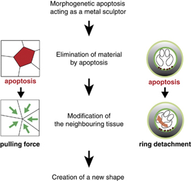
Despite considerable advances in the field of cell death research, we still know very little about how apoptosis contributes to tissue morphogenesis at the cellular level. Work on genital rotation in Drosophila and dorsal and neural closure in Drosophila and mouse suggests that apoptosis has an active role in reorganizing the cellular environment, either by inducing a rearrangement in cellular adhesion or by directly generating a tissue force. Thus, at least in some cases, sculpting through apoptosis can be viewed more like ‘metal sculpting' rather than ‘stone sculpting', where shape is created by modification rather than by the elimination of existing material (Figure 4). Here, the main purpose of cell death is not the progressive elimination of material, but rather the generation of local forces that alter tissue shape. However, at this time, the precise underlying mechanism remains elusive. It is possible that dying cells produce molecular signals to instruct neighboring cells, or that force is generated by cell detachment and/or physical elimination of cellular material. The dramatic morphological alterations of dying cells may induce a reorganization of the neighboring microenvironement that, along with biochemical signals between dying cells and neighbors, could influence cell behavior during epithelial morphogenesis. Altogether, these observations suggest that apoptosis generates a major sculpting force during morphogenesis. However, further studies are needed to determine both the molecular and mechanical signals produced by dying cells and how these might cooperate to shape tissue and organs. Here, we have described recent advances about the influence of apoptosis on the surrounding tissue during morphogenesis. It would be interesting now to determine if the force generated by apoptosis is more generally used in other morphogenetic processes involving cell migration, like in palate fusion. Similarly, further investigations are needed to determine if apoptosis is commonly used as a biological scissor during huge morphogenetic reorganization, like cardiac rotation in vertebrates.
Figure 4.
Morphogenetic apoptosis acting as a ‘metal sculptor'. Schematic representations of the cellular rearrangements taking place during dorsal closure (left) or genitalia rotation (right). Apoptosis is modifying the surrounding tissue, either by creating a force in the tissue or reducing attachment, thus creating a new shape
Acknowledgments
We thank Melanie Gettings for careful reading and suggestions on the manuscript. H Steller is an Investigator of the Howard Hughes Medical Institute. Part of this work was supported by NIH grant R01GM60124 to HS. Research in the group of M Suzanne is supported by the Agence Nationale de la Recherche Program 2010-JCJC-1208-01.
Glossary
Wg
wingless
Dpp
decapentaplegic
S1P
sphingosine 1 phosphate
S1P2
sphingosine 1 phosphate receptor
JNK
c-Jun N-teminal Kinase
115RhoGEF
GTPase-activating protein
Hid
head involution defective
MMP
Matrix metalloproteinase
Footnotes
References
- Kerr JF, Wyllie AH, Currie AR. Apoptosis: a basic biological phenomenon with wide-ranging implications in tissue kinetics. Br J Cancer. 1972;26:239–257. doi: 10.1038/bjc.1972.33. [DOI] [PMC free article] [PubMed] [Google Scholar]
- Potts MB, Cameron S. Cell lineage and cell death: Caenorhabditis elegans and cancer research. Nat Rev Cancer. 2010;11:50–58. doi: 10.1038/nrc2984. [DOI] [PubMed] [Google Scholar]
- Zakeri Z, Quaglino D, Ahuja HS. Apoptotic cell death in the mouse limb and its suppression in the hammertoe mutant. Dev Biol. 1994;165:294–297. doi: 10.1006/dbio.1994.1255. [DOI] [PubMed] [Google Scholar]
- Coleman ML, Sahai EA, Yeo M, Bosch M, Dewar A, Olson MF. Membrane blebbing during apoptosis results from caspase-mediated activation of ROCK I. Nat Cell Biol. 2001;3:339–345. doi: 10.1038/35070009. [DOI] [PubMed] [Google Scholar]
- Crawford ED, Wells JA. Caspase substrates and cellular remodeling. Ann Rev Biochem. 2011;80:1055–1087. doi: 10.1146/annurev-biochem-061809-121639. [DOI] [PubMed] [Google Scholar]
- Dix MM, Simon GM, Cravatt BF. Global mapping of the topography and magnitude of proteolytic events in apoptosis. Cell. 2008;134:679–691. doi: 10.1016/j.cell.2008.06.038. [DOI] [PMC free article] [PubMed] [Google Scholar]
- Enari M, Sakahira H, Yokoyama H, Okawa K, Iwamatsu A, Nagata S. A caspase-activated DNase that degrades DNA during apoptosis, and its inhibitor ICAD. Nature. 1998;391:43–50. doi: 10.1038/34112. [DOI] [PubMed] [Google Scholar]
- Liu X, Zou H, Slaughter C, Wang X. DFF, a heterodimeric protein that functions downstream of caspase-3 to trigger DNA fragmentation during apoptosis. Cell. 1997;89:175–184. doi: 10.1016/s0092-8674(00)80197-x. [DOI] [PubMed] [Google Scholar]
- Mahrus S, Trinidad JC, Barkan DT, Sali A, Burlingame AL, Wells JA. Global sequencing of proteolytic cleavage sites in apoptosis by specific labeling of protein N termini. Cell. 2008;134:866–876. doi: 10.1016/j.cell.2008.08.012. [DOI] [PMC free article] [PubMed] [Google Scholar]
- Sebbagh M, Renvoize C, Hamelin J, Riche N, Bertoglio J, Breard J. Caspase-3-mediated cleavage of ROCK I induces MLC phosphorylation and apoptotic membrane blebbing. Nat Cell Biol. 2001;3:346–352. doi: 10.1038/35070019. [DOI] [PubMed] [Google Scholar]
- Ndozangue-Touriguine O, Hamelin J, Breard J. Cytoskeleton and apoptosis. Biochem Pharmacol. 2008;76:11–18. doi: 10.1016/j.bcp.2008.03.016. [DOI] [PubMed] [Google Scholar]
- Fuchs Y, Steller H. Programmed cell death in animal development and disease. Cell. 2011;147:742–758. doi: 10.1016/j.cell.2011.10.033. [DOI] [PMC free article] [PubMed] [Google Scholar]
- Hernandez-Martinez R, Covarrubias L. Interdigital cell death function and regulation: new insights on an old programmed cell death model. Dev Growth Differ. 2011;53:245–258. doi: 10.1111/j.1440-169X.2010.01246.x. [DOI] [PubMed] [Google Scholar]
- Ganan Y, Macias D, Basco RD, Merino R, Hurle JM. Morphological diversity of the avian foot is related with the pattern of msx gene expression in the developing autopod. Dev Biol. 1998;196:33–41. doi: 10.1006/dbio.1997.8843. [DOI] [PubMed] [Google Scholar]
- Weatherbee SD, Behringer RR, JJt Rasweiler, Niswander LA. Interdigital webbing retention in bat wings illustrates genetic changes underlying amniote limb diversification. Proc Natl Acad Sci USA. 2006;103:15103–15107. doi: 10.1073/pnas.0604934103. [DOI] [PMC free article] [PubMed] [Google Scholar]
- Dupe V, Ghyselinck NB, Thomazy V, Nagy L, Davies PJ, Chambon P, et al. Essential roles of retinoic acid signaling in interdigital apoptosis and control of BMP-7 expression in mouse autopods. Dev Biol. 1999;208:30–43. doi: 10.1006/dbio.1998.9176. [DOI] [PubMed] [Google Scholar]
- Zhao X, Brade T, Cunningham TJ, Duester G. Retinoic acid controls expression of tissue remodeling genes Hmgn1 and Fgf18 at the digit-interdigit junction. Dev Dyn. 2010;239:665–671. doi: 10.1002/dvdy.22188. [DOI] [PMC free article] [PubMed] [Google Scholar]
- Kim WR, Sun W. Programmed cell death during postnatal development of the rodent nervous system. Dev Growth Differ. 2011;53:225–235. doi: 10.1111/j.1440-169X.2010.01226.x. [DOI] [PubMed] [Google Scholar]
- Coen L, du Pasquier D, Le Mevel S, Brown S, Tata J, Mazabraud A, et al. Xenopus Bcl-X(L) selectively protects Rohon-Beard neurons from metamorphic degeneration. Proc Natl Acad Sci USA. 2001;98:7869–7874. doi: 10.1073/pnas.141226798. [DOI] [PMC free article] [PubMed] [Google Scholar]
- Coen L, Le Blay K, Rowe I, Demeneix BA. Caspase-9 regulates apoptosis/proliferation balance during metamorphic brain remodeling in Xenopus. Proc Natl Acad Sci USA. 2007;104:8502–8507. doi: 10.1073/pnas.0608877104. [DOI] [PMC free article] [PubMed] [Google Scholar]
- Ishizuya-Oka A. Death is the major fate of medial edge epithelial cells and the cause of basal lamina degradation during palatogenesis. Development. 2003;131:15–24. doi: 10.1242/dev.00907. [DOI] [PubMed] [Google Scholar]
- Cuevo R, Covarrubias L. Death is the major fate of medial edge epithelial cells and the cause of basal lamina degradation during palatogenesis. Development. 2003;131:15–24. doi: 10.1242/dev.00907. [DOI] [PubMed] [Google Scholar]
- Lohmann I, McGinnis N, Bodmer M, McGinnis W. The Drosophila Hox gene deformed sculpts head morphology via direct regulation of the apoptosis activator reaper. Cell. 2002;110:457–466. doi: 10.1016/s0092-8674(02)00871-1. [DOI] [PubMed] [Google Scholar]
- Manjon C, Sanchez-Herrero E, Suzanne M. Sharp boundaries of Dpp signalling trigger local cell death required for Drosophila leg morphogenesis. Nat Cell Biol. 2007;9:57–63. doi: 10.1038/ncb1518. [DOI] [PubMed] [Google Scholar]
- Ravichandran KS. Beginnings of a good apoptotic meal: the find-me and eat-me signaling pathways. Immunity. 2011;35:445–455. doi: 10.1016/j.immuni.2011.09.004. [DOI] [PMC free article] [PubMed] [Google Scholar]
- Rosenblatt J, Raff MC, Cramer LP. An epithelial cell destined for apoptosis signals its neighbors to extrude it by an actin- and myosin-dependent mechanism. Curr Biol. 2001;11:1847–1857. doi: 10.1016/s0960-9822(01)00587-5. [DOI] [PubMed] [Google Scholar]
- Slattum G, McGee KM, Rosenblatt J. P115 RhoGEF and microtubules decide the direction apoptotic cells extrude from an epithelium. J Cell Biol. 2009;186:693–702. doi: 10.1083/jcb.200903079. [DOI] [PMC free article] [PubMed] [Google Scholar]
- Gu Y, Forostyan T, Sabbadini R, Rosenblatt J. Epithelial cell extrusion requires the sphingosine-1-phosphate receptor 2 pathway. J Cell Biol. 2011;193:667–676. doi: 10.1083/jcb.201010075. [DOI] [PMC free article] [PubMed] [Google Scholar]
- Fan Y, Bergmann A. Distinct mechanisms of apoptosis-induced compensatory proliferation in proliferating and differentiating tissues in the Drosophila eye. Dev Cell. 2008;14:399–410. doi: 10.1016/j.devcel.2008.01.003. [DOI] [PMC free article] [PubMed] [Google Scholar]
- Huh JR, Guo M, Hay BA. Compensatory proliferation induced by cell death in the Drosophila wing disc requires activity of the apical cell death caspase Dronc in a nonapoptotic role. Curr Biol. 2004;14:1262–1266. doi: 10.1016/j.cub.2004.06.015. [DOI] [PubMed] [Google Scholar]
- Perez-Garijo A, Martin FA, Morata G. Caspase inhibition during apoptosis causes abnormal signalling and developmental aberrations in Drosophila. Development. 2004;131:5591–5598. doi: 10.1242/dev.01432. [DOI] [PubMed] [Google Scholar]
- Perez-Garijo A, Shlevkov E, Morata G. The role of Dpp and Wg in compensatory proliferation and in the formation of hyperplastic overgrowths caused by apoptotic cells in the Drosophila wing disc. Development. 2009;136:1169–1177. doi: 10.1242/dev.034017. [DOI] [PubMed] [Google Scholar]
- Ryoo HD, Gorenc T, Steller H. Apoptotic cells can induce compensatory cell proliferation through the JNK and the Wingless signaling pathways. Dev Cell. 2004;7:491–501. doi: 10.1016/j.devcel.2004.08.019. [DOI] [PubMed] [Google Scholar]
- Bergmann A, Steller H. Apoptosis, stem cells, and tissue regeneration. Sci Signal. 2010;3:re8. doi: 10.1126/scisignal.3145re8. [DOI] [PMC free article] [PubMed] [Google Scholar]
- Martin FA, Perez-Garijo A, Morata G. Apoptosis in Drosophila: compensatory proliferation and undead cells. Int J Dev Biol. 2009;53:1341–1347. doi: 10.1387/ijdb.072447fm. [DOI] [PubMed] [Google Scholar]
- Chera S, Ghila L, Dobretz K, Wenger Y, Bauer C, Buzgariu W, et al. Apoptotic cells provide an unexpected source of Wnt3 signaling to drive hydra head regeneration. Dev Cell. 2009;17:279–289. doi: 10.1016/j.devcel.2009.07.014. [DOI] [PubMed] [Google Scholar]
- Goessling W, North TE, Loewer S, Lord AM, Lee S, Stoick-Cooper CL, et al. Genetic interaction of PGE2 and Wnt signaling regulates developmental specification of stem cells and regeneration. Cell. 2009;136:1136–1147. doi: 10.1016/j.cell.2009.01.015. [DOI] [PMC free article] [PubMed] [Google Scholar]
- Huang Q, Li F, Liu X, Li W, Shi W, Liu FF, et al. Caspase 3-mediated stimulation of tumor cell repopulation during cancer radiotherapy. Nat Med. 2011;17:860–866. doi: 10.1038/nm.2385. [DOI] [PMC free article] [PubMed] [Google Scholar]
- Li F, Huang Q, Chen J, Peng Y, Roop DR, Bedford JS, et al. Apoptotic cells activate the ‘phoenix rising' pathway to promote wound healing and tissue regeneration. Sci Signal. 2010;3:ra13. doi: 10.1126/scisignal.2000634. [DOI] [PMC free article] [PubMed] [Google Scholar]
- Yamada Y, Davis KD, Coffman CR. Programmed cell death of primordial germ cells in Drosophila is regulated by p53 and the Outsiders monocarboxylate transporter. Development. 2008;135:207–216. doi: 10.1242/dev.010389. [DOI] [PubMed] [Google Scholar]
- Toyama Y, Peralta XG, Wells AR, Kiehart DP, Edwards GS. Apoptotic force and tissue dynamics during Drosophila embryogenesis. Science. 2008;321:1683–1686. doi: 10.1126/science.1157052. [DOI] [PMC free article] [PubMed] [Google Scholar]
- Gorfinkiel N, Blanchard GB, Adams RJ, Martinez Arias A. Mechanical control of global cell behaviour during dorsal closure in Drosophila. Development. 2009;136:1889–1898. doi: 10.1242/dev.030866. [DOI] [PMC free article] [PubMed] [Google Scholar]
- Harris TJ, Sawyer JK, Peifer M. How the cytoskeleton helps build the embryonic body plan: models of morphogenesis from Drosophila. Curr Top Dev Biol. 2009;89:55–85. doi: 10.1016/S0070-2153(09)89003-0. [DOI] [PubMed] [Google Scholar]
- Knust E. Drosophila morphogenesis: movements behind the edge. Curr Biol. 1997;7:R558–R561. doi: 10.1016/s0960-9822(06)00281-8. [DOI] [PubMed] [Google Scholar]
- Kiehart DP, Galbraith CG, Edwards KA, Rickoll WL, Montague RA. Multiple forces contribute to cell sheet morphogenesis for dorsal closure in Drosophila. J Cell Biol. 2000;149:471–490. doi: 10.1083/jcb.149.2.471. [DOI] [PMC free article] [PubMed] [Google Scholar]
- Weil M, Jacobson MD, Raff MC. Is programmed cell death required for neural tube closure. Curr Biol. 1997;7:281–284. doi: 10.1016/s0960-9822(06)00125-4. [DOI] [PubMed] [Google Scholar]
- Yamaguchi Y, Shinotsuka N, Nonomura K, Takemoto K, Kuida K, Yosida H, et al. Live imaging of apoptosis in a novel transgenic mouse highlights its role in neural tube closure. J Cell Biol. 2011;195:1047–1060. doi: 10.1083/jcb.201104057. [DOI] [PMC free article] [PubMed] [Google Scholar]
- Suzanne M, Petzoldt AG, Speder P, Coutelis JB, Steller H, Noselli S. Coupling of apoptosis and L/R patterning controls stepwise organ looping. Curr Biol. 2010;20:1773–1778. doi: 10.1016/j.cub.2010.08.056. [DOI] [PMC free article] [PubMed] [Google Scholar]
- Gleichauf R. Anatomie und variabilität des geschlechtsapparates von Drosophila melanogaster (Meigen) Z Wiss Zool. 1936;148:1–66. [Google Scholar]
- Speder P, Adam G, Noselli S. Type ID unconventional myosin controls left-right asymmetry in Drosophila. Nature. 2006;440:803–807. doi: 10.1038/nature04623. [DOI] [PubMed] [Google Scholar]
- Abbott MK, Lengyel JA. Embryonic head involution and rotation of male terminalia require the Drosophila locus head involution defective. Genetics. 1991;129:783–789. doi: 10.1093/genetics/129.3.783. [DOI] [PMC free article] [PubMed] [Google Scholar]
- Grether ME, Abrams JM, Agapite J, White K, Steller H. The head involution defective gene of Drosophila melanogaster functions in programmed cell death. Genes Dev. 1995;9:1694–1708. doi: 10.1101/gad.9.14.1694. [DOI] [PubMed] [Google Scholar]
- Macias A, Romero NM, Martin F, Suarez L, Rosa AL, Morata G. PVF1/PVR signaling and apoptosis promotes the rotation and dorsal closure of the Drosophila male terminalia. Int J Dev Biol. 2004;48:1087–1094. doi: 10.1387/ijdb.041859am. [DOI] [PubMed] [Google Scholar]
- Kuranaga E, Matsunuma T, Kanuka H, Takemoto K, Koto A, Kimura K, et al. Apoptosis controls the speed of looping morphogenesis in Drosophila male terminalia. Development. 2011;138:1493–1499. doi: 10.1242/dev.058958. [DOI] [PubMed] [Google Scholar]


