Single-cell mRNA sequencing identifies subclonal heterogeneity in anti-cancer drug responses of lung adenocarcinoma cells (original) (raw)
Abstract
Background
Intra-tumoral genetic and functional heterogeneity correlates with cancer clinical prognoses. However, the mechanisms by which intra-tumoral heterogeneity impacts therapeutic outcome remain poorly understood. RNA sequencing (RNA-seq) of single tumor cells can provide comprehensive information about gene expression and single-nucleotide variations in individual tumor cells, which may allow for the translation of heterogeneous tumor cell functional responses into customized anti-cancer treatments.
Results
We isolated 34 patient-derived xenograft (PDX) tumor cells from a lung adenocarcinoma patient tumor xenograft. Individual tumor cells were subjected to single cell RNA-seq for gene expression profiling and expressed mutation profiling. Fifty tumor-specific single-nucleotide variations, including KRAS G12D, were observed to be heterogeneous in individual PDX cells. Semi-supervised clustering, based on KRAS G12D mutant expression and a risk score representing expression of 69 lung adenocarcinoma-prognostic genes, classified PDX cells into four groups. PDX cells that survived in vitro anti-cancer drug treatment displayed transcriptome signatures consistent with the group characterized by KRAS G12D and low risk score.
Conclusions
Single-cell RNA-seq on viable PDX cells identified a candidate tumor cell subgroup associated with anti-cancer drug resistance. Thus, single-cell RNA-seq is a powerful approach for identifying unique tumor cell-specific gene expression profiles which could facilitate the development of optimized clinical anti-cancer strategies.
Electronic supplementary material
The online version of this article (doi:10.1186/s13059-015-0692-3) contains supplementary material, which is available to authorized users.
Background
Identification of somatic driver mutations in cancer has led to the development of targeted therapeutics that have improved the clinical outcomes of cancer patients [1–3]. Lung adenocarcinoma (LUAD), the most common histological subtype of non-small cell lung cancer [4], is denoted by genetic alterations in the receptor tyrosine kinase (RTK)-RAS-mitogen-activated protein kinase (MAPK) pathway [2]. Companion diagnostics for hotspot mutations of EGFR, KRAS, BRAF, and ALK, which are clinically associated with specific targeted cancer therapies, are currently available for LUADs [5]. While the detection rate of currently identified actionable mutations in LUAD is over 60 % [2], efforts to catalogue all the clinically relevant genetic variations are still ongoing [6–9]. Moreover, drug resistance and disease recurrence after anti-cancer treatments require more comprehensive genomic analysis of individual LUADs [10, 11].
Although the individual cells in a tumor mass originate from a common ancestor and share early tumor-initiating genetic alterations, tumor cells frequently diverge and show heterogeneity in growth [12–14], drug resistance [15, 16], and metastatic potential [13, 14]. Intra-tumoral heterogeneity results from mutation and clonal selection dynamics during tumor growth [13, 14, 16], where individual tumor cells accumulate cell-specific genetic changes [12]. This genetic heterogeneity is significantly associated with tumor progression and the treatment outcomes of cancers [17, 18]. Therefore, monitoring intra-tumoral heterogeneity at the single-cell level would broaden our understanding of tumor recurrence mechanisms after anti-cancer treatments [19] and guide us in developing more sophisticated strategies to overcome drug resistance.
Single-cell genome profiling technology provides the highest-resolution analysis of intra-tumoral genetic heterogeneity [20–22]. Based on heterogeneity, we can identify individual cells with specific genetic alterations or genomic expression profiles that could be responsible for treatment resistance. Therefore, correlating the genotype–phenotype relationship in genetically distinct single cells can provide important new information for selecting the most appropriate clinical intervention for targeting heterogeneous LUADs [23]. For this purpose, patient-derived xenograft (PDX) cells provide a genetically and phenotypically accessible model for single cancer cell analyses of the heterogeneous histopathological, genetic, molecular, and functional characteristics of parental tumors [24, 25]. Moreover, drug-resistant tumor cells can be selected and analyzed in vitro using PDX cells.
We performed transcriptome profiling on single PDX cells from a LUAD patient to elucidate the molecular mechanisms and underlying genomic characteristics of tumor cell resistance to anti-cancer drug treatments. Single-cell transcriptome analysis uncovered heterogeneous behaviors of individual tumor cells and provided new insights into drug resistance signatures that were masked in bulk tumor analyses.
Results
Intra-tumoral genetic heterogeneity of LUAD PDX cells
Surgically removed LUAD tissue was propagated through xenograft engraftments in mice (Fig. 1a). Viable cancer cells were dissociated from the PDX tissue and primarily cultured in vitro (Figure S1a in Additional file 1). Cultured PDX cells were genomically analyzed by RNA sequencing (RNA-seq) and whole-exome sequencing (WES). Although the tumor portion in the surgical sample represented approximately 40 % of the excised tissue volume (Figure S1b in Additional file 1), multiple validated genomic analyses utilizing WES [26, 27] and expression profiles [28] indicated that human cancer cells were highly enriched (~100 %) in the PDX cells (Fig. 1b). Overall, copy number alterations and variant allele frequencies were increased in the PDX tumor, compared with the surgical specimen (Fig. 1c, d). Some mutations present in the patient tumor were lost in the PDX, suggesting that our PDX model went through a selective engraftment process [29]. The histologic characteristics of the patient tumor were well preserved in the PDX (Figure S1c in Additional file 1). The full profiles of somatic mutations in the patient tumor and PDX cells are listed in Additional file 2.
Fig. 1.
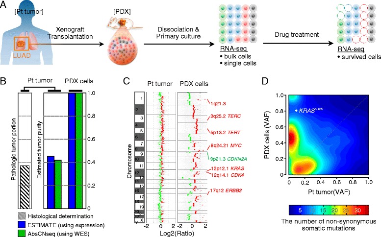
Enrichment of cancer cells in the PDX. a Schematic representation of experiments. A portion of a LUAD patient tumor (Pt tumor) was propagated by xenograft transplantation in humanized immunocompromised female NOG mice. PDX cells (PDX) were dissociated and cultured from xenograft tumors, and subjected to drug screening. b Estimated cancer cell fraction in Pt tumor and PDX cells. The fraction was quantified by histopathological examination (striped bar), or estimated based on computational analysis using expression profiles (blue) or WES data (green). c Estimated degree of normalized copy number changes in log2 ratio to matched peripheral blood for deletion (green) or amplification (red) are indicated. Representative sites of copy number changes in LUAD are labeled on the right side. d Distribution of variant allele frequencies (VAF) of the non-synonymous somatic mutations that overlap between Pt tumor and PDX cells. Color-scaled density map indicates the number of mutations
Tumor cell-enriched PDX cells (LC-PT-45) [30] were further analyzed by single-cell RNA-seq using the Fluidigm C1™ autoprep system with SMART-seq [31]. cDNAs from 34 individual PDX cells were successfully amplified. Using 100-bp paired-end sequencing, we obtained an average of 8.12 ± 2.34 million mapped reads from the captured cells (Additional file 3). Overall, 85.63 % of reads mapped to the human reference genome, which was a lower percentage than is typical for unamplified conventional RNA-seq, but comparable to other single cell RNA-seq data [31, 32]. We also sequenced 50 single H358 human lung cancer cells as cell line controls and obtained an 85.39 % mapping rate (Additional file 3). Noticeably skewed coverage at the 3’ end of transcripts, which was inversely proportional to the expression level, was observed in the single-cell RNA-seq data (Additional file 4). The use of smaller initial RNA templates for amplification is known to increase this bias [31].
Despite the sequencing bias in amplified RNAs, average gene expression in single cells correlated well with expression in bulk cells, for both H358 and PDX cells (Fig. 2a). The inter-correlation of total gene expression among the 34 individual PDX cells showed wider distribution compared with that in the 50 H358 cells (Fig. 2b), indicating moderately higher transcriptome heterogeneity. The level of transcriptome heterogeneity was also evaluated by multiple regression analysis of different sized pools (n = 5, 15, 25, 34/35, 50; randomly selected by permutation × 1000) of single cell transcriptomes to the bulk sample (Fig. 2c). The modeling demonstrated that five H358 or PDX individual cells represented >70 % of the gene expression of the whole population. When averaging increased numbers of cells, the single cell data approximated the bulk up to 85 %, suggesting that the single cell data are consistent with the bulk data (Fig. 2c). We repeated the single cell isolation and RNA-seq using 43 additional PDX cells and obtained comparable results that were highly correlated with the first data set (Fig. 2; Figure S3a–f in Additional file 5, LC-PT-45 and LC-PT-45-Re). Comparisons of gene expression data for the 43 target genes (see Additional file 6 for the gene list and Figure S3g in Additional file 5 for expression levels) between technical replicate RNA-seq sets (Figure S3h left in Additional file 5) or between RNA-seq and quantitative PCR (qPCR) analysis (Figure S3h right in Additional file 5) also demonstrated statistically significant correlation, comparable to that reported in a previous publication [33].
Fig. 2.
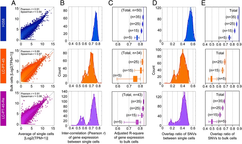
Intra-tumoral heterogeneity of PDX cells. a Scatter plots of the average gene expression of single cells (H358, n = 50; LC-PT-45, n = 34; LC-PT-45-Re, n = 43) compared with those of the corresponding bulk cells (~1 × 10⁵ cells). Black dotted lines are x = y lines with correlation coefficients (Pearson r and Spearman r) for linear fit. TPM transcripts per million. b Inter-correlation (Pearson r) between gene expression in single cells. Density plots were constructed with a kernel function fitting over the histograms. c Explanatory power (adjusted R-square) of gene expression in various numbers of single cells relative to the bulk cells was determined by multiple regression analysis with randomly selected cell numbers with permutation (×1000). d Overlap ratio of expressed single-nucleotide variations (SNV) among single cells. Density plots were constructed with a kernel function fitting over the histograms. e Overlap ratio of expressed SNVs of various numbers of single cells relative to that of the bulk cells was calculated with a randomly selected given number of cells with permutation (×1000). For boxplots in (c) and (e), box = interquartile range between the first and the third quartiles, error bars = 10th–90th percentiles
Single-cell heterogeneity of expressed single-nucleotide variants
To estimate tumor heterogeneity at the genetic mutation level, we identified expressed single-nucleotide variants (SNVs) using the single-cell RNA-seq and bulk WES data (Figure S4a in Additional file 7). After removal of potential false positive SNVs specifically found in RNA-seq using the SNPiR package [34], higher overlap ratios to bulk WES data were observed (Figure S4b middle panels in Additional file 7). Selection of SNVs found in both single cell RNA-seq and bulk WES data significantly increased the overlap ratios to dbSNP137 (Figure S4b right panels in Additional file 7). These filtered SNVs of individual PDX cells showed relatively heterogeneous expression compared with those of H358 cells in terms of the lower overlap ratios between single cells (Fig. 2d). The union of SNVs from five PDX cells (randomly selected by permutation × 1000) reflected 49 % of the expressed SNVs in the whole population, whereas those of five H358 cells represented 75 % (Fig. 2e). With increased numbers of single cells, the coverage increased up to 70 and 90 % for PDX cells (34 LC-PT-45 or 43 LC-PT-45-Re) and H358 cells, respectively.
After exclusion of germline variants by selecting only somatic SNVs from bulk WES data, expression of 50 tumor-specific non-synonymous SNVs was analyzed in individual PDX cells (Figure S4a in Additional file 7). The 50 tumor-specific SNVs showed heterogeneous expression patterns in the individual PDX cells (Fig. 3a, LC-PT-45) with numerous allele dropouts. For comparison, we plotted expression of lung cancer mutations in the H358 cell line listed in COSMIC [35] (Figure S5a in Additional file 8), which also showed variable expression patterns with more uniform coverage (Figure S5b in Additional file 8). For the PDX cells, we detected comparable mutation patterns and frequencies in the original and replicate PDX analyses (Fig. 3a; Figure S3c, f in Additional file 5, LC-PT-45 vs. LC-PT-45-Re RNA-seq). The number of reads mapped to the human genome reference were determined for individual cells to assure sequencing quality (Fig. 3b). We also performed genotyping PCR on the LC-PT-45-Re samples in parallel, which showed >70 % concordance with the RNA-seq results [Fig. 3a, LC-PT-45-Re (RNA-seq) vs. LC-PT-45-Re (genotyping PCR), and Fig. 3c; Additional file 9]. Together these data support reproducible cellular variance in SNV expression. Nevertheless, no calls and discrepant mutation calls between RNA-seq and genotyping PCR demonstrate limitations of single cell RNA-seq, which might have originated from allelic dropouts.
Fig. 3.
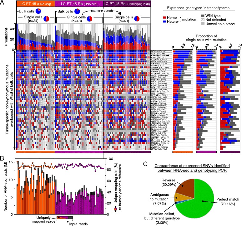
Heterogeneous expression patterns of SNVs in PDX cells. a Expressed, tumor-specific, non-synonymous somatic mutations found in more than three single cells of LC-PT-45. The replicate batch (LC-PT-45-Re) of single-cell RNA-seq and that of genotyping PCR are shown together. The bar graphs at top left show mutation events per sample; the heat maps at bottom left show mutation profiles across samples; the bar graphs at the right show normalized mutation fraction over total single cells (LC-PT-45, n = 34; LC-PT-45-Re, n = 43). b Mapping information from RNA-seq for reads mapped to the human reference genome (hg19). The bar graph (left y-axis) shows the number of RNA-seq reads and the scatter plots (right y-axis) with a connecting line show the unique mapping rate (uniquely mapped reads/input reads) in the same order as in (a). c Summary of results for the matched samples and the validated targets between RNA-seq and genotyping PCR shown in (a). See Additional file 8 for the details
Among the genes with SNVs detected in PDX cells, KRAS [1, 2], GAPVD1 [36], and JMJD1C [37] are functionally related to the RTK-RAS-MAPK signaling pathway. The hotspot KRAS G12D mutation was detected in 27 out of 34 single PDX cells (79.4 %), or 33 out of 43 PDX replicates (76.7 %). To determine whether the variable mutant allele expression was due to genetic heterogeneity, we assessed the genotypes of 12 somatic mutations at the single-cell DNA level with droplet digital PCR (ddPCR; Figure S7a in Additional file 10). When mutation rates were computed as variant allele frequencies in bulk cells or as mutant single cell fractions at both the DNA and RNA levels, they showed overall correlation (Figure S7b in Additional file 10). With respect to the KRAS mutation, all PDX cells (21 of 21) harbored the mutant allele in the single-cell DNA analysis. Of note, copy number gains (Fig. 1c) and mutant/wild-type ratios in KRAS (Figure S7c in Additional file 10) suggest that variable copy numbers of the mutant KRAS influenced the differential allele expression. These data suggest that genetic heterogeneity contributes to variable mutant allele expression. In addition, allele-biased expression may also contribute to mutant allele expression heterogeneity. Given the importance of oncogenic KRAS mutations, we defined two subpopulations in the PDX based on the expressed genotype: one with dominant KRAS G12D expression, and another without KRAS G12D expression (KRAS wild type (WT) expression).
Identification of PDX cell subgroups
To further identify subclones with possible phenotypic implications in the PDX cells, we utilized the expression profiles of 69 genes related to the clinical prognosis of LUAD patients (Additional file 11) [6] as multivariate markers to compute a risk score (RS) (Fig. 4a). A previous study [6] defined a high-RS population as those with the top 40 % of RSs (normalized RS > 0). The prognostic significance of the RS was validated in two independent public datasets from The Cancer Genome Atlas and from Korean LUAD patients (Additional file 12). Moreover, a higher RS was significantly associated with the KRAS mutation in the LUAD patient population [6] (Fig. 4b), which is consistent with a previously observed correlation of the KRAS mutation with worse clinical outcomes [5, 38].
Fig. 4.
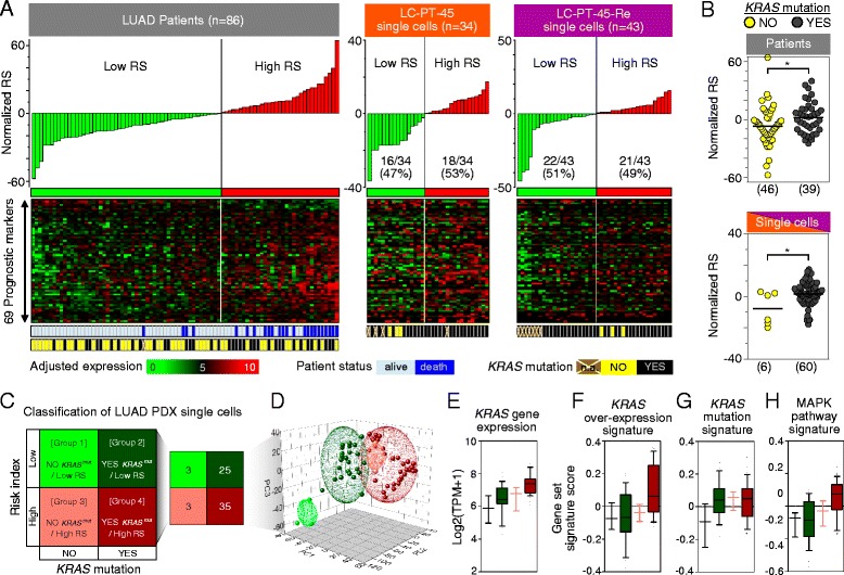
Identification of PDX cell subclones using single-cell RNA-seq data. a Top: normalized RS. Middle: heatmaps of expression of 69 prognostic markers. Bottom: KRAS mutation status of each patient (training set, n = 86) or single cell (LC-PT-45, n = 34; LC-PT-45-Re, n = 43). b Scatter plots demonstrating the effect of the KRAS mutation on the RSs of LUAD patients and PDX single cells. Horizontal lines represent the mean. *P < 0.05; **P < 0.01. c Semi-supervised clustering of single cells into four groups with estimated RS and KRAS mutant status. d Principal component analysis of the genes discriminating the subgroups. Ellipsoids were generated with standard deviations around each group. e–h Comparative features among the classified single cell subgroups. e KRAS gene expression (Log2 ratio of transcripts per million + 1). Gene set signature scores (computed by gene set variation analysis) corresponding to the KRAS over-expression signature [39] (f), KRAS mutation signature [40] (g), and MAPK pathway signature (gene sets from BioCarta) (h). For the boxplots in (e–h), boxes = the interquartile range between the first and third quartiles, and error bars = 10th–90th percentiles
Interestingly, individual PDX cells were calculated to have a wide RS distribution (Fig. 4a). Eighteen out of the 34 PDX cells or 21 out of 43 of the replicate samples were determined to be high-RS. We combined the replicate PDX RNA-seq data for further analysis and found that PDX cells with KRAS G12D expression tend to have a higher RS (Fig. 4b). The finding is consistent with those of LUAD patients in clinical studies [6]. Altogether, semi-supervised clustering based on the expression of the KRAS mutation and RS classified the PDX cells into four groups: group 1, no KRAS G12D (KRAS WT)/low RS (n = 3); group 2, KRAS G12D/low RS (n = 25); group 3, no KRAS G12D (KRAS WT)/high RS (n = 3); and group 4, KRAS G12D/high RS (n = 35) (Fig. 4c).
These four groups displayed characteristic gene expression profiles that likely reflect the different phenotypes among individual PDX cells (Figure S9a in Additional file 13). In particular, group 4 had enhanced gene expression signatures associated with KRAS overexpression and activation of the RAS-MAPK signaling pathway [39, 40] (Fig. 4f, h), which correlated well with KRAS mutational status. Group 4 PDX cells also showed significantly higher cell cycle gene mRNA expression (Figure S9c in Additional file 13) [41]. In contrast, despite having the KRAS mutation signature (Fig. 4g), group 2 cells had lower KRAS expression levels and KRAS overexpression signatures (Fig. 4e, f), lower RAS-MAPK signaling pathway activation status (Fig. 4h), and reduced expression of cell cycle-related genes (Figure S9c in Additional file 13).
The distinct gene expression signatures among the four groups were visualized by a principal component analysis (PCA) plot using genes exclusively expressed by each group, with a criterion of at least a twofold change in transcripts per million (TPM) ratio with statistical significance (_t_-test P < 0.05; Fig. 4d). Although group 2 cells showed a lower RAS-MAPK signaling pathway activation status, they had significantly upregulated expression of ion channel transport pathway-related genes (Figure S9b in Additional file 13), which has been implicated in the drug resistance mechanism [10].
Phenotypic interpretation of PDX cell subgroups
The results above indicated that, in the PDX cell population, there is a specific subgroup (group 4) that is predicted to be more aggressive than the other groups. This subset is characterized by a high RS, KRAS mutation, RAS-MAPK signaling pathway activation, and upregulation of cell cycle-related genes. To determine whether individual cells associate with tumor phenotypic aggressiveness, such as drug resistance, we screened the in vitro sensitivity of the PDX cells against a panel of 25 anti-cancer agents used in non-small cell lung cancer treatment (Additional file 14). The PDX cells were highly sensitive to a variety of drug treatments, including docetaxel, and molecular pathway targeting agents. Among the identified agents, we focused on the MEK1/2 inhibitor selumetinib, and the phosphatidylinositide 3-kinase (PI3K) inhibitors BKM120 and BEZ235 (PI3K/mTOR), because of their potential clinical benefits [42, 43]. Other cytotoxic drugs (e.g., carboplatin, and the Notch inhibitor DAPT) did not show any effects (Fig. 5a). Although docetaxel, BKM120, BEZ235, and selumetinib showed tumoricidal effects, some PDX cells survived the three days of treatment with these drugs when utilized at their reported IC50.
Fig. 5.
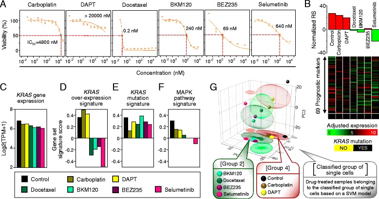
Interpretation of drug responses using single-cell signatures. a Dose response curves for the four selected anti-cancer compounds (cytotoxic compounds carboplatin and docetaxel; molecular targeting compounds DAPT, BKM120, BEZ235, and selumetinib). b Normalized RSs (top) and adjusted-expression of the 69 prognostic markers (middle) with KRAS mutant expression (bottom) for the control and drug-treated PDX cells. c–f Comparative features among the control and drug-treated PDX cells. c KRAS gene expression (Log2 ratio of TPM + 1). Gene set signature scores (computed by gene set variation analysis) corresponding to the KRAS over-expression signature [39] (d), KRAS mutation signature [40] (e), and MAPK pathway signature (gene sets from BioCarta) (f). g Results from the principal component (PC) analysis on single cells along with the control and drug-treated PDX cells. Ellipsoids correspond to the single cell subgroups (group 1, light green; group 2, dark green; group 3, light red; group 4, dark red), with the control and drug-treated PDX cells projected on the _PC1_-PC2 plane. Using single cell subgroups as a training set, classification support vector machine type 1 (C-SVM classification) was applied to a test set of the control and drug-treated PDX cells
When evaluated as a bulk population, PDX cells manifested group 4-like characteristics with high RS and KRAS G12D. Ineffective treatments with carboplatin or DAPT did not alter these properties of the group (Fig. 5b–g). However, those PDX cells that survived the docetaxel, BKM120, BEZ235, or selumetinib treatments showed group 2-like gene expression signatures: low RS (Fig. 5b), slight decrease in total KRAS expression levels (Fig. 5c), down-regulation of gene expression signatures associated with KRAS overexpression (Fig. 5d), preservation of the mutant KRAS G12D expression signature (Fig. 5e), and down-regulation of RAS-MAPK signaling pathway activation (Fig. 5f). Moreover, upregulation of ion channel transport genes (Figure S9b in Additional file 13) and downregulation of cell cycle-related genes (Figure S9c in Additional file 13) were observed in these treatment groups. The overall gene expression signature represented by PCA confirmed the group 2 cell-like properties of the drug-resistant PDX cells, in a support vector machine (SVM) model (Fig. 5g). Altogether, these results suggest that the drug-resistant population was cell-cycle quiescent and with possibly higher transporter activity for the anti-cancer drugs.
We further determined whether the group 2-like population conveyed the low risk gene expression signature after anti-cancer drug treatment with selumetinib (Figure S11a in Additional file 15). Interestingly, the low RS of surviving PDX cells was gradually reverted to a high RS after drug removal (Figure S11b in Additional file 15). The KRAS over-expression signature (Fig. S11d in Additional file 15) and MAPK pathway activation (Figure S11f in Additional file 15) recovered as well. By contrast, the level of total KRAS expression (Figure S11c in Additional file 15) and mutational status (Figure S11e in Additional file 15) were not altered by drug removal. The possible mechanisms of the dynamic nature of these gene expression signatures, such as epigenetic regulation or recovery of heterogeneity by clonal proliferation, need to be further elucidated.
Validation of analytical procedures in an independent lung cancer PDX case
To validate our strategy of using single cell RNA-seq data for subgroup identification, we used an independent set of PDX cells derived from a lung cancer-brain metastasis (LC-MBT-15) [30]. The LC-MBT-15 PDX harbors an insertional mutation in EGFR exon 20, a well-known driver mutation in LUAD conferring resistance to reversible EGFR inhibitors [44, 45]. Single cells from LC-MBT-15 had less heterogeneous transcriptome and SNV expression compared with the KRAS mutant PDX cells (Figure S12a–e in Additional file 16), which might have been caused by extensive clonal selection during serial anti-cancer treatments before PDX establishment (see the patient description in “Materials and methods”). Nonetheless, the LC-MBT-15 single cells were still clustered into two subgroups by RS, similar to the original PDX case (Figure S12f, i in Additional file 16). In contrast to the KRAS G12D mutation, the EGFR mutation was modestly detected and showed no preferential expression in the high RS group (Figure S12g, h in Additional file 16).
Drug screening on LC-MBT-15 cells was performed using 28 lung cancer drugs (Additional file 17). LC-MBT-15 cells were highly sensitive to the irreversible EGFR/HER2 inhibitor afatinib and the c-Met inhibitor tivantinib but were resistant to the reversible EGFR inhibitor erlotinib. When gene expression profiles for the drug-resistant populations were analyzed 3 days later, PCA of the single cells and application of a SVM model for drug-treated populations revealed that the drug-resistant populations shared the gene expression signature of the low RS group (Figure S12j, l in Additional file 16). Interestingly, upregulation of ion channel transport genes was also noted in the drug-resistant populations (Figure S12k in Additional file 16), similar to the low risk group single cells. These results are consistent with the original LC-PT-45 PDX case, and further support the observation that (1) single cell profiles of a population reveal cells with drug-resistant signatures and (2) the drug-resistant population may come from a subset with higher transporter activity and low cell proliferation activity.
Discussion
Single-cell genome analysis enables measurement of the extent of intra-tumoral heterogeneity, which may provide clues for solving problems such as cancer recurrence, metastasis, and drug resistance [46]. Single-cell RNA-seq can provide integrative information on both gene expression and somatic SNVs, which makes it a comprehensive tool to connect a cell’s genotype with its expression profile and phenotype. We used tumor cell-enriched LUAD PDX cells to define genomic signatures of individual tumor cells, and then verified the applicability of translating this information into biological cancer cell phenotypes such as drug responses.
When interpreting single cell RNA-seq data, the data quality needs to be considered, because of the high magnitude of amplification in the sequencing process. Sequence errors can be incorporated during the reverse transcription, cDNA amplification, and library construction processes, causing false positive mutation calls. RNA editing and monoallelic expression can also cause discrepancies between SNV calls from RNA and DNA sequencing. In this study, we focused on the RNA-seq SNVs that were simultaneously detected by WES and identified in more than three single cells. This approach would minimize the probability of false positive SNV calls. On the other hand, false negative SNV calls could result from missing reads at the mutant position in both DNA and RNA sequencing, which might be misinterpreted as biological heterogeneity [47]. Various approaches such as Nuc-seq, which increases the starting material by using G2/M phase cells, are reported to increase the genome coverage up to 91 % for DNA sequencing [48]. For the RNA-seq-based genotype analysis, mutations in rare transcripts are most prone to the dropout events, suggesting that RNA-seq is suitable for genotyping highly expressed oncogenic driver mutations.
Despite limitations in the accuracy of single-cell RNA-seq, in this study we observed good correlations between the merged single-cell data and the bulk cell data at both the gene expression and expressed SNV levels. Once the number of single cells exceeded 30, the averaged expression levels and consensus SNVs largely recapitulated the data from bulk populations. Significant correlations were also detected between replicate RNA-seq analyses and with the PCR-based genotyping method. While these concordant results and overall high expression level of KRAS support the validity of the KRAS mutation calls in RNA, 12–16 % of cells had insufficient RNA read counts at the mutant position, resulting in ambiguous calls. Downsizing sequencing data further increased the number of ambiguous calls (data not shown), indicating that a sufficient number of reads is critical in the RNA-based mutation analysis. This is in contrast to the gene expression analysis, which required only 0.5 million reads for the transcriptome estimation [33].
We isolated single PDX cells from a _KRAS_-driven tumor, which represents 25–33 % of LUADs [2, 3]. Comparison of the patient tumor and PDX cells revealed a significant enrichment of KRAS mutant tumor areas in the PDX, indicating that this PDX is a good model in which to study _KRAS_-driven tumors. However, we observed loss of some mutations as well as increased variant allele frequencies of many shared mutations and de novo mutations, possibly resulting from the expansion of subclones [29]. These subclones might be undergoing proliferation due to clonal selection and adaptation in the PDX, leading to a transient increase in genetic variation of the sample. In the longer term, the selection would have diminished the level of tumor heterogeneity originally present in the patient tumor. Therefore, use of freshly isolated tumor cells is warranted for the accurate estimation of tumor heterogeneity in future studies.
Because activating KRAS mutations are associated with poor LUAD prognosis and due to the current lack of reliable targeting agents [5, 38], it is a clinical challenge to find efficient treatment strategies for _KRAS_-driven cancers. According to the KRAS mutation status, the PDX cells analyzed as a bulk population showed clinically unfavorable genomic characteristics when the RS was calculated from the signature of 69 prognostic genes [6]. However, single-cell RNA-seq of PDX cells revealed intra-tumoral heterogeneity in terms of the KRAS mutant and RS gene expression characteristics. Having individual tumor cells that display intra-tumorally heterogeneous molecular signatures that are prognostic in LUAD patients is an interesting attribute. Similar findings were reported in other single-cell or multi-regional studies in glioblastoma [32], in which single cells from the same tumor were classified into multiple subtypes. Moreover, glioblastoma patients with mixed subtype cells manifested worse prognoses [32], suggesting the prognostic value of defining intra-tumoral heterogeneity.
The intra-tumoral heterogeneity might be driven by DNA mutations as well as by epigenetic and regulatory mechanisms. In this study we identified individual cells with variable mutant KRAS gene expression and RSs. Both genetic and non-genetic factors likely contributed to specify the subpopulations. The gene expression signatures might be driven by genomic profiles, including KRAS, and other environmental factors, including the drug treatment. The gradual reversion of drug-resistant signatures after drug withdrawal (Additional file 15) suggests that non-genetic regulatory mechanisms could be involved in the specification. To devise effective anti-cancer treatment strategies, we need to understand the underlying mechanisms whereby transcriptome heterogeneity is maintained in the tumor.
According to the prognostic value of the activating KRAS mutation and RS, PDX cells with KRAS G12D expression and high RS would be expected to be drug resistant. Moreover, as a whole population, the PDX cells had a high KRAS G12D variant allele frequency and high RS that masked the no KRAS G12D (KRAS WT) and/or low RS cell types. The use of tumoricidal anti-cancer drugs with different mechanisms of action (cytotoxic and targeting specific signaling pathways) dramatically changed the gene expression features of the PDX cells in this study from KRAS G12D plus high RS to KRAS G12D plus low RS. The result was counterintuitive, since high RS is significantly associated with worse prognosis of LUAD patients. However, in an independent PDX case, cells with a low RS also survived in vitro anti-cancer treatments, supporting the validity of the unexpected results.
The unexpected results indicate that (1) tumor cells with activated KRAS signatures were drug targets, but the KRAS mutation itself was not a target, and (2) the actual tumor population responsible for drug resistance might be masked by dominant genomic characteristics within a bulk population. In this study, the cells that survived the effective treatments retained the KRAS mutation but seemed to stay in a dormant state without activating KRAS signaling. Interestingly, the molecular signatures of this group indicated upregulation of genes involved in the ion channel transport and P-type ATPases, which might play key roles in drug resistance [10]. Whether this potentially drug-resistant population is indeed a pre-existing tumor subclone or dynamically changes gene expression signatures in response to drug treatments needs to be addressed by future studies.
Conclusions
This study demonstrates that gene expression and somatic SNVs of single tumor cells could be retrieved simultaneously by single-cell RNA-seq. Furthermore, the genomic data obtained could be used to elucidate potentially drug-resistant subclones and to generate hypotheses on the molecular mechanisms of treatment resistance that are masked in the whole cancer cell population.
Materials and methods
Patient samples and PDX cells
This study was carried out in accordance with the principles of the Declaration of Helsinki, and approved by The Samsung Medical Center (Seoul, Korea) Institutional Review Board (no. 2010-04-004). Participants in this study gave written informed consent for research and publication of the results. Surgical specimens were acquired from a 60-year-old male patient who underwent surgical resection of a 37-mm irregular primary lung lesion in the right middle lobe (LC-PT-45), and from a 57-year-old female patient who underwent surgical resection of a metachronous brain metastasis (LC-MBT-15). The LC-PT-45 tumor was taken in a treatment-naïve status whereas the LC-MBT-15 tumor was taken after standard chemotherapy and erlotinib treatments. Pathologic examination of the primary tumors revealed a poorly differentiated lung adenocarcinoma based on the World Health Organization criteria [49]. The PDX cells were isolated and cultured in vitro as described previously [24, 30, 50]. Briefly, surgically removed tumor tissues were directly injected into the subrenal space of 6–8-week-old humanized immunocompromised female NOG (NOD/Shi- SCID/IL-2Rγ-null) mice (Orient Bio, Seongnam, Korea). Xenograft tumors were taken from the mice for PDX cell culture and validated by short tandem repeat DNA fingerprinting as having been derived from the original tumor. We used PDX cells at fewer than three in vitro passages for single-cell RNA-seq and drug screening. Animal care and handling was performed according to the National Institute of Health Guide for the Care and Use of Laboratory Animals (NIH publication no.80-23, revised 1978).
Drug screening with PDX cells
Dissociated PDX cells were cultured in neurobasal media-A supplemented with N2 (×1/2; Life Technologies, Carlsbad, CA, USA), B27 (×1/2; GIBCO, San Diego, CA, USA), basic fibroblast growth factor (bFGF; 25 ng/mL; R&D Systems, Minneapolis, MN, USA), epidermal growth factor (EGF; 25 ng/mL; R&D Systems), neuregulin 1 (NRG; 10 ng/mL; R&D Systems), and insulin-like growth factor 1(IGF1; 100 ng/mL; R&D Systems). The cells grown in these serum-free sphere culture conditions were seeded in 384-well plates (500 cells/well), and treated with a drug library (Selleck, Houston, TX, USA). The drug library was composed of targeted agents and cytotoxic chemotherapeutics, which were included in the clinical guideline or current clinical trial for the treatment of non-small cell lung cancer. After 3 days of incubation at 37 °C in a 5 % CO2 humidified incubator, cell viability was analyzed using an adenosine triphosphate monitoring system based on firefly luciferase (ATPlite™ 1step; PerkinElmer, Waltham, CA, USA). Test concentrations for each drug were empirically determined to produce a clinically relevant spectrum of drug activity. Dose response curves and corresponding half maximal (50 %) inhibitory concentration values (IC50) were calculated using the S+ Chip Analyzer (Samsung Electro-Mechanics, Suwon, Korea) [51].
WES and data processing
Genomic DNA was extracted from PDX cells using the QIAamp® DNA Mini kit (Qiagen, Hilden, Germany) or QIAamp DNA Blood Maxi Kit (Qiagen). Exomes were captured using the SureSelect XT Human All Exon V5 kit (Agilent Technologies, Inc., Santa Clara, CA, USA). The sequencing library was constructed and analyzed by the HiSeq 2000 or 2500 systems (Illumina, San Diego, CA, USA) using the 100-bp paired-end mode of the TruSeq Rapid PE Cluster kit and TruSeq Rapid SBS kit (Illumina). Mean target coverage for exome data was 153.4 ± 26.99 × .
Exome-sequencing reads were aligned to the hg19 reference genome using BWA-0.7.10 [52]. Putative duplications were marked by Picard-1.93 software [53]. Sites potentially harboring small insertions or deletions were realigned, and SNVs were called by applying GATK-3.2 [54] ‘HaplotypeCaller’ with known variant sites identified from phase I of the 1000 Genomes Project [55] and dbSNP-137 [56], using default option parameters. Then, called variants were evaluated to obtain highly accurate call sets through a two-stage processing step of ‘VariantRecalibrator’ and ‘ApplyRecalibration’, using default option parameters. To detect somatic mutations with increased sensitivity both in lower and higher allele frequencies [57], we used the caller programs of MuTect-1.1.5 [58] and VarScan2 [59].
Estimation of copy number variation from WES was performed using the ExomeCNV software package [26] in default quantification mode. Circular binary segmentation was applied to determine the neighboring regions of DNA that exhibited a statistically significant difference in copy number. The output was also applied to infer tumor purity using AbsCNseq [27].
Isolation of single cells and RNA-seq
We used the C1™ Single-Cell Auto Prep System (Fluidigm, San Francisco, CA, USA) with the SMARTer kit (Clontech, Mountain View, CA, USA). For the original experiment, 44 cells were captured as a single isolate on a C1 array chip for mRNA sequencing (17–25 μm) as determined by microscopic examination, and 34 passed the required criteria for cDNA quantity and quality as measured with a Qubit® 2.0 Fluorometer (Life Technologies) and 2100 Bioanalyzer (Agilent). RNA from bulk cell samples was also amplified using a SMARTer kit with 10 ng of starting material. Libraries were generated using the Nextera XT DNA Sample Prep Kit (Illumina) and sequenced on the HiSeq 2500 using the 100-bp paired-end mode of the TruSeq Rapid PE Cluster kit and TruSeq Rapid SBS kit.
RNA-seq data processing
RNA-seq reads were aligned to the human genome reference (hg19) together with splice junction information of each sample using the two-pass default mode of STAR_2.4.0d [60]. Gene expression was quantified by implementing RSEM v.1.2.18 [61] in default mode with Genecode v.19 [62] annotation, and calculated as the sum of isoform expression. Pre-processing steps for RNA-seq reads before calling variants were optimized by deduplication, splitting reads into exon segments, hard-clipping any sequences overhanging the intronic regions, realigning reads and recalibration using GATK-3.2 [54]. Then, variants were called by ‘HaplotypeCaller’ mode with option parameters of (−R hg19.fa --genotyping_mode DISCOVERY -recoverDanglingHeads -dontUseSoftClippedBases --dbsnp dbsnp_137.hg19.vcf -stand_emit_conf 20 -stand_call_conf 20 -nct 4). Highly accurate variants were filtered by applying ‘VariantFiltration’ (option parameters: −F hg19.fa -window 35 -cluster 3 -filterName FS -filter “FS > 30.0” -filterName QD -filter “QD < 2.0” \). After removal of variant call quality Q < 20, further filtering was applied to SNVs that were considered to be potential false positives in RNA-seq by SNPiR [34]. We regarded only those SNVs which overlapped with WES as true positives. The overall process of calling and filtering the variants is summarized in Figure S4a in Additional file 7.
Computing RS using multivariate markers
RSs were regression coefficients calculated by a linear combination of the expression values of the prognosis markers using a training set [6] of LUAD patients. Prognosis markers were also derived from the previous report [6] that classified LUAD patients according to gene expression profiles of the suggested markers, and 69 genes were ultimately chosen by overlapping our data sets after gene filtering of zero expression across all single cells. These filtered genes (Additional file 11) were validated as prognosis markers with independent LUAD datasets from The Cancer Genome Atlas [2] and from a Korean LUAD cohort [63]. Batch effects on gene expression between independent datasets were removed by means of ComBat [64]. Regression coefficients and P values of the training set were estimated using univariate Cox proportional hazards regression modeling and ordered by P values. To partition patient samples into high- and low-RS-based groups upon computation of response score, we applied a 60th percentile cutoff as described in Beer et al_._ [6]. Survival analysis was performed using the R Survival package [65] and validated through Kaplan-Meier survival curves with log-rank testing (training set, P = 1.04 × 10⁻⁶; validation set, P = 9.25 × 10⁻³) (Figure S8b in Additional file 12).
To classify the control and drug-treated PDX cells into semi-supervised clustered single cells (LC-PT-45, Fig. 4; LC-MBT-15, Additional file 16: Figure S12), a classification SVM type 1 (C-SVM classification) model was applied using the R package e1071 [66].
Gene set signature activation analysis
To characterize gene expression features of a subgroup compared with the other groups among the classified single cells, we utilized the GSEA-P program with default mode searching for significantly enriched gene set signatures [67]. Applied gene sets were derived from the three major curated pathway databases of KEGG, REACTOME, and BIOCARTA in MSigDB v.4.0 [68]. To estimate the gene set activation status of a single sample, gene set variation analysis [69] was applied in default mode.
Validating gene expression and expressed SNVs at the RNA level by qPCR
Gene expression and expressed SNVs were assessed by qPCR or SNP type PCR across single cells using a Biomark HD system (Fluidigm). cDNAs obtained from the C1 array for mRNA sequencing chip were subjected to specific target amplification following the manufacturer’s recommendations. For the gene expression qPCR, Delta Gene Assay (Fluidigm) with EvaGreen second generation dsDNA binding dye was performed for gene sets selected from the RS genes (Additional file 6). To compare correlations between RNA-seq and qPCR platforms for the selected 43 gene expression, mean fold change over median expression was calculated as in the previous study [33]. Validation of expressed SNVs at the RNA level was carried out using a SNP Type Assay (Fluidigm) with locus-specific primer sequences. Primers were designed using D3™ software (Fluidigm), and sequences are available in Additional file 6.
Validating genomic variants at the DNA level by ddPCR
PDX cells were labeled with 6-carboxyfluorescein succinimidyl ester (Life Technologies) and sorted into single cells using a FACSAria™ III flow cytometer (BD Biosciences, CA, USA). Wells with a single green fluorescence signal were manually inspected and selected for amplification of genomic DNA with a GenomiPhi V2 DNA Amplification Kit (GE Healthcare, Little Chalfont, UK). The mutant alleles were detected using ddPCR Supermix for Probes reagents (Bio-Rad, Hercules, CA, USA) implemented using a QX200 ddPCR system, following the manufacturer’s protocols. The negative signal of droplets was normalized with a vehicle control, and the numbers of wild-type or mutation alleles in droplets were estimated in a Poisson distribution. Variant allele frequency was calculated by counting copies of mutation alleles over the total number of detected alleles. We regarded genotypes of detected variants as homozygous when the variant allele frequency was higher than 90 %. Sequences of the primers used in ddPCR are available in Additional file 6.
Statistical analysis of single-cell gene expression
Linear regression was applied to scatter plots of the averaged single cells over the pooled-cell samples in Fig. 2a with zero intercepts. The inter-correlation distribution between single cells was calculated as Pearson’s and Spearman’s correlation coefficients, and plotted as a density plot with a kernel function fitting over the histograms (Fig. 2b). Multiple regression analysis estimated how many single cells hypothetically accounted for the pooled cell fraction. Single-cell samples were randomly chosen with the given number and the adjusted R2 (Fig. 2c) and the overlap ratio (Fig. 2e) were determined 1000 times with permutation. The differences in normalized RS, gene expression, and gene set activation score between single-cell subgroups were tested using two-tailed Student’s _t_-tests.
Data access
The data reported in this paper have been deposited at the Samsung Genome Institute (SGI) data repository [70] and at the NCBI Gene Expression Omnibus (GEO) under accession number GSE69405.
Acknowledgements
This research was supported by the Bio & Medical Technology Development Program of the National Research Foundation funded by Ministry of Science and Technology (2012M3A9B2029132 to W-YP) and the Korea Health Technology R&D Project through the Korea Health Industry Development Institute (KHIDI), funded by the Ministry of Health & Welfare, Republic of Korea (HI13C2096 to W-YP). This study was supported by a grant of the Korea Health technology R&D Project, Ministry of Health & Welfare, Republic of Korea (A120446 to KMJ).
Abbreviations
bp
base pair
ddPCR
droplet digital PCR
LUAD
lung adenocarcinoma
MAPK
mitogen-activated protein kinase
PCA
principal component analysis
PCR
polymerase chain reaction
PDX
patient-derived xenograft
PI3K
phosphatidylinositide 3-kinase
qPCR
quantitative PCR
RNA-seq
RNA sequencing
RS
risk score
RTK
receptor tyrosine kinase
SNV
single-nucleotide variation
SVM
support vector machine
TPM
transcripts per million
WES
whole-exome sequencing
WT
wild type
Additional files
Footnotes
Kyu-Tae Kim, Hye Won Lee and Hae-Ock Lee contributed equally to this work.
Competing interests
The authors declare that they have no competing interests.
Authors’ contributions
K-TK, SCK, WSC and HHE performed the statistical and bioinformatics analyses; K-TK, H-OL, and HHE performed single cell experiments; HWL, D-HN and YJS generated PDX models and performed drug screening; K-TK, HWL, H-OL, KMJ and W-YP wrote the manuscript; JHK, KMJ and W-YP designed and guided the study. All authors read and approved the final manuscript.
Contributor Information
Kyu-Tae Kim, Email: kimqtae@snu.ac.kr.
Hye Won Lee, Email: nsproper@naver.com.
Hae-Ock Lee, Email: haeock.lee@samsung.com.
Sang Cheol Kim, Email: sang.cheol.kim@samsung.com.
Yun Jee Seo, Email: yunjee.seo@gmail.com.
Woosung Chung, Email: cws1021@skku.edu.
Hye Hyeon Eum, Email: smflsdkdl@snu.ac.kr.
Do-Hyun Nam, Email: nsnam@skku.edu.
Junhyong Kim, Email: junhyong@sas.upenn.edu.
Kyeung Min Joo, Email: kmjoo@skku.edu.
Woong-Yang Park, Email: woongyang.park@samsung.com.
References
- 1.Ding L, Getz G, Wheeler DA, Mardis ER, McLellan MD, Cibulskis K, et al. Somatic mutations affect key pathways in lung adenocarcinoma. Nature. 2008;455:1069–75. doi: 10.1038/nature07423. [DOI] [PMC free article] [PubMed] [Google Scholar]
- 2.The Cancer Genome Atlas Research Network Comprehensive molecular profiling of lung adenocarcinoma. Nature. 2014;511:543–50. doi: 10.1038/nature13385. [DOI] [PMC free article] [PubMed] [Google Scholar]
- 3.Imielinski M, Berger AH, Hammerman PS, Hernandez B, Pugh TJ, Hodis E, et al. Mapping the hallmarks of lung adenocarcinoma with massively parallel sequencing. Cell. 2012;150:1107–20. doi: 10.1016/j.cell.2012.08.029. [DOI] [PMC free article] [PubMed] [Google Scholar]
- 4.Youlden DR, Cramb SM, Baade PD. The international epidemiology of lung cancer: geographical distribution and secular trends. J Thorac Oncol. 2008;3:819–31. doi: 10.1097/JTO.0b013e31818020eb. [DOI] [PubMed] [Google Scholar]
- 5.Lindeman NI, Cagle PT, Beasley MB, Chitale DA, Dacic S, Giaccone G, et al. Molecular testing guideline for selection of lung cancer patients for EGFR and ALK tyrosine kinase inhibitors: guideline from the College of American Pathologists, International Association for the Study of Lung Cancer, and Association for Molecular Pathology. J Thorac Oncol. 2013;8:823–59. doi: 10.1097/JTO.0b013e318290868f. [DOI] [PMC free article] [PubMed] [Google Scholar]
- 6.Beer DG, Kardia SL, Huang CC, Giordano TJ, Levin AM, Misek DE, et al. Gene-expression profiles predict survival of patients with lung adenocarcinoma. Nat Med. 2002;8:816–24. doi: 10.1038/nm733. [DOI] [PubMed] [Google Scholar]
- 7.Chen HY, Yu SL, Chen CH, Chang GC, Chen CY, Yuan A, et al. A five-gene signature and clinical outcome in non-small-cell lung cancer. N Engl J Med. 2007;356:11–20. doi: 10.1056/NEJMoa060096. [DOI] [PubMed] [Google Scholar]
- 8.Lau SK, Boutros PC, Pintilie M, Blackhall FH, Zhu CQ, Strumpf D, et al. Three-gene prognostic classifier for early-stage non small-cell lung cancer. J Clin Oncol. 2007;25:5562–9. doi: 10.1200/JCO.2007.12.0352. [DOI] [PubMed] [Google Scholar]
- 9.Yu SL, Chen HY, Chang GC, Chen CY, Chen HW, Singh S, et al. MicroRNA signature predicts survival and relapse in lung cancer. Cancer Cell. 2008;13:48–57. doi: 10.1016/j.ccr.2007.12.008. [DOI] [PubMed] [Google Scholar]
- 10.Willers H, Azzoli CG, Santivasi WL, Xia F. Basic mechanisms of therapeutic resistance to radiation and chemotherapy in lung cancer. Cancer J. 2013;19:200–7. doi: 10.1097/PPO.0b013e318292e4e3. [DOI] [PMC free article] [PubMed] [Google Scholar]
- 11.Spaans JN, Goss GD. Drug resistance to molecular targeted therapy and its consequences for treatment decisions in non-small-cell lung cancer. Front Oncol. 2014;4:190. doi: 10.3389/fonc.2014.00190. [DOI] [PMC free article] [PubMed] [Google Scholar]
- 12.Navin N, Krasnitz A, Rodgers L, Cook K, Meth J, Kendall J, et al. Inferring tumor progression from genomic heterogeneity. Genome Res. 2010;20:68–80. doi: 10.1101/gr.099622.109. [DOI] [PMC free article] [PubMed] [Google Scholar]
- 13.Bashashati A, Ha G, Tone A, Ding J, Prentice LM, Roth A, et al. Distinct evolutionary trajectories of primary high-grade serous ovarian cancers revealed through spatial mutational profiling. J Pathol. 2013;231:21–34. doi: 10.1002/path.4230. [DOI] [PMC free article] [PubMed] [Google Scholar]
- 14.Gerlinger M, Rowan AJ, Horswell S, Larkin J, Endesfelder D, Gronroos E, et al. Intratumor heterogeneity and branched evolution revealed by multiregion sequencing. N Engl J Med. 2012;366:883–92. doi: 10.1056/NEJMoa1113205. [DOI] [PMC free article] [PubMed] [Google Scholar]
- 15.Dawson SJ, Tsui DW, Murtaza M, Biggs H, Rueda OM, Chin SF, et al. Analysis of circulating tumor DNA to monitor metastatic breast cancer. N Engl J Med. 2013;368:1199–209. doi: 10.1056/NEJMoa1213261. [DOI] [PubMed] [Google Scholar]
- 16.Keats JJ, Chesi M, Egan JB, Garbitt VM, Palmer SE, Braggio E, et al. Clonal competition with alternating dominance in multiple myeloma. Blood. 2012;120:1067–76. doi: 10.1182/blood-2012-01-405985. [DOI] [PMC free article] [PubMed] [Google Scholar]
- 17.Mroz EA, Tward AD, Pickering CR, Myers JN, Ferris RL, Rocco JW. High intratumor genetic heterogeneity is related to worse outcome in patients with head and neck squamous cell carcinoma. Cancer. 2013;119:3034–42. doi: 10.1002/cncr.28150. [DOI] [PMC free article] [PubMed] [Google Scholar]
- 18.Jamal-Hanjani M, Hackshaw A, Ngai Y, Shaw J, Dive C, Quezada S, et al. Tracking genomic cancer evolution for precision medicine: the lung TRACERx study. PLoS Biol. 2014;12:e1001906. doi: 10.1371/journal.pbio.1001906. [DOI] [PMC free article] [PubMed] [Google Scholar]
- 19.Klco JM, Spencer DH, Miller CA, Griffith M, Lamprecht TL, O’Laughlin M, et al. Functional heterogeneity of genetically defined subclones in acute myeloid leukemia. Cancer Cell. 2014;25:379–92. doi: 10.1016/j.ccr.2014.01.031. [DOI] [PMC free article] [PubMed] [Google Scholar]
- 20.Xu X, Hou Y, Yin X, Bao L, Tang A, Song L, et al. Single-cell exome sequencing reveals single-nucleotide mutation characteristics of a kidney tumor. Cell. 2012;148:886–95. doi: 10.1016/j.cell.2012.02.025. [DOI] [PMC free article] [PubMed] [Google Scholar]
- 21.Hou Y, Song L, Zhu P, Zhang B, Tao Y, Xu X, et al. Single-cell exome sequencing and monoclonal evolution of a JAK2-negative myeloproliferative neoplasm. Cell. 2012;148:873–85. doi: 10.1016/j.cell.2012.02.028. [DOI] [PubMed] [Google Scholar]
- 22.Navin N, Kendall J, Troge J, Andrews P, Rodgers L, McIndoo J, et al. Tumour evolution inferred by single-cell sequencing. Nature. 2011;472:90–4. doi: 10.1038/nature09807. [DOI] [PMC free article] [PubMed] [Google Scholar]
- 23.Kreso A, O’Brien CA, van Galen P, Gan OI, Notta F, Brown AM, et al. Variable clonal repopulation dynamics influence chemotherapy response in colorectal cancer. Science. 2013;339:543–8. doi: 10.1126/science.1227670. [DOI] [PMC free article] [PubMed] [Google Scholar]
- 24.Joo KM, Kim J, Jin J, Kim M, Seol HJ, Muradov J, et al. Patient-specific orthotopic glioblastoma xenograft models recapitulate the histopathology and biology of human glioblastomas in situ. Cell Rep. 2013;3:260–73. doi: 10.1016/j.celrep.2012.12.013. [DOI] [PubMed] [Google Scholar]
- 25.Ding L, Ellis MJ, Li S, Larson DE, Chen K, Wallis JW, et al. Genome remodelling in a basal-like breast cancer metastasis and xenograft. Nature. 2010;464:999–1005. doi: 10.1038/nature08989. [DOI] [PMC free article] [PubMed] [Google Scholar]
- 26.Sathirapongsasuti JF, Lee H, Horst BA, Brunner G, Cochran AJ, Binder S, et al. Exome sequencing-based copy-number variation and loss of heterozygosity detection: ExomeCNV. Bioinformatics. 2011;27:2648–54. doi: 10.1093/bioinformatics/btr462. [DOI] [PMC free article] [PubMed] [Google Scholar]
- 27.Bao L, Pu M, Messer K. AbsCN-seq: a statistical method to estimate tumor purity, ploidy and absolute copy numbers from next-generation sequencing data. Bioinformatics. 2014. Epub ahead of print. [DOI] [PMC free article] [PubMed]
- 28.Yoshihara K, Shahmoradgoli M, Martinez E, Vegesna R, Kim H, Torres-Garcia W, et al. Inferring tumour purity and stromal and immune cell admixture from expression data. Nat Commun. 2013;4:2612. doi: 10.1038/ncomms3612. [DOI] [PMC free article] [PubMed] [Google Scholar]
- 29.Eirew P, Steif A, Khattra J, Ha G, Yap D, Farahani H, et al. Dynamics of genomic clones in breast cancer patient xenografts at single-cell resolution. Nature. 2015;518:422–6. doi: 10.1038/nature13952. [DOI] [PMC free article] [PubMed] [Google Scholar]
- 30.Lee HW, Lee JI, Lee SJ, Cho HJ, Song HJ, Jeong DE, Seo YJ, Shin S, Joung JG, Kwon YJ, et al. Patient-derived xenografts from non-small cell lung cancer brain metastases are valuable translational platforms for the development of personalized targeted therapy. Clin Cancer Res. 2014;21:1172–82. doi: 10.1158/1078-0432.CCR-14-1589. [DOI] [PubMed] [Google Scholar]
- 31.Ramskold D, Luo S, Wang YC, Li R, Deng Q, Faridani OR, et al. Full-length mRNA-seq from single-cell levels of RNA and individual circulating tumor cells. Nat Biotechnol. 2012;30:777–82. doi: 10.1038/nbt.2282. [DOI] [PMC free article] [PubMed] [Google Scholar]
- 32.Patel AP, Tirosh I, Trombetta JJ, Shalek AK, Gillespie SM, Wakimoto H, et al. Single-cell RNA-seq highlights intratumoral heterogeneity in primary glioblastoma. Science. 2014;344:1396–401. doi: 10.1126/science.1254257. [DOI] [PMC free article] [PubMed] [Google Scholar]
- 33.Wu AR, Neff NF, Kalisky T, Dalerba P, Treutlein B, Rothenberg ME, et al. Quantitative assessment of single-cell RNA-sequencing methods. Nat Methods. 2014;11:41–6. doi: 10.1038/nmeth.2694. [DOI] [PMC free article] [PubMed] [Google Scholar]
- 34.Piskol R, Ramaswami G, Li JB. Reliable identification of genomic variants from RNA-seq data. Am J Hum Genet. 2013;93:641–51. doi: 10.1016/j.ajhg.2013.08.008. [DOI] [PMC free article] [PubMed] [Google Scholar]
- 35.Forbes SA, Tang G, Bindal N, Bamford S, Dawson E, Cole C, et al. COSMIC (the Catalogue of Somatic Mutations in Cancer): a resource to investigate acquired mutations in human cancer. Nucleic Acids Res. 2010;38:D652–7. doi: 10.1093/nar/gkp995. [DOI] [PMC free article] [PubMed] [Google Scholar]
- 36.Hunker CM, Galvis A, Kruk I, Giambini H, Veisaga ML, Barbieri MA. Rab5-activating protein 6, a novel endosomal protein with a role in endocytosis. Biochem Biophys Res Commun. 2006;340:967–75. doi: 10.1016/j.bbrc.2005.12.099. [DOI] [PubMed] [Google Scholar]
- 37.Wang L, Yamaguchi S, Burstein MD, Terashima K, Chang K, Ng HK, et al. Novel somatic and germline mutations in intracranial germ cell tumours. Nature. 2014;511:241–5. doi: 10.1038/nature13296. [DOI] [PMC free article] [PubMed] [Google Scholar]
- 38.Sonobe M, Kobayashi M, Ishikawa M, Kikuchi R, Nakayama E, Takahashi T, et al. Impact of KRAS and EGFR gene mutations on recurrence and survival in patients with surgically resected lung adenocarcinomas. Ann Surg Oncol. 2012;19:S347–54. doi: 10.1245/s10434-011-1799-8. [DOI] [PubMed] [Google Scholar]
- 39.Barbie DA, Tamayo P, Boehm JS, Kim SY, Moody SE, Dunn IF, et al. Systematic RNA interference reveals that oncogenic KRAS-driven cancers require TBK1. Nature. 2009;462:108–12. doi: 10.1038/nature08460. [DOI] [PMC free article] [PubMed] [Google Scholar]
- 40.Sweet-Cordero A, Mukherjee S, Subramanian A, You H, Roix JJ, Ladd-Acosta C, et al. An oncogenic KRAS2 expression signature identified by cross-species gene-expression analysis. Nat Genet. 2005;37:48–55. doi: 10.1038/ng1490. [DOI] [PubMed] [Google Scholar]
- 41.Huang J, Wu S, Barrera J, Matthews K, Pan D. The Hippo signaling pathway coordinately regulates cell proliferation and apoptosis by inactivating Yorkie, the Drosophila Homolog of YAP. Cell. 2005;122:421–34. doi: 10.1016/j.cell.2005.06.007. [DOI] [PubMed] [Google Scholar]
- 42.Engelman JA, Chen L, Tan X, Crosby K, Guimaraes AR, Upadhyay R, et al. Effective use of PI3K and MEK inhibitors to treat mutant Kras G12D and PIK3CA H1047R murine lung cancers. Nat Med. 2008;14:1351–6. doi: 10.1038/nm.1890. [DOI] [PMC free article] [PubMed] [Google Scholar]
- 43.Janne PA, Shaw AT, Pereira JR, Jeannin G, Vansteenkiste J, Barrios C, et al. Selumetinib plus docetaxel for KRAS-mutant advanced non-small-cell lung cancer: a randomised, multicentre, placebo-controlled, phase 2 study. Lancet Oncol. 2013;14:38–47. doi: 10.1016/S1470-2045(12)70489-8. [DOI] [PubMed] [Google Scholar]
- 44.Greulich H, Chen TH, Feng W, Janne PA, Alvarez JV, Zappaterra M, et al. Oncogenic transformation by inhibitor-sensitive and -resistant EGFR mutants. PLoS Med. 2005;2:e313. doi: 10.1371/journal.pmed.0020313. [DOI] [PMC free article] [PubMed] [Google Scholar]
- 45.Wu JY, Wu SG, Yang CH, Gow CH, Chang YL, Yu CJ, et al. Lung cancer with epidermal growth factor receptor exon 20 mutations is associated with poor gefitinib treatment response. Clin Cancer Res. 2008;14:4877–82. doi: 10.1158/1078-0432.CCR-07-5123. [DOI] [PubMed] [Google Scholar]
- 46.Burrell RA, McGranahan N, Bartek J, Swanton C. The causes and consequences of genetic heterogeneity in cancer evolution. Nature. 2013;501:338–45. doi: 10.1038/nature12625. [DOI] [PubMed] [Google Scholar]
- 47.Navin NE. Cancer genomics: one cell at a time. Genome Biol. 2014;15:452. doi: 10.1186/s13059-014-0452-9. [DOI] [PMC free article] [PubMed] [Google Scholar]
- 48.Wang Y, Waters J, Leung ML, Unruh A, Roh W, Shi X, et al. Clonal evolution in breast cancer revealed by single nucleus genome sequencing. Nature. 2014;512:155–60. doi: 10.1038/nature13600. [DOI] [PMC free article] [PubMed] [Google Scholar]
- 49.Beasley MB, Brambilla E, Travis WD. The 2004 World Health Organization classification of lung tumors. Semin Roentgenol. 2005;40:90–7. doi: 10.1053/j.ro.2005.01.001. [DOI] [PubMed] [Google Scholar]
- 50.Joo KM, Kim SY, Jin X, Song SY, Kong DS, Lee JI, et al. Clinical and biological implications of CD133-positive and CD133-negative cells in glioblastomas. Lab Invest. 2008;88:808–15. doi: 10.1038/labinvest.2008.57. [DOI] [PubMed] [Google Scholar]
- 51.Lee DW, Choi YS, Seo YJ, Lee MY, Jeon SY, Ku B, et al. High-throughput screening (HTS) of anticancer drug efficacy on a micropillar/microwell chip platform. Anal Chem. 2014;86:535–42. doi: 10.1021/ac402546b. [DOI] [PubMed] [Google Scholar]
- 52.Li H, Durbin R. Fast and accurate short read alignment with Burrows-Wheeler transform. Bioinformatics. 2009;25:1754–60. doi: 10.1093/bioinformatics/btp324. [DOI] [PMC free article] [PubMed] [Google Scholar]
- 53.Picard. http://sourceforge.net/projects/picard/files/picard-tools/1.118/.
- 54.DePristo MA, Banks E, Poplin R, Garimella KV, Maguire JR, Hartl C, et al. A framework for variation discovery and genotyping using next-generation DNA sequencing data. Nat Genet. 2011;43:491–8. doi: 10.1038/ng.806. [DOI] [PMC free article] [PubMed] [Google Scholar]
- 55.1000 Genomes. http://www.1000genomes.org/.
- 56.Sherry ST, Ward MH, Kholodov M, Baker J, Phan L, Smigielski EM, et al. dbSNP: the NCBI database of genetic variation. Nucleic Acids Res. 2001;29:308–11. doi: 10.1093/nar/29.1.308. [DOI] [PMC free article] [PubMed] [Google Scholar]
- 57.Wang Q, Jia P, Li F, Chen H, Ji H, Hucks D, et al. Detecting somatic point mutations in cancer genome sequencing data: a comparison of mutation callers. Genome Med. 2013;5:91. doi: 10.1186/gm495. [DOI] [PMC free article] [PubMed] [Google Scholar]
- 58.Cibulskis K, Lawrence MS, Carter SL, Sivachenko A, Jaffe D, Sougnez C, et al. Sensitive detection of somatic point mutations in impure and heterogeneous cancer samples. Nat Biotechnol. 2013;31:213–9. doi: 10.1038/nbt.2514. [DOI] [PMC free article] [PubMed] [Google Scholar]
- 59.Koboldt DC, Zhang Q, Larson DE, Shen D, McLellan MD, Lin L, et al. VarScan 2: somatic mutation and copy number alteration discovery in cancer by exome sequencing. Genome Res. 2012;22:568–76. doi: 10.1101/gr.129684.111. [DOI] [PMC free article] [PubMed] [Google Scholar]
- 60.Dobin A, Davis CA, Schlesinger F, Drenkow J, Zaleski C, Jha S, et al. STAR: ultrafast universal RNA-seq aligner. Bioinformatics. 2013;29:15–21. doi: 10.1093/bioinformatics/bts635. [DOI] [PMC free article] [PubMed] [Google Scholar]
- 61.Li B, Dewey CN. RSEM: accurate transcript quantification from RNA-Seq data with or without a reference genome. BMC Bioinformatics. 2011;12:323. doi: 10.1186/1471-2105-12-323. [DOI] [PMC free article] [PubMed] [Google Scholar]
- 62.Harrow J, Frankish A, Gonzalez JM, Tapanari E, Diekhans M, Kokocinski F, et al. GENCODE: the reference human genome annotation for The ENCODE Project. Genome Res. 2012;22:1760–74. doi: 10.1101/gr.135350.111. [DOI] [PMC free article] [PubMed] [Google Scholar]
- 63.Lee ES, Son DS, Kim SH, Lee J, Jo J, Han J, et al. Prediction of recurrence-free survival in postoperative non-small cell lung cancer patients by using an integrated model of clinical information and gene expression. Clin Cancer Res. 2008;14:7397–404. doi: 10.1158/1078-0432.CCR-07-4937. [DOI] [PubMed] [Google Scholar]
- 64.Johnson WE, Li C, Rabinovic A. Adjusting batch effects in microarray expression data using empirical Bayes methods. Biostatistics. 2007;8:118–27. doi: 10.1093/biostatistics/kxj037. [DOI] [PubMed] [Google Scholar]
- 65.R package survival. http://cran.r-project.org/web/packages/survival/index.html.
- 66.R package e1071. http://cran.r-project.org/web/packages/e1071/index.html
- 67.Subramanian A, Kuehn H, Gould J, Tamayo P, Mesirov JP. GSEA-P: a desktop application for Gene Set Enrichment Analysis. Bioinformatics. 2007;23:3251–3. doi: 10.1093/bioinformatics/btm369. [DOI] [PubMed] [Google Scholar]
- 68.Subrammanian A, Tamayo P, Mootha VK, Mukherjee S, Ebert BL, Gillette MA, et al. Gene set enrichment analysis: a knowledge-based approach for interpreting genome-wide expression profiles. Proc Natl Acad Sci U S A. 2005;102:15545–50. doi: 10.1073/pnas.0506580102. [DOI] [PMC free article] [PubMed] [Google Scholar]
- 69.Hanzelmann S, Castelo R, Guinney J. GSVA: gene set variation analysis for microarray and RNA-seq data. BMC Bioinformatics. 2013;14:7. doi: 10.1186/1471-2105-14-7. [DOI] [PMC free article] [PubMed] [Google Scholar]
- 70.Samsung Genome Institute. SingleCell/LUAD_Project. http://tbi.skku.edu/SGI/SingleCell/LUAD_Project.