Riboswitches in Eubacteria Sense the Second Messenger Cyclic Di-GMP (original) (raw)
. Author manuscript; available in PMC: 2017 Feb 13.
Published in final edited form as: Science. 2008 Jul 18;321(5887):411–413. doi: 10.1126/science.1159519
Abstract
Cyclic di-GMP is a circular RNA dinucleotide that functions as a second messenger in diverse species of bacteria to trigger wide-ranging physiological changes, including cell differentiation, conversion between motile and biofilm lifestyles, and virulence gene expression. However, the mechanisms used by cyclic di-GMP to regulate gene expression have remained a mystery. We demonstrate that cyclic di-GMP in many bacterial species is sensed by a riboswitch class in mRNA that controls the expression of genes involved in numerous fundamental cellular processes. A variety of cyclic di-GMP regulons are revealed, including some riboswitches associated with virulence gene expression, pili formation, and flagellar organelle biosynthesis. In addition, sequences matching the consensus for cyclic di-GMP riboswitches are present in the genome of a bacteriophage.
The second messenger cyclic di-GMP (1–4) (fig. S1) is formed from two guanosine-5′-triphosphate molecules by diguanylate cyclase (DGC) enzymes. Once formed, the compound is degraded selectively by phosphodiesterase (PDE) enzymes that contain either EAL or HD-GYP amino acid domains (see also SOM). The activities of these synthesis and degradation enzymes are triggered by various stimuli and modulate cellular cyclic di-GMP concentrations (4) to signal physiological changes. Cyclic di-GMP can be bound by some DGC proteins to allosterically repress its own synthesis (5, 6). The only other protein targets known are G. xylinus cellulose synthase (7, 8), Pseudomonas aeruginosa PelD protein (9) and PilZ domain proteins (10). However, cyclic di-GMP binding presumably affects only the pathways in which these proteins participate, and therefore these interactions cannot fully explain its global cellular effects (3, 11).
It has been hypothesized (3) that the existence of cyclic di-GMP riboswitches could explain how this second messenger controls the transcription and translation of many genes. Riboswitches are mRNA domains that control gene expression in response to changing concentrations of their target ligand (12, 13). We have discovered a highly-conserved RNA domain called GEMM (14) residing immediately upstream of the open reading frames (ORFs) for DGC and PDE proteins in some organisms and residing upstream of some genes that are likely controlled by cyclic di-GMP. The high conservation and genomic distributions of GEMM RNAs are characteristic of riboswitches.
Most GEMM RNAs conform to one of two similar architectures termed Type 1 and Type 2 (fig. S2) that are distinguished by the presence of specific tetraloop and tetraloop receptor sequences (see SOM). Both types carry two base paired regions (P1 and P2) that exhibit extensive covariation in the 503 representatives identified (14). The genome of the pathogenic bacterium Vibrio cholerae carries two sequences for Type 1 GEMM RNAs (fig. S3). One (Vc1) resides upstream of the gbpA gene, and a second (Vc2) resides upstream of a gene (VC1722) homologous to tfoX (14, 15).
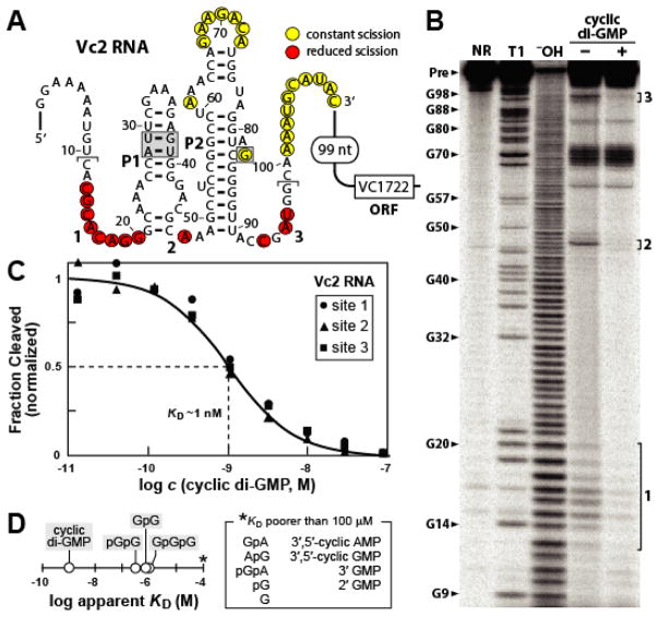
Biochemical and genetic analyses were carried out with both GEMM RNAs to determine whether they function as aptamers for cyclic di-GMP. A 110-nucleotide Vc2 RNA construct (110 Vc2; Fig. 1A) was subjected to in-line probing (16) in the absence or presence of 100 μM cyclic di-GMP (Fig. 1B). Despite the constrained structure of the second messenger, cyclic di-GMP exhibits a half life for spontaneous degradation of no poorer than ~150 days under in-line probing conditions (fig. S4). Therefore, the biologically relevant ligand for this aptamer class most likely is cyclic di-GMP and not the breakdown products of this second messenger. The changing patterns of spontaneous cleavage at the base of stems P1 and P2 suggest that the conserved nucleotides near the regions undergoing structural modulation (labeled 1 through 3) are important for ligand binding. Similar results were obtained for a construct encompassing the Vc1 RNA and for representative cyclic di-GMP aptamers from Bacillus cereus and Clostridium difficile.
Fig. 1.
A cyclic di-GMP aptamer from V. cholerae. (A) Sequence and structure of the Vc2 RNA from V. cholerae chromosome 2, and its proximity to the ORF of VC1722. Nucleotides shown correspond to the 110 Vc2 RNA construct. Bold numbers identify regions of ligand-mediated structure modulation as observed in B. Brackets identify the minimal 5′ or 3′ terminus (when the opposing terminus for 110 Vc2 RNA is retained) that exhibits structural modulation when tested with 10 nM cyclic di-GMP. Nucleotides in shaded boxes were mutated for studies depicted in Fig. 2A. (B) PAGE separation of RNA products generated by in-line probing of 5′ 32P-labeled 110 Vc2 RNA. NR (no reaction); T1 (partial digest with RNase T1); −OH (partial digest with alkali). RNA was incubated in the absence (−) or presence (+) of 100 μM cyclic di-GMP. (C) Plot of the normalized fraction of 110 Vc2 aptamer cleaved versus cyclic di-GMP concentration. Sites of structural modulation are as depicted in B. (D) Comparison of _K_D values exhibited by 110 Vc2 aptamer for cyclic di-GMP (fig. S5) and various analogs. G (guanosine); pG, pGpG, pGpA (5′ phosphorylated mono- and dinucleotides); GpGpG (trinucleotide), AMP and GMP (adenosine- and guanosine monophosphate, respectively).
The 110 Vc2 RNA appears to form a one-to-one saturable complex with a dissociation constant (_K_D) of approximately 1 nM for cyclic di-GMP (Fig. 1C and fig. S5). This interaction is nearly three orders of magnitude tighter than the affinity measured for cyclic di-GMP binding by an Escherichia coli PilZ protein domain (_K_D = 840 nM; 10). Furthermore, various analogs of the second messenger are strongly discriminated against by the Vc2 aptamer (Fig. 1D). The linear breakdown product of cyclic di-GMP by EAL PDEs (1–3), pGpG, is bound by the aptamer nearly three orders of magnitude less tightly. Similarly, the pG (5′ GMP) product of HD-GYP PDEs is not bound by the aptamer even at a concentration that is five orders of magnitude higher than the _K_D for cyclic di-GMP.
Bacterial riboswitches typically carry a conserved aptamer located immediately upstream of a less well-conserved expression platform. Most expression platforms in bacteria form structures that control transcription termination or translation initiation (17). Similar architectures are associated with many GEMM RNA representatives (see SOM), suggesting that cyclic di-GMP aptamers are likely to be components of riboswitches. Four 5′ UTRs from V. cholerae, B. cereus, and C. difficile were examined for riboswitch function. DNAs encompassing Vc2, its predicted native promoter, and the adjoining expression platform were used to prepare translational fusions with the E. coli lacZ reporter gene. Transformed E. coli cells carrying the aptamer-reporter fusion constructs were grown in liquid medium and assayed for β-galactosidase activity.
The reporter construct carrying the wild-type (WT) Vc2 aptamer (Fig. 1A and Fig. 2A) exhibits a high level of gene expression (Fig. 2B). In contrast, constructs M1 and M3 that carry mutations disrupting P1 or altering an otherwise strictly conserved nucleotide in the P2 bulge express the reporter gene at less than 10% of WT. Furthermore, an aptamer that carries four compensatory mutations (M2) that restore the structure of P1 exhibits near-WT gene expression. These results parallel the ligand-binding activities of these RNAs and suggest that the riboswitch integrating the Vc2 aptamer functions as an “on” switch. Similarly, transcriptional fusions were prepared for cyclic di-GMP riboswitches from B. cereus (Bc1 and Bc2) and C. difficile (Cd1) (fig. S6), and cloned into B. subtilis. The expression patterns for WT and equivalent M3 variants are consistent with “on” switch function for Bc1 and “off” switch function for Bc2 and Cd1 (Fig. 2B).
Fig. 2.
Representative cyclic di-GMP aptamers are components of gene control elements. (A) Reporter fusion constructs carry wild-type (WT) or mutant (M1 through M3) riboswitches from V. cholerae (Vc2) in E. coli, or carry the equivalent WT and M3 riboswitches from B. cereus (Bc1 and Bc2) or C. difficile (Cd1) (fig. S6) in B. subtilis. (B) β-galactosidase reporter gene assays for reporter fusion constructs described in A. Maximum Miller units measured for the four aptamer representatives were 436, 47, 5 and 51, respectively. (C) B. subtilis cells carrying a β-galactosidase reporter construct fused to a wild-type (WT) Cd1 riboswitch and transformed with a plasmid lacking (−) or carrying a normal (+) or mutant (E170A) V. cholerae vieA gene encoding an EAL phosphodiesterase (PDE).
Modulation of gene expression by cyclic di-GMP riboswitches was established by examining “off” switch action of the Cd1 RNA while manipulating the cellular concentration of the second messenger in by expression of V. cholerae VieA, an EAL phosphodiesterase (PDE) that is expected to lower the cellular concentration of cyclic di-GMP by promoting hydrolysis of the second messenger to its linear pGpG form (18). The Cd1 riboswitch is located in the 5′ UTR of a large operon that encodes for the proteins required to construct the entire flagella apparatus of this species (see Table S1). This arrangement suggests that some bacteria use a riboswitch to sense changes in cyclic di-GMP concentrations and regulate organelle biosynthesis to control motile versus sessile lifestyle transformations.
Transformed B. subtilis cells carrying a transcriptional fusion of the WT Cd1 riboswitch with lacZ, and grown on agar plates with X-gal, exhibit low β-galactosidase activity when co-transformed with either the empty plasmid vector or the vector coding for the inactive E170A VieA mutant (Fig. 2C). In contrast, robust β-galactosidase activity is evident in cells that are co-transformed with the reporter construct and the vector coding for VieA PDE. A similar trend is observed when these transformed cells are assayed from liquid culture, whereas a reporter construct carrying the M3 mutation in the Cd1 aptamer remains largely unaffected by VieA activity (fig. S7). Moreover, cyclic di-GMP causes transcription termination of the Cd1 RNA in an in vitro transcription assay (fig. S8). These observations indicate that C. difficile uses the Cd1 riboswitch to regulate transcription of the flagellar protein operon to controls motility in response to cyclic di-GMP signaling.
The VC1722 gene associated with the Vc2 riboswitch codes for a protein similar to a known transcription factor that is important for competence (15). It has been previously shown that a V. cholerae mutant producing a rugose phenotype has elevated cyclic di-GMP levels and exhibits higher expression of the VC1722 mRNA (19, 20). This is consistent with our data indicating that the Vc2 riboswitch associated with the VC1722 mRNA is a genetic “on” switch that yields higher gene expression when cyclic di-GMP concentrations are elevated (Fig. 2C).
The remaining V. cholerae cyclic di-GMP riboswitch (Vc1; fig. S3) is associated with the gbpA gene, which codes for a sugar-binding protein reported to be the key determinant permitting the bacterium to colonize mammalian intestines, leading to cholera disease (21). It has also been shown that V. cholerae lowers its cyclic di-GMP levels when colonizing mammalian host intestines (34). The V. cholerae vieA gene used in our study to demonstrate Cd1 riboswitch response to changing cyclic di-GMP levels (Fig. 2C) is known to be essential for bacterial infection. Thus, it is possible that a reduction in cyclic di-GMP levels brought about by the action of VieA is sensed by the Vc1 riboswitch to facilitate expression of GbpA and V. cholerae infection.
Some organisms have a strikingly high number of cyclic di-GMP riboswitch representatives (fig. S3 and fig. S9 through S12). Geobacter uraniumreducens, with 30 representatives identified, has the largest number of cyclic di-GMP aptamer RNAs among bacterial species whose genomes have been sequenced. These are distributed upstream of 25 different transcriptional units, with five RNAs carrying tandem cyclic di-GMP aptamers. Also intriguing is the identification of cyclic di-GMP riboswitch representatives residing within PhiCD119 bacteriophage DNA that are integrated within the C. difficile genome. The riboswitch sequence, located within the lysis module of the bacteriophage genome, is also evident in DNA packaged into bacteriophage particles. Although more than 20 metabolite-sensing riboswitch classes have been reported, and thousands of representatives of these classes have been identified, the cyclic di-GMP RNAs are the only examples of bacteriophage-associated riboswitches found after exhaustive bioinformatics searches. Perhaps viruses have little need for sensing fundamental metabolic products, but might gain an evolutionary advantage by monitoring the physiological transformations of bacterial cells brought about by changing concentrations of the second messenger cyclic di-GMP.
Acknowledgments
We thank Dianali Rivera for assistance with experiments analyzing c-di-GMP stability and integrity. We also acknowledge the efforts of the FT-ICR Mass Spectrometry Resource of the Keck Biotechnology Resource Laboratory and we thank Nicholas Carriero and Robert Bjornson for assisting with our use of the Yale Life Sciences High Performance Computing Center (NIH grant RR19895-02). This work was supported by NIH grants (R33 DK07027 and GM 068819) to R.R.B and by an NHLBI grant (HV28186). E.R.L. was supported by a grant (T32GM007223) from the National Institute of General Medical Sciences. J.N.K. was supported by a predoctoral fellowship from the NSF. RNA research in the Breaker laboratory is also supported by the Howard Hughes Medical Institute.
References and Notes
- 1.Jenal U, Malone J. Annu Rev Genet. 2006;40:385. doi: 10.1146/annurev.genet.40.110405.090423. [DOI] [PubMed] [Google Scholar]
- 2.Ryan RP, Fouhy Y, Lucey JF, Maxwell Dow J. J Bacteriol. 2006;188:8327. doi: 10.1128/JB.01079-06. [DOI] [PMC free article] [PubMed] [Google Scholar]
- 3.Tamayo R, Pratt JT, Camilli A. Annu Rev Microbiol. 2007;61:131. doi: 10.1146/annurev.micro.61.080706.093426. [DOI] [PMC free article] [PubMed] [Google Scholar]
- 4.Simm R, Morr M, Kader A, Nimtz M, Römling U. Mol Microbiol. 2004;53:1123. doi: 10.1111/j.1365-2958.2004.04206.x. [DOI] [PubMed] [Google Scholar]
- 5.Chan C, et al. Proc Natl Acad Sci USA. 2004;101:17084. doi: 10.1073/pnas.0406134101. [DOI] [PMC free article] [PubMed] [Google Scholar]
- 6.Wassman P, et al. Structure. 2007;15:915. doi: 10.1016/j.str.2007.06.016. [DOI] [PubMed] [Google Scholar]
- 7.Ross P, et al. FEBS Lett. 1985;186:191. doi: 10.1016/0014-5793(85)80706-7. [DOI] [PubMed] [Google Scholar]
- 8.Ross P, et al. Nature. 1987;325:279. doi: 10.1038/325279a0. [DOI] [PubMed] [Google Scholar]
- 9.Lee VT, Matewish JM, Kessler JL, Hyodo M, Jayakawa Y, Lory S. Mol Microbiol. 2007;65:1474. doi: 10.1111/j.1365-2958.2007.05879.x. [DOI] [PMC free article] [PubMed] [Google Scholar]
- 10.Ryjenkov DA, Simm R, Römling U, Gomelsky M. J Biol Chem. 2006;281:30310. doi: 10.1074/jbc.C600179200. [DOI] [PubMed] [Google Scholar]
- 11.Wolfe AJ, Visick KL. J Bacteriol. 2008;190:463. doi: 10.1128/JB.01418-07. [DOI] [PMC free article] [PubMed] [Google Scholar]
- 12.Winkler WC, Breaker RR. Annu Rev Microbiol. 2005;59:487. doi: 10.1146/annurev.micro.59.030804.121336. [DOI] [PubMed] [Google Scholar]
- 13.Breaker R. Science. 2008;319:1795. doi: 10.1126/science.1152621. [DOI] [PubMed] [Google Scholar]
- 14.Weinberg Z, et al. Nucleic Acids Res. 2007;35:4809. doi: 10.1093/nar/gkm487. [DOI] [PMC free article] [PubMed] [Google Scholar]
- 15.Meibom KL, Blokesch M, Dolganov NA, Wu CY, Schoolnik GK. Science. 2005;310:1824. doi: 10.1126/science.1120096. [DOI] [PubMed] [Google Scholar]
- 16.Soukup GA, Breaker RR. RNA. 1999;5:1308. doi: 10.1017/s1355838299990891. [DOI] [PMC free article] [PubMed] [Google Scholar]
- 17.Barrick JE, Breaker RR. Genome Biol. 2007;8:R239. doi: 10.1186/gb-2007-8-11-r239. [DOI] [PMC free article] [PubMed] [Google Scholar]
- 18.Tamayo R, Tischler AD, Camilli A. J Biol Chem. 2005;280:33324. doi: 10.1074/jbc.M506500200. [DOI] [PMC free article] [PubMed] [Google Scholar]
- 19.Lim B, Beyhan S, Meir J, Yildiz FH. Mol Microbiol. 2006;60:331. doi: 10.1111/j.1365-2958.2006.05106.x. [DOI] [PubMed] [Google Scholar]
- 20.Beyhan S, Yildiz FH. Mol Microbiol. 2007;63:995. doi: 10.1111/j.1365-2958.2006.05568.x. [DOI] [PubMed] [Google Scholar]
- 21.Kirn TJ, Jude BA, Taylor RK. Science. 2005;438:863. doi: 10.1038/nature04249. [DOI] [PubMed] [Google Scholar]
- 22.Govind R, Fralick JA, Rolfe RD. J Bacteriol. 2006;188:2568. doi: 10.1128/JB.188.7.2568-2577.2006. [DOI] [PMC free article] [PubMed] [Google Scholar]

