Developmental Decrease in NMDA Receptor Desensitization Associated with Shift to Synapse and Interaction with Postsynaptic Density-95 (original) (raw)
Abstract
NMDA receptors (NMDARs) play a crucial role in neuronal development, synaptic plasticity, and excitotoxicity; therefore, regulation of NMDAR function is important in both physiological and pathological conditions. Previous studies indicate that the NMDAR-mediated synaptic current decay rate increases during development because of a switch in receptor subunit composition, contributing to developmental changes in plasticity. To test whether NMDAR desensitization also changes during development, we recorded whole-cell NMDA-evoked currents in cultured rat hippocampal neurons. We found that glycine-independent desensitization of NMDARs decreases during development. This decrease was not dependent on a switch in subunit composition or differential receptor sensitivity to agonist-, Ca2+-, or Zn2+-induced increase in desensitization. Instead, several lines of evidence indicated that the developmental decrease in desensitization was tightly correlated with synaptic localization of the receptor, suggesting that association of NMDARs with proteins selectively expressed at synapses in mature neurons might account for developmental alterations in desensitization. Accordingly, we tested the role of interactions between PSD-95 (postsynaptic density-95) and NMDARs in regulating receptor desensitization. Overexpression of PSD-95 reduced NMDAR desensitization in immature neurons, whereas agents that interfere with synaptic targeting of PSD-95, or induce movement of NMDARs away from synapses and uncouple the receptor from PSD-95, increased NMDAR desensitization in mature neurons. We conclude that synaptic localization and association with PSD-95 increases stability of hippocampal neuronal NMDAR responses to sustained agonist exposure. Our results elucidate an additional mechanism for differentially regulating NMDAR function in neurons of different developmental stages or the response of subpopulations of NMDARs in a single neuron.
Keywords: NMDA receptor, desensitization, PSD-95, patch-clamp recording, hippocampal pyramidal neurons, neuronal cultures
Introduction
Fast excitatory synaptic transmission in the mammalian CNS is mediated predominantly by three types of ionotropic glutamate receptors, including AMPA, NMDA, and kainate receptors (Monaghan et al., 1989). Extensive studies have established that NMDA receptors (NMDARs) play a crucial role in neuronal development and synaptic plasticity and that overactivation of NMDARs leads to excitotoxicity (Dingledine et al., 1999).
Activation of NMDARs is limited by a number of processes, including peak current rundown, Ca2+-dependent inactivation, glycine-dependent desensitization, and glycine-independent desensitization (for review, see McBain and Mayer, 1994; Dingledine et al., 1999). Regulation of NMDAR activity by these processes is believed to be important in both physiological and pathological conditions. For example, manipulations that suppress Ca2+-dependent rundown of NMDARs enhance NMDA- or glutamate-induced excitotoxicity (Furukawa et al., 1995; Abdel-Hamid and Baimbridge, 1997; Furukawa et al., 1997), whereas conditions that promote Ca2+-dependent rundown protect from glutamate- or oxygen-glucose deprivation-induced excitotoxicity (Furukawa et al., 1995; Sattler et al., 2000). As well, NMDAR desensitization during repetitive firing contributes to shaping synaptic responses and neuronal activity (Tong et al., 1995; Jones and Westbrook, 1996). Rapid desensitization of non-NMDARs is protective against AMPA-induced excitotoxicity (Zorumski et al., 1990; May and Robison, 1993; Brorson et al., 1995; Raymond et al., 1996). Desensitization and inactivation of NMDARs may also be protective during a sustained glutamate insult by limiting calcium influx.
NMDARs are tetrameric complexes of two NR1 subunits in combination with NR2A, 2B, 2C, 2D, or NR3 (Dingledine et al., 1999). NMDAR glycine-independent desensitization, which is determined by two domains flanking the putative agonist-binding domain in the N terminus of the NR2 subunit (Krupp et al., 1998; Villarroel et al., 1998), is subtype dependent. Desensitization of NR1/NR2A is greater than NR1/NR2B (Krupp et al., 1996), whereas NR1/NR2C and NR1/NR2D do not desensitize at all when expressed in heterologous cells (Krupp et al., 1998; Villarroel et al., 1998). Moreover, this form of desensitization is regulated by calcineurin (Tong and Jahr, 1994; Tong et al., 1995), which causes a time-dependent increase in desensitization (Sather et al., 1992; Tong and Jahr, 1994; Tong et al., 1995) through direct dephosphorylation of serine residues in the NR2 C-terminal tail (Krupp et al., 2002). Zinc also regulates glycine-independent desensitization via an allosteric interaction between the zinc-binding and ligand-binding domains (Zheng et al., 2001).
Here we report a developmental decrease in glycine-independent desensitization of NMDARs in cultured hippocampal neurons. Although expression of the two major NR2 subunits in the hippocampal region, 2A and 2B, is developmentally regulated, with 2B subunits dominating early and 2A subunits replacing 2B later in development (Williams et al., 1993; Hollmann and Heinemann, 1994; Monyer et al., 1994; Zhong et al., 1994; Li et al., 1998; Rao et al., 1998; Stocca and Vicini, 1998; Tovar and Westbrook, 1999; Barria and Malinow, 2002), the change in desensitization was not dependent on the switch in subunit composition, nor was it dependent on altered agonist, Ca2+, or zinc sensitivity. Instead, we found that the progressive decrease in extent of glycine-independent desensitization during development correlated well with increasing localization of NMDARs at synapses and their association with PSD-95 (postsynaptic density-95), a scaffolding protein highly expressed in the PSD. We propose that the coupling of NMDARs with other signaling proteins via PSD-95 that occurs at synapses mediates the developmental change in desensitization.
Materials and Methods
Primary neuronal cultures and transfection. Embryonic hippocampal cultures were prepared as described previously (Li et al., 2002). Briefly, rat hippocampi were dissected from 17- to 18-d-old embryos and subsequently dissociated using papain and trituration with fire-polished glass pipettes. Cells were plated in Neurobasal medium supplemented with B27 and grown on poly-d-lysine-coated coverslips at a density of ∼300-400 cells/mm2. Cultures were maintained in a humidified atmosphere with 5%CO2. One-half of the medium was changed twice every week. Neurons were transfected with either PSD-95-green fluorescent protein (GFP) or GFP construct by lipid-mediated gene transfer kit (_N_-[1-(2,3-dioleoyloxy)propyl]-_N,N,N_-trimethylammonium methyl-sulfate) as described previously (Craven et al., 1999) or by Effectene Transfection Reagent (Qiagen, Hilden, Germany). Briefly, neurons were transfected at 2-4 d in vitro (DIV). For transfection of eight wells (in a 24-well plate), 2 μg of DNA was mixed with 150 μl of EC buffer and 16 μl of enhancer. After 5 min of incubation at room temperature, 25 μl of Effectene was added. The resulting solution was mixed gently and incubated for another 10 min at room temperature. Fresh Neurobasal medium (1 ml) was then added to the solution. A total of 135 μl of this DNA-Effectene mix solution was added into each well of neurons containing 200 μl of medium and incubated at 37°C for 3-5 hr. DNA-Effectene mix solution was then replaced with 0.5 ml of medium (containing 50% conditioned and 50% fresh medium) for each well. Neurons were used for patch-clamp recording 2-3 d after transfection.
Microisland cultures were made as described previously (Bekkers and Stevens, 1991; Li et al., 2002). Briefly, glass coverslips were coated with 0.15% agarose and allowed to dry. A solution of poly-d-lysine (0.5 mg/ml) and collagen (1.9 mg/ml; Upstate Biotechnology, Waltham, MA) was then sprayed on the agarose background to form microdots. After growth of the glial feeder layers on the microdots, the CA1-CA3 region of hippocampi from postnatal day 0-3 rats were removed, enzymatically (20 U/ml papain) and mechanically dissociated, and then plated. Cells were grown in a solution based on Neurobasal A medium (Invitrogen, Burlington, Ontario, Canada) supplemented with 2.5% fetal calf serum, 2% B27, 20 mm glucose, 0.5 mm glutamine, and 0.2% penicillin-streptomycin stock.
HEK293 cell culture and transfection. HEK293 cells (CRL 1573; American Type Culture Collection, Rockville, MD) were maintained as we described previously (Chen et al., 1997). Cells were transfected using the calcium phosphate precipitation method (Chen et al., 1997) with a total of 12 μg of plasmid DNA/10 cm plate. Cells were transfected with equal amounts of cDNAs encoding NR1-1a, NR2 (A or B), and GFP to allow for identification of transfected cells during recording.
Electrophysiology. Neurons from embryonic rat hippocampal cultures were used for recording at 4-7 DIV, 10-11 DIV, or >13 DIV. Autaptic cultures were used for recording at 11-14 DIV. Patch-clamp recording in the whole-cell mode was performed using either the nystatin perforated-patch method to maintain intracellular contents, as described previously (Li et al., 2002), or conventional whole-cell patch-clamp recording method (Hamill et al., 1981). For the conventional whole-cell recording, the electrode solution contained 115 mm Cs-methanesulfonate, 10 mm HEPES, 20 mm K2-creatine phosphate, 50 U/ml creatine phosphokinase, 4 mm MgATP, and 10 mm BAPTA, pH 7.26 (KOH), 310 mOsm. For recording on autaptic neurons, the electrode solution contained 112.5 mm K-methanesulfonate, 8 mm NaCl, 5 mm MgATP, 20 mm HEPES, 0.2 mm BAPTA, 20 mm K2-creatine phosphate, and 50 U/ml creatine phosphokinase, pH 7.2.
The cells were continuously superfused with the external recording solution. For HEK293 cells, the external recording solution contained the following (in mm): 145 NaCl, 5.4 KCl, 11 glucose, 2 CaCl2, and 10 HEPES, pH 7.3. For neurons the external recording solution contained the following (in mm): 167 NaCl, 2.4 KCl, 10 HEPES, 10 glucose, 2 CaCl2, and 0 or 1 MgCl2, pH 7.3 (325 mOsm). TTX (300 nm), strychnine (2 μm), and glycine (30 or 100 μm as indicated) were added just before use. TTX was omitted for autaptic culture recording. As described previously (Chen et al., 1997), agonist or drug application was gravity fed to the cells using a θ-tube. NMDA (100 μm or 1-2 mm as indicated) was included only in the agonist side of the θ-tube, which contained the same solution as the external recording solution, except sometimes with reduced CaCl2 concentration as indicated (0.2 mm CaCl2 for conventional whole-cell recording; 0 mm CaCl2 plus 100 μm EGTA with 1 mm MgCl2 for nystatin patch recording). For recording on autaptic neurons, EPSCs were evoked by depolarizing the cells from -65 to 0 mV for 1.5 msec, and the whole-cell responses were evoked by applying 1 mm NMDA in the external solution in which CaCl2 was replaced with 1 mm SrCl2 to minimize Ca2+-dependent inactivation (Legendre et al., 1993; Krupp et al., 1996). All other drugs, when used as indicated, were included in both the control and agonist side of the θ-tube. Rapid switching between the two solutions was achieved by computer-controlled solenoid-driven valves. The recording protocol consisted of 10 sec agonist applications at a rate of one per minute, unless otherwise indicated.
All recordings were made in voltage-clamp mode. Data were acquired using the Axopatch 200B patch-clamp amplifier (Axon Instruments, Foster City, CA). Currents were filtered at 5 kHz and digitized at 10 kHz. pClamp 6.1 or 8.1 software (Axon Instruments) was used for data acquisition and analysis. Access resistance and cell capacitance were monitored by applying a brief, 5 mV hyperpolarizing voltage step just before each agonist application (Li et al., 2002). The series resistance was compensated 60-70%. The membrane holding potential was -70 mV (for nystatin patch recording) or -65 mV (for conventional whole-cell recording) when Mg2+ was not present in the recording solutions and was +40 mV when 1 mm Mg2+ was present.
Immunocytochemistry and quantification. To characterize the subcellular localization of NMDARs and the expression pattern of overexpressed PSD-95-GFP in cultured hippocampal neurons, cells were double labeled with antibodies against the presynaptic marker synaptophysin and NR1, or GFP and NR1, or GFP and PSD-95. Briefly, neuronal cultures were fixed with methanol at -20°C for 10 min. After washing with PBS, the cells were incubated with 1% BSA for 30 min. Primary antibodies, including mouse monoclonal NR1 antibody (1:500; Chemicon, Temecula, CA), rabbit polyclonal synaptophysin antibody (1:1000; Zymed Laboratories, San Francisco, CA), mouse monoclonal PSD-95 (5 μg/ml; Chemicon), chicken (1:2000; Chemicon) or rabbit (1:100; Affinity BioReagents, Golden, CO) GFP antibodies were then incubated with the cultures at room temperature for 1 hr. After washing with PBS, the cultures were further incubated with green fluorescent anti-rabbit, mouse, or chicken (Alexa 488; 1:1000; Molecular Probes, Eugene, OR) and red fluorescent anti-mouse or rabbit (Cy3; 1:200; Jackson ImmunoResearch, West Grove, PA) secondary antibodies at room temperature for 1 hr.
Images were acquired using a CCD camera affixed to a Zeiss (Oberkochen, Germany) inverted microscope with AxioVision software and displayed in Adobe Photoshop 5.5 (Adobe Systems, San Jose, CA). To quantify the synaptic localization of NMDARs in different conditions, we counted the number of synaptic receptor clusters per dendritic length as described previously (Crump et al., 2001). Briefly, images were exported into Northern Eclipse (Empix Imaging, Missasauga, Ontario, Canada) and custom-written software routines (O. Prange and A. E. El-Husseini, unpublished observations) were applied for image analysis. Only puncta with intensity at least twofold greater than the diffuse dendritic staining were chosen. Synaptic NMDAR clusters were defined by overlap with synaptophysin-positive puncta and counted per dendritic length. The resulting data were analyzed with Origin software (Microcal Software, Northampton, MA). For quantification, 5-10 neurons from two to three different batches of cultures and experiments for each condition were randomly chosen on the basis of healthy morphology.
Coimmunoprecipatition. Sister hippocampal neuronal cultures of 14-17 DIV in 10 cm dishes were treated with 2-bromopalmitate, vehicle, or picrotoxin (PTX) as indicated in Results and then scraped into a PBS-based harvest buffer containing 1 mm EGTA, 1 mm EDTA, 1 mm PMSF, 2 μg/ml aprotinin, 20 μg/ml pepstatin A, and 20 μg/ml leupeptin. For one 10 cm dish, 1 ml of harvest buffer was used. After centrifugation, the pellet was lysed and solubilized in harvest buffer containing 0.1% SDS and 0.8% Triton X-100 (0.5 ml final volume). One-tenth of the lysate was reserved for input loading, and the remainder was incubated with protein-A and protein-G beads, each 20 μl, for 1 hr at 4°C to remove any proteins that nonspecifically bind to the beads. After brief centrifugation, the supernatant was reserved and incubated with 10 μg of rabbit polyclonal anti-NR2A antibody (Upstate Biotechnology) for 1 hr at 4°C. Protein-A and protein-G beads, each 30 μl, were then added to the solution and incubated at 4°C for 1 hr. The beads were washed three times with a Tris wash buffer containing 50 mm Tris, pH 7.4, 150 mm NaCl, 1 mm EDTA, 1 mm EGTA, and 1% Triton X-100. The proteins were eluted from the beads and denatured by boiling in loading buffer for 5 min and then resolved by SDS-PAGE as described previously (Chen et al., 1999). Proteins from each of the different conditions (vehicle, 2-bromopalmitate, and PTX) were loaded to the same gel. After transfer, membranes were probed with antibodies against the NMDAR NR2A subunit (1:1000 of affinity-purified rabbit polyclonal; a generous gift from Dr. Richard Huganir, Johns Hopkins Medical School, Baltimore, MD) and PSD-95 (mouse monocolonal; 5 μg/ml; Chemicon). The resulting blots were quantified by densitometric analysis (Li et al., 2002). The amount of PSD-95 coimmunoprecipated with NR2A was determined as the PSD-95 to NR2A band-density ratio.
Materials. Genistein, bpV(phen), protein kinase inhibitor (PKI), and KN93 were from Calbiochem-Novabiochem (San Diego, CA). Latrunculin A or B was from Biomol (Plymouth Meeting, PA). 2-Bromopalmitate was a gift from Dr. A. E. El-Husseini (University of British Columbia, Vancouver, British Columbia, Canada**)**. All other chemicals were purchased from Sigma (St. Louis, MO). The PSD-95-GFP and PSD-95(C3,5S)-GFP constructs were gifts from Dr. D. S. Bredt (University of California at San Francisco, San Francisco, CA) and were described previously (Craven et al., 1999). NR1-1a (Dingledine et al., 1999) and NR2B cDNAs were gifts from Dr. Nakanishi (Kyoto University, Kyoto, Japan), and NR2A (ϵ1) was a gift from Dr. Mishina (Tokyo University, Tokyo, Japan). These constructs were described previously (Raymond et al., 1996; Chen et al., 1997). Tissue culture reagents were obtained from Invitrogen.
Data analysis. Results are presented as mean ± SE. Sets of different results were compared using one-way ANOVA or Student's t test as appropriate, and significant differences were determined at the 95% confidence intervals. Three to 10 responses of each cell were averaged for estimation of steady-state to peak current (_I_ss/_I_p) ratio. In some cases, only the first response was measured because the _I_ss/_I_p ratio was quite stable over time during our recording conditions.
Results
Neuronal NMDARs show developmental change in desensitization independent of subunit composition
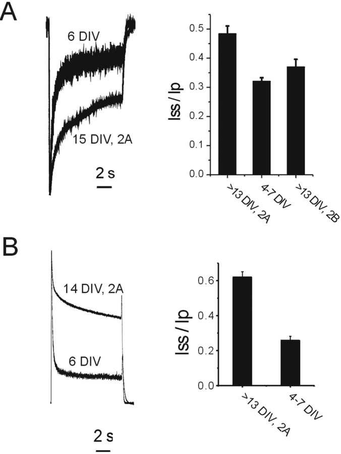
NMDAR glycine-independent desensitization is involved in limiting receptor activation during repeated or sustained exposure to glutamate (McBain and Mayer, 1994; Tong et al., 1995). To identify factors regulating this process in cultured hippocampal neurons, we measured desensitization of NMDAR currents in response to 10 sec applications of 100 μm NMDA in the continuous presence of 30 μm glycine, using the nystatin perforated-patch recording technique to preserve intracellular contents and endogenous signaling mechanisms. Because NMDAR subunit composition is an important determinant of receptor function and modulation, we began by comparing desensitization of 2A-containing NMDARs (recorded from >13 DIV neurons bathed in the NR1/NR2B-subtype-selective antagonist ifenprodil; this population would include both NR1/NR2A and NR1/NR2B/NR2A) to that of NMDARs in 4-7 DIV neurons, which mainly express NR1/NR2B, and to 2B-subtype NMDARs isolated pharmacologically in >13 DIV neurons. We showed previously that ifenprodil blocked 76% of the NMDAR-mediated current recorded from 4-7 DIV neurons (Li et al., 2002), suggesting that those neurons indeed mainly express 2B-subtype NMDARs, as reported by others (Tovar and Westbrook, 1999). To isolate 2B-subtype NMDARs in >13 DIV neurons, we used a combination of (+)-5-methyl-10,11-dihydro-5H-dibenzo [a,d] cyclohepten-5,10-imine maleate (MK-801) and ifenprodil (Li et al., 2002). In the presence of both ifenprodil and MK-801, repeated applications of NMDA resulted in selective activation of 2A-containing receptors that were then irreversibly blocked by MK-801. We then removed ifenprodil and MK-801 and washed extensively with control solution to recover the 2B-subtype NMDAR current. Strikingly, the 2B-subtype NMDARs in both 4-7 DIV and >13 DIV neurons showed a higher degree of glycine-independent desensitization compared with 2A-containing NMDARs in >13 DIV neurons when recorded in 2 mm Ca2+ (Fig. 1_A_). To minimize differences in apparent desensitization attributable to Ca2+-dependent processes, we made recordings in the absence of external Ca2+ and at positive holding potentials (to minimize any residual Ca2+ entry). Also, because the 2B-subtype is more sensitive to agonist than the 2A-subtype of NMDARs (Dingledine et al., 1999) and NMDA is less potent than glutamate (Sather et al., 1992), we repeated our experiment using 2 mm NMDA to saturate both subtypes of receptors. Under these conditions, desensitization was still markedly more extensive for 2B-subtype NMDARs in 4-7 DIV neurons than for 2A-containing NMDARs in >13 DIV (Fig. 1_B_). These data indicate that Ca2+-dependent processes and differential sensitivity to NMDA cannot explain the difference in NMDAR desensitization.
Figure 1.
Neuronal 2B-subtype NMDARs show more glycine-independent desensitization than 2A-containing NMDARs. A, Left, Representative responses mediated by NMDARs in a 6 DIV neuron or a 15 DIV neuron in the presence of 10 μm ifenprodil, in response to 100 μm NMDA (30 μm glycine) in the presence of 2 mm calcium. Right, Pooled data showing that NMDARs in 4-7 DIV neurons or 2B-subtype NMDARs in >13 DIV neurons have higher degree of desensitization than 2A-containing NMDARs in >13 DIV neurons. _V_H of 65 mV. Recordings were made from n = 12, 12, and 7 different neurons for the three different groups: >13 DIV, 2A; 4-7 DIV; and >13 DIV, 2B, respectively. B, Same as A, except Ca2+ was replaced by 1 mm Mg2+, holding potential was +40 mV, [NMDA] was 2 mm, and [glycine] was 100 μm. All recordings were made using the nystatin perforated recording technique.
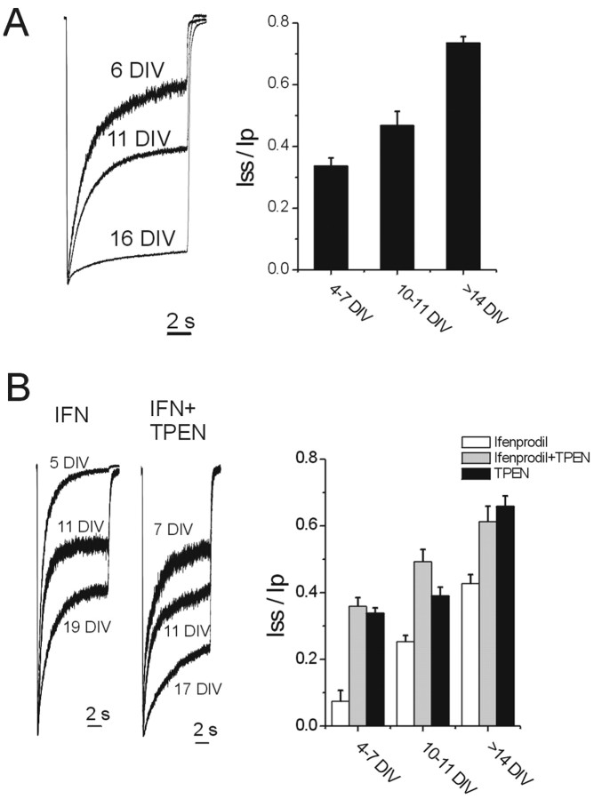
To define the macroscopic kinetics of the NMDAR response more precisely and further investigate the factors regulating differences in neuronal NMDAR desensitization during development, we also used conventional whole-cell recording. In these experiments, the external Ca2+ was kept low (0.2-1 mm), and the intracellular pipette solution contained 10 mm BAPTA to minimize Ca2+-dependent inactivation (Zheng et al., 2001). Also, saturating concentrations of NMDA (1 mm) and glycine (100 μm) were used to isolate glycine-independent desensitization from other mechanisms. Under these conditions, neuronal NMDARs showed a clear decrease in desensitization (increase in _I_ss/_I_p) during development (Fig. 2_A_). The _I_ss/_I_p ratio was 34 ± 3% (n = 25), 47 ± 5% (n = 12), and 74 ± 2% (n = 30) for neurons of 4-7, 10-11, and >14 DIV, respectively (p < 0.0001 by one-way ANOVA followed by Bonferroni's multiple comparisons test). The decrease in desensitization with development was not attributable to a decrease in NMDA-evoked current amplitude, because mean peak current amplitude was much smaller for immature neurons (2234 ± 248 pA; n = 12) than for mature neurons (5802 ± 512 pA; n = 16).
Figure 2.
Developmental change in NMDAR desensitization is independent of subunit composition and zinc sensitivity. A, Left, Representative responses of NMDARs in 6, 11, and 16 DIV neurons in response to 1 mm NMDA (100 μm glycine) in 0.2 mm extracellular Ca2+. Electrode solution contained 10 mm BAPTA. Recordings were made using conventional whole-cell patch-clamp technique with VH of -65 mV. Currents were normalized for comparison of desensitization. Right, Quantification of desensitization showing a developmental change. B, Recording conditions were identical to A, except that 3 μm ifenprodil (IFN) and/or 2 μm TPEN was added to external solution. Inclusion of 2 μm TPEN to remove ambient zinc in recording solution did not significantly change NMDAR desensitization compared with control condition (p > 0.05 at each development stage compared with data shown in A). Ifenprodil increased desensitization (left and right panels), an effect reversed by inclusion of TPEN (middle and right panels), but the trend toward decreased desensitization with maturation was preserved. Recordings were made from the following: n = 5, 12, and 5 different neurons of 4-7, 10-11, and >14 DIV, respectively, for ifenprodil plus TPEN; n = 6, 4, and 5 of 4-7, 10-11, and >14 DIV, respectively, for TPEN alone; and n = 5, 5, and 16 of 4-7, 10-11, and >14, respectively, for ifenprodil alone.
Zinc in the submicromolar range has been shown to increase NMDAR desensitization for recombinant receptors expressed in HEK293 cells (Chen et al., 1997; Zheng et al., 2001). Because salt solutions often contain submicromolar zinc as a result of contamination (Paoletti et al., 1997), we wondered whether a change in sensitivity to ambient zinc during development might explain our results. To test this possibility, we added 2 μm_N,N,N_′,_N_′-tetrakis-(2-pyridylmethyl)-ethylenediamine (TPEN) to chelate ambient zinc in the external solution (Paoletti et al., 1997). In contrast to results for recombinant NMDARs in HEK293 cells, chelation of zinc did not decrease the desensitization of hippocampal neuronal NMDARs at any developmental stage (Fig. 2, compare _I_ss/_I_p for TPEN condition in B with that in A), and the progressive increase in _I_ss/_I_p with development was preserved. Thus, the developmental change in desensitization of neuronal NMDARs is not attributable to altered receptor sensitivity to zinc.
To determine whether NR2A-containing NMDARs also undergo the developmental change in desensitization, we repeated the experiments in the presence of the NR2B-subtype-selective inhibitor ifenprodil (3 μm). Although ifenprodil increased desensitization at each developmental stage (all _I_ss/_I_p ratios were lower), the developmental decrease in desensitization was still apparent (Fig. 2_B_). The _I_ss/_I_p ratio was 7 ± 3% (n = 5), 25 ± 2% (n = 5), and 43 ± 3% (n = 16) for neurons of 4-7, 10-11, and >14 DIV, respectively (p < 0.0001 by one-way ANOVA followed by Bonferroni's multiple comparisons test). An increase in NMDAR desensitization in the presence of ifenprodil has also been reported for recombinant receptors expressed in HEK293 cells (Zheng et al., 2001). In our experiments, increased desensitization in the presence of ifenprodil was abolished for all developmental stages examined by addition of 2 μm TPEN (Fig. 2_B_), suggesting that NMDARs are more sensitive to ambient zinc in the presence of ifenprodil. Because relative differences in desensitization were still observed in the presence of ifenprodil, we concluded that developmental changes in neuronal NMDAR desensitization were not dependent on a switch in subunit composition.
NMDAR desensitization is regulated by receptor subcellular localization
One major difference between NMDARs in 4-7 DIV and those in >13 DIV neurons is their subcellular localization. NMDARs expressed in immature neurons are mainly extrasynaptic, whereas the majority are localized to synapses in more mature neurons (Li et al., 1998; Rao et al., 1998; Tovar and Westbrook, 1999). It is possible that synaptic localization renders NMDARs more resistant to desensitization. This explanation would also be consistent with our results showing that 2B-subtype NMDARs in >13 DIV neurons desensitize more extensively than 2A-subtype NMDARs (Fig. 1_A_), because the majority of NR2A subunits are incorporated into synapses whereas NR2B subunits continue to predominate at extrasynaptic sites (Li et al., 1998; Stocca and Vicini, 1998; Tovar and Westbrook, 1999; Barria and Malinow, 2002; Li et al., 2002).
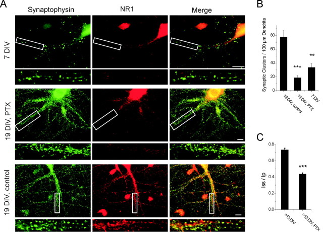
To test whether extrasynaptic NMDARs desensitize more extensively than synaptic NMDARs, we chronically treated >13 DIV neurons with PTX, which decreases inhibitory input leading to robust bursting (Murphy et al., 1992). As a result, NMDAR activation is chronically increased, eventually leading to a decreased number of synaptic NMDARs (Crump et al., 2001). Treatment with 100 μm PTX for 1 week indeed depleted NMDARs from synapses (and from the dendrite, in general) in >13 DIV neurons, as assessed by immunocytochemical colocalization with synaptophysin (Fig. 3_A,B_), and also markedly increased NMDAR desensitization (Fig. 3_C_). Note that treatment with PTX also disrupted the interaction between NMDARs and PSD-95 (see Fig. 5_A_ and text below).
Figure 3.
Synaptic localization of NMDARs increases with development and is decreased by picrotoxin treatment. A, Representative photomicrographs of double labeling of NR1 (red) and synaptophysin (green) in cultured hippocampal neurons of 7 DIV, 19 DIV chronically treated with PTX, and 19 DIV treated with vehicle (control), respectively. Scale bars, 10 μm. White boxes outline regions shown at higher gain directly below each photomicrograph. B, Quantification of the number of synaptic NR1 clusters per 100 μm dendrite for different conditions (19 DIV, control, 78 ± 9; 19 DIV, PTX, 19 ± 3; 7 DIV, 36 ± 6; **p < 0.01, ***_p_ < 0.001 compared with control condition). _C_, Recordings were made, as described in Figure 2_A_, from >13 DIV neurons treated with vehicle alone (>13 DIV) or picrotoxin (>13 DIV, PTX). _I_ss/_I_p, 74 ± 2 and 44 ± 2%, n = 30 and 12, for control and PTX group, respectively; ***p < 0.0001.
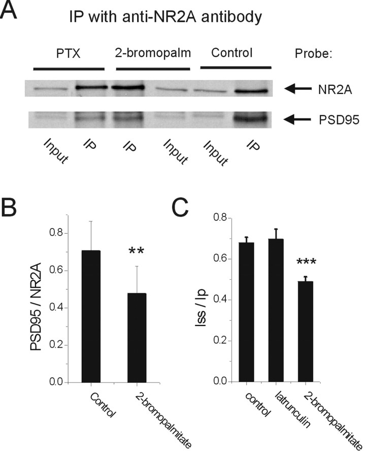
Figure 5.
Inhibition of palmitoylation disrupts the interaction between NMDARs and PSD-95 and increases NMDAR desensitization in mature neurons. A, Representative Western blot showing coimmunoprecipation of NR2A and PSD-95. Sister cultures of >13 DIV were treated with different conditions, including PTX, 2-bromopalmitate, or control (vehicle only). An anti-NR2A antibody was used for immunoprecipitation (IP), and the resulting blots were probed with both NR2A and PSD-95 antibodies. B, The band density of PSD-95 and NR2A were quantified using densitometry and presented as the PSD-95 to NR2A band density ratio (**p < 0.01 by paired _t_ test; _n_ = 5 separate experiments with different batches of neurons). _C_, Treatment with 2-bromopalmitate to disperse PSD-95, but not treatment with latrunculin to depolymerize F-actin or with vehicle only (control), increased NMDAR desensitization in >13 DIV neurons. _I_ss/_I_p was 49 ± 2 and 68 ± 3%, n = 5 and 12 different neurons for 2-bromopalmitate and control, respectively; ***p < 0.001 by unpaired _t_ test. _I_ss/_I_p was 70 ± 5%, _n_ = 11 for latrunculin-treated neurons; _p_ > 0.05 compared with control. Recording conditions were identical to those described in Figure 2_A_.
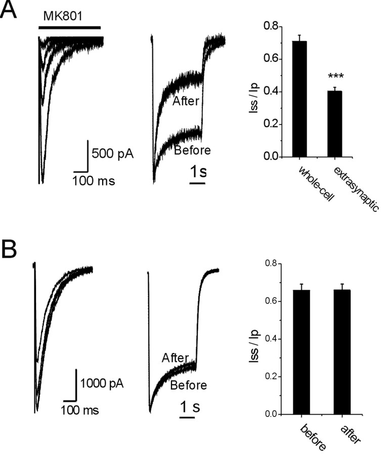
To test within a single cell whether extrasynaptic NMDARs desensitize more readily than synaptic NMDARs, we used single-cell microisland cultures (Bekkers and Stevens, 1991; Li et al., 2002) to directly compare extrasynaptic NMDARs with total surface NMDARs. Neurons with autaptic synapses in 11-14 DIV cultures were stimulated with short depolarizations to generate action potentials in the presence of MK-801 (20 μm). Under these conditions, synaptic NMDARs were selectively activated and blocked after 57 ± 6 stimuli (n = 7) (Fig. 4_A_). Desensitization (assessed by exogenous application of NMDA) before and after blockade of the synaptic NMDARs in a representative neuron is shown in Figure 4_A_ (middle). Responses mediated by extrasynaptic NMDARs (spared from activity-dependent MK-801 block) clearly showed a higher degree of desensitization compared with whole-cell responses mediated by the combined pool of synaptic and extrasynaptic NMDARs (Fig. 4_A_, middle and right). Approximately 60% of the NMDARs in these autaptic neurons were synaptic, because the extrasynaptic NMDAR-mediated response was 39 ± 4% of the initial whole-cell response (n = 7). On the other hand, when the same number of synaptic stimuli was applied in the absence of MK-801, there was no change in NMDA-evoked current desensitization (Fig. 4_B_), indicating that the increase in desensitization was not a nonspecific effect of repeated synaptic activation or attributable to prolonged whole-cell recording.
Figure 4.
Extrasynaptic NMDARs show more extensive desensitization than synaptic NMDARs. A, Left, NMDAR-mediated synaptic responses were recorded from an autaptic neuron in the presence of 20 μm MK-801. CNQX (5 μm) and 2 mm Ca2+ were present in the extracellular recording solution. Standard whole-cell voltage clamp was used. First, 10th, 20th, and 44th responses in the presence of MK-801 are shown. Middle, Responses of the same neuron to 1 mm NMDA (100 μm glycine) before and after blockade of synaptic NMDARs. Ca2+ was replaced with 1 mm Sr2+. Currents were normalized for comparison of desensitization. _V_H of -65 mV. Right, Quantification showing that extrasynaptic NMDAR-mediated responses had higher degree of desensitization than whole-cell responses. _I_ss/_I_p, 41 ± 2 and 71 ± 4% for extrasynaptic and whole-cell responses, respectively; n = 8; ***p < 0.0001; paired _t_ test. _B_, Same as _A_, except MK-801 was omitted during the synaptic activation. Left, First, 10th, 20th, and 60th synaptic NMDAR responses are shown. Middle, Responses of the same neuron to 1 mm NMDA (100 μm glycine) before and after synaptic activation. Currents were normalized for comparison of desensitization. Right, Quantification showing no change in NMDA-evoked current desensitization after repeated activation of synaptic NMDARs (_p_ > 0.05; n = 6; paired t test).
Effect of PSD-95 on NMDAR desensitization
Unlike extrasynaptic NMDARs, synaptic NMDARs are directly associated with a variety of proteins that are enriched in the PSD compartment, including the major scaffolding protein PSD-95 (Kornau et al., 1995; Niethammer et al., 1996). To determine whether PSD-95 can play a role in mediating regulation of NMDAR desensitization, we first treated mature (>13 DIV) neurons with 2-bromopalmitate, a palmitoylation inhibitor that can disperse PSD-95 away from synapses without affecting synaptic clustering of NMDARs (El-Husseini et al., 2002). To confirm that treatment with 2-bromopalmitate results in dissociation of NMDARs from PSD-95, we treated neurons with 100 μm 2-bromopalmitate for 10 hr and then examined the interaction between NMDARs and PSD-95 using immunoprecipitation with an anti-NR2A antibody. As expected, 2-bromopalmitate treatment significantly decreased the amount of PSD-95 coimmunoprecipitated with NR2A compared with control (vehicle-only treatment) (Fig. 5_A,B_). Chronic treatment with picrotoxin also markedly decreased the interaction of NMDARs with PSD-95 (Fig. 5_A_) (n = 2 different experiments), consistent with the immunocytochemical data (Fig. 3_A,B_). Incubation with 100 μm 2-bromopalmitate for 6-10 hr before recording significantly increased NMDAR desensitization (Fig. 5_C_). On the other hand, treatment with latrunculin B (5 μm) to depolymerize F-actin, which disperses both NMDARs and PSD-95 away from synapses in which they remain stably associated (Allison et al., 1998; Sattler et al., 2000), had no effect on desensitization (Fig. 5_C_); similarly, latrunculin A had no effect, and data were combined. These results suggest that association with PSD-95 and not necessarily synaptic localization is required to make NMDARs in mature neurons more resistant to desensitization.
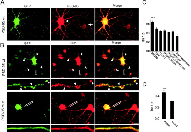
To further test the effects of PSD-95, we overexpressed in immature (4-7 DIV) neurons wild-type PSD-95 or mutant PSD-95(C3,5S), both fused with GFP. As has been shown previously (Craven et al., 1999), overexpressed PSD-95-GFP (wild-type) forms discrete puncta on the dendrites (Fig. 6_A,B_, wt). Most of the PSD-95 puncta colocalize with clusters of NMDARs (Fig. 6_B_, top panel). On the other hand, the PSD-95(C3,5S)-GFP mutant, which cannot be palmitoylated (Craven et al., 1999), shows diffuse staining throughout the cell (Fig. 6_B_, bottom panel, mut). The level of PSD-95 expression in PSD-95-GFP-transfected neurons was 9 ± 2-fold (measured in n = 5 different neurons) that of endogenous PSD-95 in untransfected cells, as determined by immunostaining with a PSD-95 antibody and comparing the signal intensity of transfected (identified by GFP staining) and nearby untransfected neurons (Fig. 6_A_). Overexpression of PSD-95-GFP wild-type, but not the (C3,5S) mutant, decreased NMDAR desensitization (p < 0.001 by one-way ANOVA followed by Bonferroni's multiple comparisons test), and the effect of PSD-95-GFP overexpression was abolished by treatment with 2-bromopalmitate before recording (Fig. 6_C_) (_I_ss/_I_p: PSD-95, 65 ± 2%, _n_ = 8; GFP, 50 ± 2%, _n_ = 8; control, 45 ± 2%, _n_ = 7; PSD-95 mutant, 46 ± 4%, _n_ = 7; PSD-95 with 2-bromopalmitate, 42 ± 3%, _n_ = 7). Similar to >14 DIV neurons, latrunculin treatment did not change NMDAR desensitization for 4-7 DIV neurons (Fig. 6_C_) (_I_ss/_I_p, 44 ± 3%; n = 7). Treatment with 2-bromopalmitate slightly, but not significantly, increased desensitization compared with other groups (Fig. 6_C_) (_I_ss/_I_p, 34 ± 3%; n = 7; p > 0.05), suggesting that the endogenous level of PSD-95 interacting with NMDARs was low at this early stage of development.
Figure 6.
Overexpression of PSD-95 decreases NMDAR desensitization in immature neurons. A, Left, A representative image of a 7 DIV neuron transfected with PSD-95-GFP and stained with anti-GFP antibody. Middle, The same neuron is shown stained with anti-PSD-95 antibody (arrowhead). An adjacent untransfected neuron (arrow) is also shown. Note that the level of PSD-95 signal is much higher for the transfected neuron (for quantification, see Results). B, Neurons were transfected with either PSD-95-GFP wild type (wt) or the (C3,5S) mutant (mut) and stained with both GFP (left) and NR1 (middle) antibodies. Note that the PSD-95 wild type shows punctate staining along the dendrites and many of the puncta colocalize with the NR1 staining (arrow heads), whereas the (C3,5S) mutant shows only diffuse staining. Scale bars, 10 μm. White boxes outline regions shown at higher gain directly below each photomicrograph. C, Overexpression in 4-7 DIV neurons of PSD-95 wild type decreased NMDAR desensitization compared with overexpression of GFP alone, untransfected (control), overexpression of PSD-95 mutant, overexpression of PSD-95 wild type treated with 2-bromopalmitate, neurons treated with latrunculin, or neurons treated with 2-bromopalmitate. One-way ANOVA followed by Bonferroni's multiple comparisons test showed that the _I_ss/_I_p value for the PSD-95 wild-type group was significantly greater than all other groups (***p < 0.001), whereas all the other groups were not significantly different from each other. Recording conditions were identical to those used in Figure 2_A_. D, As in C, 4-7 DIV neurons transfected with PSD-95 wild type were compared with untransfected neurons of the same age, except that current responses were evoked by 100 μm NMDA (30 μm glycine) in the presence of 2 mm Ca2+, and nystatin perforated-patch recording technique was used (_V_H of -65 mV). **p < 0.01.
To determine whether, under more physiological conditions (with Ca2+-dependent desensitization intact), overexpression of PSD-95 can still change the kinetics of NMDAR-mediated responses, we recorded currents evoked by 100 μm NMDA and 30 μm glycine in the presence of 2 mm calcium using the nystatin patch-clamp method. Again, overexpression of PSD-95 significantly decreased NMDAR desensitization (Fig. 6_D_) (_I_ss/_I_p, 47 ± 4 and 34 ± 2% for PSD-95-transfected and control nontransfected group, respectively; n = 7 for both groups; p < 0.01). Consistent with a previous study (El-Husseini et al., 2000), overexpression of PSD-95 did not significantly change the amplitude of the peak current mediated by NMDARs (PSD-95, 1532 ± 507 pA, _n_ = 8; GFP, 1687 ± 260 pA, _n_ = 8; _p_ > 0.05). Together, these data suggest that association of NMDARs with PSD-95 is necessary to mediate a dramatic decrease in receptor desensitization during development.
Modulation of NMDAR desensitization by protein kinase-phosphatase activity
Because PSD-95 associates with a variety of signaling molecules (Sheng and Pak, 2000), it is possible that its effect was exerted by recruiting kinases and/or phosphatases to the NMDAR complex, thereby altering the balance between phosphorylation and dephosphorylation of either the NMDAR itself or a closely associated protein. To test this hypothesis, we examined the effects of kinases and phosphatases that have been implicated in regulating NMDAR function.
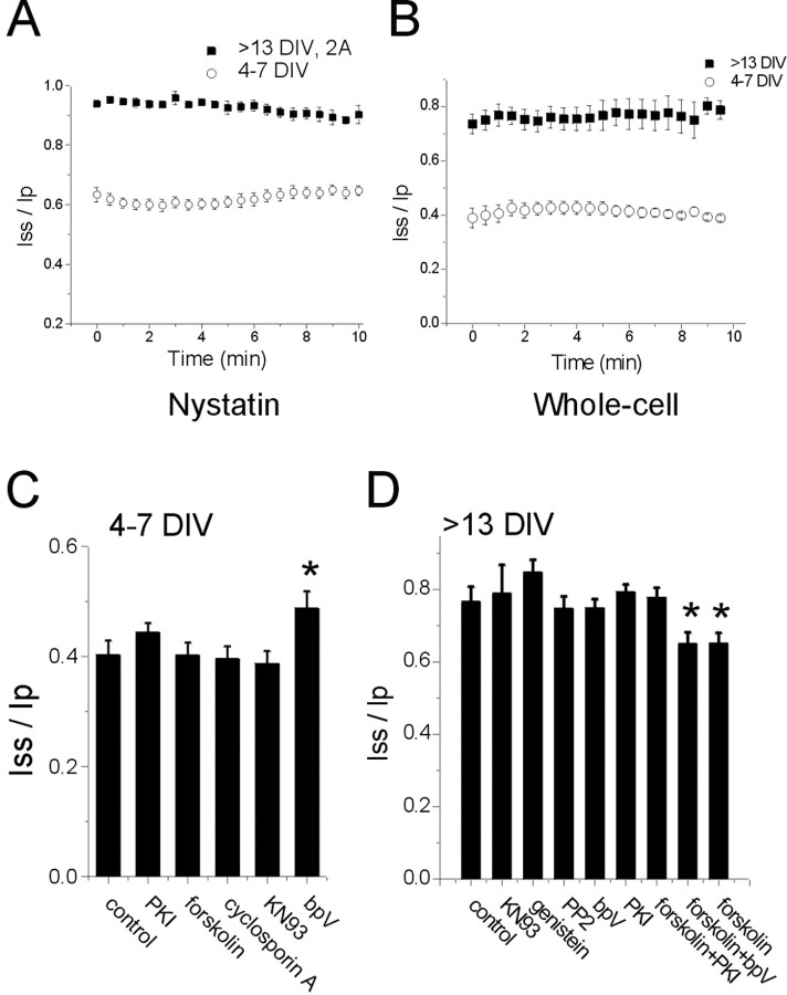
Glycine-independent desensitization is promoted by calcineurin activity (Lieberman and Mody, 1994; Tong and Jahr, 1994; Tong et al., 1995), an effect that appears as a time-dependent increase in desensitization associated with prolonged whole-cell dialysis and an increase in intracellular Ca2+ (Sather et al., 1992; Tong and Jahr, 1994; Krupp et al., 2002). Using either the nystatin perforated or conventional whole-cell patch-clamp recording method, we found that the _I_ss/_I_p ratio was stable over time for both 4-7 and >13 DIV neurons (Fig. 7_A,B_), suggesting that calcineurin did not play a large role in regulating NMDAR desensitization under our experimental conditions. This result was not surprising because we used conditions that promoted maintenance of low intracellular Ca2+, thus minimizing calcineurin activity. In support of this conclusion, application of a calcineurin inhibitor cyclosporin A (500 nm) for 5-15 min had no effect on NMDAR desensitization in 4-7 DIV neurons (Fig. 7_C_) (_I_ss/_I_p, 40 ± 3%, n = 13 and 40 ± 2%, n = 6 for control and cyclosporin A, respectively; p > 0.05). Previous studies also showed that the effect of calcineurin on NMDAR desensitization was inhibited by blocking Ca2+ entry (Lieberman and Mody, 1994) and that, under conditions of high intracellular Ca2+ buffering (10 mm EGTA), calcineurin-mediated dephosphorylation was slow and did not appreciably contribute to fast synaptic NMDAR desensitization (Raman et al., 1996).
Figure 7.
NMDAR desensitization is regulated by phosphorylation-dephosphorylation. A, Desensitization of NMDARs in either 4-7 or >13 DIV neurons was stable over time, suggesting that calcineurin did not regulate desensitization. Recordings were made using the nystatin perforated-patch recording technique, with _V_H of +40 mV in the absence of Ca2+. Currents were evoked by 100 μm NMDA (30 μm glycine). Ifenprodil at 10 μm was included for recordings from >13 DIV neurons. B, Same as A, except that recordings were made using conventional whole-cell voltage clamp (_V_H of -65 mV) with 10 mm BAPTA in the electrode solution and 0.2 mm external Ca2+ in the absence of ifenprodil, and currents were evoked by 1 mm NMDA (100 μm glycine). C, Treatment of 4-7 DIV neurons with bpV(phen) decreased NMDAR desensitization, but treatment with PKI, forskolin, cyclosporin A, or KN93 did not. D, Treatment of >13 DIV neurons with forskolin increased NMDAR desensitization, but treatment with genistein, PP2, bpV(phen), or KN93 did not. The effect of forskolin was blocked by coincubation with PKI but not bpV(phen). *p < 0.05 versus control group by unpaired t test. Recording conditions in C and D were identical to those in B.
To address further the role of protein phosphorylation in regulating NMDAR desensitization, we applied a variety of protein kinase and phosphatase inhibitors-activators to neuronal cultures. For NMDARs in 4-7 DIV neurons, application of forskolin (50 μm, 5-15 min), a protein kinase A (PKA) activator that augments NMDAR-mediated responses by overcoming the effect of calcineurin (Raman et al., 1996), or the PKA inhibitor PKI (Raman et al., 1996), had no effect on _I_ss/_I_p (Fig. 7_C_). Similarly, a Ca2+/calmodulin kinase II inhibitor KN93 (10 μm, 5-15 min) and a tyrosine phosphatase inhibitor bpV(phen) (100 μm, 5-15 min) had no significant effect on desensitization when control and all five treatment groups were compared using one-way ANOVA followed by Bonferroni's multiple comparisons test (Fig. 7_C_) (p > 0.05 for comparison among all groups). On the other hand, the tyrosine phosphatase inhibitor bpV(phen) resulted in a small decrease in NMDAR desensitization that was significantly different when compared directly with the control (vehicle treatment) group alone (_I_ss/_I_p, 49 ± 3%, n = 12 and 40 ± 3%, n = 13 for bpV(phen) and control groups, respectively; p < 0.05; unpaired t test).
Results for mature (>13 DIV) neurons were similar to those of experiments with immature neurons. Treatment of >13 DIV neurons with either the tyrosine kinase inhibitor genistein (50 μm, 5-15 min) or Src family tyrosine kinase inhibitor PP2 (10 μm, 5-15 min), or KN93, bpV(phen), forskolin, or PKI all failed to significantly affect NMDAR desensitization, using the one-way ANOVA followed by Bonferroni's multiple comparisons test for all seven groups (p > 0.05) (Fig. 7_D_). However, incubation with forskolin resulted in a small increase in NMDAR desensitization that was significantly different when compared directly with the control group alone, and its effect was blocked by coincubation with PKI (Fig. 7_D_) (_I_ss/_I_p, 77 ± 4%, 65 ± 3%, and 78 ± 3%; n = 13, 11, and 7 for control, forskolin, and forskolin plus PKI, respectively; p < 0.05 between forskolin and control, or between forskolin and forskolin plus PKI, by unpaired _t_ test). Coincubation with bpV(phen) did not block the effect of forskolin on desensitization (Fig. 7_D_) [_I_ss/_I_p, 65 ± 3%; _n_ = 7; _p_ < 0.05 between forskolin plus bpV(phen) and control; _p_ > 0.05 between forskolin alone and forskolin plus bpV(phen)], suggesting that the effect of forskolin occurred through the activation of PKA and not via cross-talk between PKA and tyrosine kinases or phosphatases as proposed by a recent study (Woodward, 2002). Together, these data indicate that activity of the protein kinases and phosphatases tested in our experiments cannot account for the dramatic difference in extent of NMDAR desensitization observed in immature versus mature neurons.
Discussion
Here, we report a developmental decrease in NMDAR desensitization that was not dependent on a switch in NMDAR subunit composition or change in sensitivity to ambient zinc but was tightly correlated with a change in receptor subcellular localization. A major difference between synaptic and extrasynaptic NMDARs is that the former colocalize and interact with many signaling proteins that are concentrated within the PSD compartment (Ziff, 1997). Of the many PSD proteins that can potentially modulate NMDAR properties, PSD-95 has gained the most attention. PSD-95 interacts with the C-terminal E_T/S_DV sequences of NMDAR NR2 subunits through its first two PDZ (postsynaptic density-95/Discs large/zona occludens-1) domains (Kornau et al., 1995). In cultured hippocampal neurons, PSD-95 clusters colocalize with synaptic, but not nonsynaptic, NMDARs (Rao et al., 1998). The effects of PSD-95 on NMDARs are diverse. For example, PSD-95 can cluster NMDARs expressed in COS-7 cells (Kim et al., 1996), couple NMDARs to Src family tyrosine kinases and thus promote receptor tyrosine phosphorylation (Tezuka et al., 1999; Liao et al., 2000), couple NMDARs to nitric oxide sythase and thus to nitric oxide toxicity (Sattler et al., 1999), increase NMDAR expression (Yamada et al., 1999; Rutter and Stephenson, 2000), decrease NMDAR sensitivity to glutamate (Yamada et al., 1999; Rutter and Stephenson, 2000), and inhibit the potentiating effects of PKC and insulin on NMDARs (Yamada et al., 1999; Liao et al., 2000).
We provide several lines of evidence to suggest that colocalization of PSD-95 with NMDARs can primarily account for the developmental change in NMDAR desensitization. It is known that PSD-95 is coclustered with NMDARs primarily at synapses (Rao et al., 1998), where we showed that NMDARs exhibit little desensitization. Furthermore, inhibition of palmitoylation in mature neurons increased NMDAR desensitization. Inhibitors of PSD-95 palmitoylation have been shown to decrease PSD-95 clusters at synapses and increase diffuse immunostaining of PSD-95 throughout dendritic shafts and the soma, indicating a shift from synaptic to nonsynaptic localization and also away from membranes to the cytoplasm (El-Husseini et al., 2002). However, NMDARs remain clustered at synapses under these conditions (El-Husseini et al., 2002), suggesting that NMDARs and PSD-95 become “uncoupled.” Consistent with this, we found that inhibition of PSD-95 palmitoylation disrupted the interaction between NMDARs and PSD-95 assessed by a coimmunoprecipitation assay. Similarly, chronic PTX treatment of mature neurons resulted in a decrease in synaptic NMDARs and dissociation from PSD-95 (Figs. 3, 5) (Crump et al., 2001). Under these conditions, we found that NMDAR desensitization was increased markedly to levels not significantly different from those found in immature (4-7 DIV) neurons. However, because 2-bromopalmitate is a general palmitoylation inhibitor (El-Husseini et al., 2002), we cannot rule out the possibility that inhibition of palmitoylation of other PSD proteins may also contribute to the regulation of NMDAR desensitization. Given the dramatic effect of PSD-95 overexpression in immature neurons, the contribution from other proteins, if there is any, should be low. On the other hand, treatment with latrunculin, resulting in actin depolymerization, causes movement of both NMDARs and PSD-95 away from synapses, in which the two remain coclustered (Allison et al., 1998). Consistent with this, latrunculin treatment of either mature or immature neurons had no effect on NMDAR desensitization. Finally, when PSD-95 expression was increased ninefold and observed to colocalize with NMDAR clusters in transfected immature neurons, NMDAR desensitization was dramatically reduced and not significantly different from that found in mature (>13 DIV) neurons. Although NMDAR clusters were not quantitatively compared between PSD-95-transfected and control neurons, there was no visually apparent difference in NMDAR staining pattern between the two groups. The effect of wild-type PSD-95 overexpression in immature neurons was also abolished by inhibition of palmitoylation. Also, overexpression of a mutant PSD-95 that cannot be palmitoylated was ineffective in changing desensitization, suggesting that palmitoylation and proper targeting of PSD-95 is important for its regulation of NMDAR function.
It should be noted that treatments that altered NMDAR desensitization in mature neurons also disrupted association of PSD-95 and NMDARs by shifting one or both away from synapses rather than changing overall expression levels of PSD-95. It is interesting that, in a previous study, suppression of PSD-95 protein levels in cultured cortical neurons using an oligonucleotide antisense approach did not result in a change in macroscopic kinetics of NMDAR-mediated currents (Sattler et al., 1999). It is possible that those results were attributable to compensation by other signaling molecules during chronic suppression of PSD-95 expression. Furthermore, treatment of cultured hippocampal neurons with a peptide to disrupt interaction of PSD-95 and NR2B did not alter the time course or amplitude of the NMDA-evoked calcium response (Aarts et al., 2002). However, because the interaction between NR2A and PSD-95 remained intact under those conditions (Aarts et al., 2002), and because NR2A is preferentially targeted to synapses, NMDAR-PSD-95 interactions would have remained primarily intact at synapses, and therefore no increase in desensitization would be expected.
NMDARs in 4-7 DIV neurons are mostly extrasynaptic and do not interact with PSD-95. Overexpression of PSD-95 in those neurons may regulate NMDAR desensitization by recruiting tyrosine kinases and promoting tyrosine phosphorylation of NMDARs (Tezuka et al., 1999; Liao et al., 2000). This is supported by the fact that inhibition of tyrosine phosphatase activity by bpV(phen) decreased NMDAR desensitization in 4-7 DIV neurons (Fig. 7_C_). Consistently, bpV(phen) also decreased the desensitization of recombinant NMDARs expressed in HEK293 cells (Krupp et al., 2002). It must be emphasized, however, that an altered balance of tyrosine phosphorylation attributable to PSD-95 recruitment of protein tyrosine kinases may not fully explain the difference in NMDAR desensitization between immature and mature neurons, because this difference is large compared with the effect of tyrosine phosphatase inhibition on NMDAR desensitization in immature neurons. In addition, desensitization of NMDARs in >13 DIV neurons was not influenced by incubation with either PP2 or genistein, agents that inhibit tyrosine kinases, suggesting that synaptic NMDARs are resistant to tyrosine dephosphorylation or else that factors other than tyrosine kinases-phosphatases contribute to regulation of NMDAR desensitization in mature neurons versus immature neurons.
PSD-95 can also induce the assembly of signaling molecules other than tyrosine kinases in the PSD to regulate synaptic NMDAR function. For example, PKA is targeted to NMDARs via an interaction between PSD-95 and AKAP (A-kinase anchoring protein) (Colledge et al., 2000), and we found that activation of PKA by forskolin resulted in a small increase in desensitization of NMDARs in >13 DIV neurons. This effect of PKA was somewhat surprising, because previous studies indicate that PKA activation can overcome the effects of calcineurin (Raman et al., 1996) and prevent the calcineurin-dependent increase in desensitization (Krupp et al., 2002). However, the effects of PKA on NMDAR function are complicated; both upregulation (Blank et al., 1997; Westphal et al., 1999) and downregulation (Woodward, 2002) of NMDAR function have been reported. Moreover, mutation in NR2A of putative phosphorylation sites serine 900 (S900A) and serine 929 (S929A) resulted in an increase and decrease of desensitization, respectively (Krupp et al., 2002). These studies suggest that phosphorylation of NR2A by PKA at different sites can have opposite effects. Notably, we found that PKA or its inhibitor did not regulate NMDAR desensitization in 4-7 DIV neurons, in which the large majority of NMDARs are extrasynaptic. This may be explained by recent studies showing that regulation of glutamate receptors by PKA requires targeting of PKA to the receptors by “bridging” proteins, including yotiao, PSD-95/AKAP, and SAP97 (synapse-associated protein 97)/AKAP, all of which are enriched in the PSD (Lin et al., 1998; Westphal et al., 1999; Colledge et al., 2000).
A developmental change in the decay of single NMDAR-mediated synaptic responses has been reported to depend on a switch in subunit composition (Carmignoto and Vicini, 1992; Hestrin, 1992; Monyer et al., 1994; Flint et al., 1997). Here we showed that decay of whole- cell NMDAR current in response to sustained activation is also regulated developmentally, and this change is associated with recruitment of NMDARs to synapses during development. We reported previously that the activity of extrasynaptic NMDARs is more readily downregulated through a process called rundown compared with synaptic NMDARs (Li et al., 2002). Together with the current findings, our results indicate that NMDAR function within a single neuron is fine-tuned on the basis of receptor subcellular localization. Thus, synaptic NMDAR activity, which is resistant to downregulation by either rundown or desensitization, can maintain high-fidelity transmission at synapses to fulfill its role in synaptic plasticity during repetitive firing. Activity of extrasynaptic NMDARs, on the other hand, is readily shut down by desensitization and/or rundown to limit neuronal damage during pathological conditions such as ischemia, in which sustained glutamate release and spillover can occur. Differential regulation of synaptic and nonsynaptic NMDAR activity may, in part, underlie the different roles these two groups of receptors play in excitotoxicity (Sattler et al., 1999; Hardingham et al., 2002).
Footnotes
This work was supported by operating grants from the Heart and Stroke Foundation of British Columbia and Yukon (L.A.R.) and Canadian Institutes for Health Research (CIHR) Grant MOP-49586 (T.H.M., L.A.R.). L.A.R. and T.H.M. hold CIHR Investigator and Michael Smith Foundation for Health Research Senior Scholar Awards. We thank T. Luo and L. Zhang for excellent technical assistance and Dr. A. E. El-Husseini for helpful discussions and advice.
Correspondence should be addressed to Dr. Lynn A. Raymond, Department of Psychiatry, University of British Columbia, 4N3-2255 Wesbrook Mall, Vancouver, British Columbia, Canada V6T 1Z3. E-mail: lynnr@interchange.ubc.ca.
Copyright © 2003 Society for Neuroscience 0270-6474/03/2311244-•$15.00/0
References
- Aarts M, Liu Y, Liu L, Besshoh S, Arundine M, Gurd JW, Wang YT, Salter MW, Tymianski M ( 2002) Treatment of ischemic brain damage by perturbing NMDA receptor- PSD-95 protein interactions. Science 298: 846-850. [DOI] [PubMed] [Google Scholar]
- Abdel-Hamid KM, Baimbridge KG ( 1997) The effects of artificial calcium buffers on calcium responses and glutamate-mediated excitotoxicity in cultured hippocampal neurons. Neuroscience 81: 673-687. [DOI] [PubMed] [Google Scholar]
- Allison DW, Gelfand VI, Spector I, Craig AM ( 1998) Role of actin in anchoring postsynaptic receptors in cultured hippocampal neurons: differential attachment of NMDA versus AMPA receptors. J Neurosci 18: 2423-2436. [DOI] [PMC free article] [PubMed] [Google Scholar]
- Barria A, Malinow R ( 2002) Subunit-specific NMDA receptor trafficking to synapses. Neuron 35: 345-353. [DOI] [PubMed] [Google Scholar]
- Bekkers JM, Stevens CF ( 1991) Excitatory and inhibitory autaptic currents in isolated hippocampal neurons maintained in cell culture. Proc Natl Acad Sci USA 88: 7834-7838. [DOI] [PMC free article] [PubMed] [Google Scholar]
- Blank T, Nijholt I, Teichert U, Kugler H, Behrsing H, Fienberg A, Greengard P, Spiess J ( 1997) The phosphoprotein DARPP-32 mediates cAMP-dependent potentiation of striatal _N_-methyl-d-aspartate responses. Proc Natl Acad Sci USA 94: 14859-14864. [DOI] [PMC free article] [PubMed] [Google Scholar]
- Brorson JR, Manzolillo PA, Gibbons SJ, Miller RJ ( 1995) AMPA receptor desensitization predicts the selective vulnerability of cerebellar Purkinje cells to excitotoxicity. J Neurosci 15: 4515-4524. [DOI] [PMC free article] [PubMed] [Google Scholar]
- Carmignoto G, Vicini S ( 1992) Activity-dependent decrease in NMDA receptor responses during development of the visual cortex. Science 258: 1007-1011. [DOI] [PubMed] [Google Scholar]
- Chen N, Moshaver A, Raymond LA ( 1997) Differential sensitivity of recombinant _N_-methyl-d-aspartate receptor subtypes to zinc inhibition. Mol Pharmacol 51: 1015-1023. [DOI] [PubMed] [Google Scholar]
- Chen N, Luo T, Raymond LA ( 1999) Subtype-dependence of NMDA receptor channel open probability. J Neurosci 19: 6844-6854. [DOI] [PMC free article] [PubMed] [Google Scholar]
- Colledge M, Dean RA, Scott GK, Langeberg LK, Huganir RL, Scott JD ( 2000) Targeting of PKA to glutamate receptors through a MAGUK-AKAP complex. Neuron 27: 107-119. [DOI] [PubMed] [Google Scholar]
- Craven SE, El-Husseini AE, Bredt DS ( 1999) Synaptic targeting of the postsynaptic density protein PSD-95 mediated by lipid and protein motifs. Neuron 22: 497-509. [DOI] [PubMed] [Google Scholar]
- Crump FT, Dillman KS, Craig AM ( 2001) cAMP-dependent protein kinase mediates activity-regulated synaptic targeting of NMDA receptors. J Neurosci 21: 5079-5088. [DOI] [PMC free article] [PubMed] [Google Scholar]
- Dingledine R, Borges K, Bowie D, Traynelis SF ( 1999) The glutamate receptor ion channels. Pharmacol Rev 51: 7-61. [PubMed] [Google Scholar]
- El-Husseini AE, Schnell E, Chetkovich DM, Nicoll RA, Bredt DS ( 2000) PSD-95 involvement in maturation of excitatory synapses. Science 290: 1364-1368. [PubMed] [Google Scholar]
- El-Husseini Ael-D, Schnell E, Dakoji S, Sweeney N, Zhou Q, Prange O, Gauthier-Campbell C, Aguilera-Moreno A, Nicoll RA, Bredt DS ( 2002) Synaptic strength regulated by palmitate cycling on PSD-95. Cell 108: 849-863. [DOI] [PubMed] [Google Scholar]
- Flint AC, Maisch US, Weishaupt JH, Kriegstein AR, Monyer H ( 1997) NR2A subunit expression shortens NMDA receptor synaptic currents in developing neocortex. J Neurosci 17: 2469-2476. [DOI] [PMC free article] [PubMed] [Google Scholar]
- Furukawa K, Smith-Swintosky VL, Mattson MP ( 1995) Evidence that actin depolymerization protects hippocampal neurons against excitotoxicity by stabilizing [Ca2+]iExp Neurol 133: 153-163. [DOI] [PubMed] [Google Scholar]
- Furukawa K, Fu W, Li Y, Witke W, Kwiatkowski DJ, Mattson MP ( 1997) The actin-severing protein gelsolin modulates calcium channel and NMDA receptor activities and vulnerability to excitotoxicity in hippocampal neurons. J Neurosci 17: 8178-8186. [DOI] [PMC free article] [PubMed] [Google Scholar]
- Hamill OP, Marty A, Neher E, Sakmann B, Sigworth FJ ( 1981) Improved patch-clamp techniques for high-resolution current recording from cells and cell-free membrane patches. Pflügers Arch 391: 85-100. [DOI] [PubMed] [Google Scholar]
- Hardingham GE, Fukunaga Y, Bading H ( 2002) Extrasynaptic NMDARs oppose synaptic NMDARs by triggering CREB shut-off and cell death pathways. Nat Neurosci 5: 405-414. [DOI] [PubMed] [Google Scholar]
- Hestrin S ( 1992) Developmental regulation of NMDA receptor-mediated synaptic currents at a central synapse. Nature 357: 686-689. [DOI] [PubMed] [Google Scholar]
- Hollmann M, Heinemann S ( 1994) Cloned glutamate receptors. Annu Rev Neurosci 17: 31-108. [DOI] [PubMed] [Google Scholar]
- Jones MV, Westbrook GL ( 1996) The impact of receptor desensitization on fast synaptic transmission. Trends Neurosci 19: 96-101. [DOI] [PubMed] [Google Scholar]
- Kim E, Cho KO, Rothschild A, Sheng M ( 1996) Heteromultimerization and NMDA receptor-clustering activity of Chapsyn-110, a member of the PSD-95 family of proteins. Neuron 17: 103-113. [DOI] [PubMed] [Google Scholar]
- Kornau HC, Schenker LT, Kennedy MB, Seeburg PH ( 1995) Domain interaction between NMDA receptor subunits and the postsynaptic density protein PSD-95. Science 269: 1737-1740. [DOI] [PubMed] [Google Scholar]
- Krupp JJ, Vissel B, Heinemann SF, Westbrook GL ( 1996) Calcium-dependent inactivation of recombinant _N_-methyl-d-aspartate receptors is NR2 subunit specific. Mol Pharmacol 50: 1680-1688. [PubMed] [Google Scholar]
- Krupp JJ, Vissel B, Heinemann SF, Westbrook GL ( 1998) N-terminal domains in the NR2 subunit control desensitization of NMDA receptors. Neuron 20: 317-327. [DOI] [PubMed] [Google Scholar]
- Krupp JJ, Vissel B, Thomas CG, Heinemann SF, Westbrook GL ( 2002) Calcineurin acts via the C-terminus of NR2A to modulate desensitization of NMDA receptors. Neuropharmacology 42: 593-602. [DOI] [PubMed] [Google Scholar]
- Legendre P, Rosenmund C, Westbrook GL ( 1993) Inactivation of NMDA channels in cultured hippocampal neurons by intracellular calcium. J Neurosci 13: 674-684. [DOI] [PMC free article] [PubMed] [Google Scholar]
- Li B, Chen N, Luo T, Otsu Y, Murphy TH, Raymond LA ( 2002) Differential regulation of synaptic and extra-synaptic NMDA receptors. Nat Neurosci 5: 833-834. [DOI] [PubMed] [Google Scholar]
- Li JH, Wang YH, Wolfe BB, Krueger KE, Corsi L, Stocca G, Vicini S ( 1998) Developmental changes in localization of NMDA receptor subunits in primary cultures of cortical neurons. Eur J Neurosci 10: 1704-1715. [DOI] [PubMed] [Google Scholar]
- Liao GY, Kreitzer MA, Sweetman BJ, Leonard JP ( 2000) The postsynaptic density protein PSD-95 differentially regulates insulin- and Src-mediated current modulation of mouse NMDA receptors expressed in Xenopus oocytes. J Neurochem 75: 282-287. [DOI] [PubMed] [Google Scholar]
- Lieberman DN, Mody I ( 1994) Regulation of NMDA channel function by endogenous Ca2+-dependent phosphatase. Nature 369: 235-239. [DOI] [PubMed] [Google Scholar]
- Lin JW, Wyszynski M, Madhavan R, Sealock R, Kim JU, Sheng M ( 1998) Yotiao, a novel protein of neuromuscular junction and brain that interacts with specific splice variants of NMDA receptor subunit NR1. J Neurosci 18: 2017-2027. [DOI] [PMC free article] [PubMed] [Google Scholar]
- May PC, Robison PM ( 1993) Cyclothiazide treatment unmasks AMPA excitotoxicity in rat primary hippocampal cultures. J Neurochem 60: 1171-1174. [DOI] [PubMed] [Google Scholar]
- McBain CJ, Mayer ML ( 1994) _N_-methyl-d-aspartic acid receptor structure and function. Physiol Rev 74: 723-760. [DOI] [PubMed] [Google Scholar]
- Monaghan DT, Bridges RJ, Cotman CW ( 1989) The excitatory amino acid receptors: their classes, pharmacology, and distinct properties in the function of the central nervous system. Annu Rev Pharmacol Toxicol 29: 365-402. [DOI] [PubMed] [Google Scholar]
- Monyer H, Burnashev N, Laurie DJ, Sakmann B, Seeburg PH ( 1994) Developmental and regional expression in the rat brain and functional properties of four NMDA receptors. Neuron 12: 529-540. [DOI] [PubMed] [Google Scholar]
- Murphy TH, Blatter LA, Wier WG, Baraban JM ( 1992) Spontaneous synchronous synaptic calcium transients in cultured cortical neurons. J Neurosci 12: 4834-4845. [DOI] [PMC free article] [PubMed] [Google Scholar]
- Niethammer M, Kim E, Sheng M ( 1996) Interaction between the C terminus of NMDA receptor subunits and multiple members of the PSD-95 family of membrane-associated guanylate kinases. J Neurosci 16: 2157-2163. [DOI] [PMC free article] [PubMed] [Google Scholar]
- Paoletti P, Ascher P, Neyton J ( 1997) High-affinity zinc inhibition of NMDA NR1-NR2A receptors. J Neurosci 17: 5711-5725. [DOI] [PMC free article] [PubMed] [Google Scholar]
- Raman IM, Tong G, Jahr CE ( 1996) Beta-adrenergic regulation of synaptic NMDA receptors by cAMP-dependent protein kinase. Neuron 16: 415-421. [DOI] [PubMed] [Google Scholar]
- Rao A, Kim E, Sheng M, Craig AM ( 1998) Heterogeneity in the molecular composition of excitatory postsynaptic sites during development of hippocampal neurons in culture. J Neurosci 18: 1217-1229. [DOI] [PMC free article] [PubMed] [Google Scholar]
- Raymond LA, Moshaver A, Tingley WG, Huganir RL ( 1996) Glutamate receptor ion channel properties predict vulnerability to cytotoxicity in a transfected nonneuronal cell line. Mol Cell Neurosci 7: 102-115. [DOI] [PubMed] [Google Scholar]
- Rutter AR, Stephenson FA ( 2000) Coexpression of postsynaptic density-95 protein with NMDAReceptors results in enhanced receptor expression together with a decreased sensitivity to l-glutamate. J Neurochem 75: 2501-2510. [DOI] [PubMed] [Google Scholar]
- Sather W, Dieudonne S, MacDonald JF, Ascher P ( 1992) Activation and desensitization of _N_-methyl-d-aspartate receptors in nucleated outside-out patches from mouse neurones. J Physiol (Lond) 450: 643-672. [DOI] [PMC free article] [PubMed] [Google Scholar]
- Sattler R, Xiong Z, Lu WY, Hafner M, MacDonald JF, Tymianski M ( 1999) Specific coupling of NMDA receptor activation to nitric oxide neurotoxicity by PSD-95 protein. Science 284: 1845-1848. [DOI] [PubMed] [Google Scholar]
- Sattler R, Xiong Z, Lu WY, MacDonald JF, Tymianski M ( 2000) Distinct roles of synaptic and extrasynaptic NMDA receptors in excitotoxicity. J Neurosci 20: 22-33. [DOI] [PMC free article] [PubMed] [Google Scholar]
- Sheng M, Pak DT ( 2000) Ligand-gated ion channel interactions with cytoskeletal and signaling proteins. Annu Rev Physiol 62: 755-778. [DOI] [PubMed] [Google Scholar]
- Stocca G, Vicini S ( 1998) Increased contribution of NR2A subunit to synaptic NMDA receptors in developing rat cortical neurons. J Physiol (Lond) 507: 13-24. [DOI] [PMC free article] [PubMed] [Google Scholar]
- Tezuka T, Umemori H, Akiyama T, Nakanishi S, Yamamoto T ( 1999) PSD-95 promotes Fyn-mediated tyrosine phosphorylation of the _N_-methyl-d-aspartate receptor subunit NR2A. Proc Natl Acad Sci USA 96: 435-440. [DOI] [PMC free article] [PubMed] [Google Scholar]
- Tong G, Jahr CE ( 1994) Regulation of glycine-insensitive desensitization of the NMDA receptor in outside-out patches. J Neurophysiol 72: 754-761. [DOI] [PubMed] [Google Scholar]
- Tong G, Shepherd D, Jahr CE ( 1995) Synaptic desensitization of NMDA receptors by calcineurin. Science 267: 1510-1512. [DOI] [PubMed] [Google Scholar]
- Tovar KR, Westbrook GL ( 1999) The incorporation of NMDA receptors with a distinct subunit composition at nascent hippocampal synapses _in vitro_J Neurosci 19: 4180-4188. [DOI] [PMC free article] [PubMed] [Google Scholar]
- Villarroel A, Regalado MP, Lerma J ( 1998) Glycine-independent NMDA receptor desensitization: localization of structural determinants. Neuron 20: 329-339. [DOI] [PubMed] [Google Scholar]
- Westphal RS, Tavalin SJ, Lin JW, Alto NM, Fraser ID, Langeberg LK, Sheng M, Scott JD ( 1999) Regulation of NMDA receptors by an associated phosphatase-kinase signaling complex. Science 285: 93-96. [DOI] [PubMed] [Google Scholar]
- Williams K, Russell SL, Shen YM, Molinoff PB ( 1993) Developmental switch in the expression of NMDA receptors occurs in vivo and in vitro. Neuron 10: 267-278. [DOI] [PubMed] [Google Scholar]
- Woodward JJ ( 2002) Prostacyclin-induced rundown of _N_-methyl-d-aspartate receptor currents in HEK293 cells is protein kinase A-dependent and NR2 subunit-selective. J Neurochem 80: 598-604. [DOI] [PubMed] [Google Scholar]
- Yamada Y, Chochi Y, Takamiya K, Sobue K, Inui M ( 1999) Modulation of the channel activity of the epsilon2/zeta1-subtype _N_-methyl-d-aspartate receptor by PSD-95. J Biol Chem 274: 6647-6652. [DOI] [PubMed] [Google Scholar]
- Zheng F, Erreger K, Low CM, Banke T, Lee CJ, Conn PJ, Traynelis SF ( 2001) Allosteric interaction between the amino terminal domain and the ligand binding domain of NR2A. Nat Neurosci 4: 894-901. [DOI] [PubMed] [Google Scholar]
- Zhong J, Russell SL, Pritchett DB, Molinoff PB, Williams K ( 1994) Expression of mRNAs encoding subunits of the _N_-methyl-d-aspartate receptor in cultured cortical neurons. Mol Pharmacol 45: 846-853. [PubMed] [Google Scholar]
- Ziff EB ( 1997) Enlightening the postsynaptic density. Neuron 19: 1163-1174. [DOI] [PubMed] [Google Scholar]
- Zorumski CF, Thio LL, Clark GD, Clifford DB ( 1990) Blockade of desensitization augments quisqualate excitotoxicity in hippocampal neurons. Neuron 5: 61-66. [DOI] [PubMed] [Google Scholar]
