Cyclin I activates Cdk5 and regulates expression of Bcl-2 and Bcl-XL in postmitotic mouse cells (original) (raw)
Abstract
Cyclin I is an atypical cyclin because it is most abundant in postmitotic cells. We previously showed that cyclin I does not regulate proliferation, but rather controls survival of podocytes, terminally differentiated epithelial cells that are essential for the structural and functional integrity of kidney glomeruli. Here, we investigated the mechanism by which cyclin I safeguards against apoptosis and found that cyclin I bound and activated cyclin-dependent kinase 5 (Cdk5) in isolated mouse podocytes and neurons. Cdk5 activity was reduced in glomeruli and brain lysates from cyclin I–deficient mice, and inhibition of Cdk5 increased in vitro the susceptibility to apoptosis in response to cellular damage. In addition, levels of the prosurvival proteins Bcl-2 and Bcl-XL were reduced in podocytes and neurons from cyclin I–deficient mice, and restoration of Bcl-2 or Bcl-XL expression prevented injury-induced apoptosis. Furthermore, we found that levels of phosphorylated MEK1/2 and ERK1/2 were decreased in cyclin I–deficient podocytes and that inhibition of MEK1/2 restored Bcl2 and Bcl-XL protein levels. Of interest, this pathway was also defective in mice with experimental glomerulonephritis. Taken together, these data suggest that a cyclin I–Cdk5 complex forms a critical antiapoptotic factor in terminally differentiated cells that functions via MAPK signaling to modulate levels of the prosurvival proteins Bcl-2 and Bcl-XL.
Introduction
Neurons and kidney podocytes share many characteristics, including being postmitotic and terminally differentiated cells (1). As such, they typically do not reengage the cell cycle, leading to a limited capacity to proliferate. Indeed, beyond the developing brain and kidney, there is very little evidence supporting neuronal and podocyte mitosis and thus proliferation under normal or diseased states. Thus, following cell injury characterized by apoptosis such as occurs in neurodegenerative disease and ischemic brain injury (2, 3) as well as in diabetic and nondiabetic kidney diseases (4–6), respectively, neuronal or podocyte number decreases. Reduced cellular number then leads to organ dysfunction. A decrease in podocyte number underlies the development of proteinuria and glomerulosclerosis in experimental and human disease (7, 8). Moreover, there is a significant correlation with reduced podocyte number and decreased kidney function (9). Neurons and podocytes are therefore dependent on critical biological pathways to maintain and enhance their survival to minimize cell death following injury in disease. This may be distinct from nonterminally differentiated cells that can readily restore cell number because of their high proliferative capacity.
Although cell-cycle proteins were originally considered to govern cell proliferation, there is a large body of research showing that specific cell-cycle proteins also function to maintain cell survival and that this biological role is distinct from that of proliferation (reviewed in ref. 10). To this end, cyclin-dependent kinase 5 (Cdk5) is required for the survival of neurons (reviewed in ref. 11), and we have recently reported on the prosurvival function of cyclin I in podocytes (12). Interestingly, cyclin I and Cdk5 are both expressed in terminally differentiated podocytes and neurons. Cyclin I, first cloned from human forebrain cortex, displays highest sequence homology to cyclins G1 and G2 (13), and in contrast to other cyclins, its mRNA levels do not oscillate during the cell cycle (12, 14, 15). It does not regulate proliferation or cell differentiation, and cyclin I–null mice do not show any spontaneous phenotypic abnormalities (12).
In these studies, we have investigated the mechanisms by which cyclin I protects postmitotic cells from apoptosis. We show that cyclin I binds and activates Cdk5 and that cyclin I–Cdk5 determines the level of activity of MEK1/2. These kinases in turn govern expression of the prosurvival proteins Bcl-2 and Bcl-XL. Furthermore, we show that p35-Cdk5, which is also present in podocytes and neurons, also promotes cell survival but that this occurs via a pathway distinct from the one activated by cyclin I–Cdk5. In vivo studies were also performed to validate the cell-culture studies.
Results
Cyclin I binds and activates constitutively expressed Cdk5 in terminally differentiated cells.
Podocytes are terminally differentiated and highly specialized epithelial cells in the kidney glomerulus, also known as the filtering unit. They function to limit proteinuria and are critical for the normal shape of the glomerulus. We have recently reported that Cdk5, similar to cyclin I, is abundantly expressed in podocytes (16). Moreover, our data also showed that cyclin I and Cdk5 immunostaining colocalized in podocytes of normal kidney glomeruli (data not shown) and in normal brain cells (data not shown). In contrast, neither cyclin I nor Cdk5 immunostaining were present in the glomerular endothelial cells nor in the fibroblast-like mesangial cells. These results suggest that within the glomerulus, cyclin I and Cdk5 are specific to podocytes, which provided the rationale to determine whether cyclin I binds to and activates endogenous Cdk5 in podocytes.
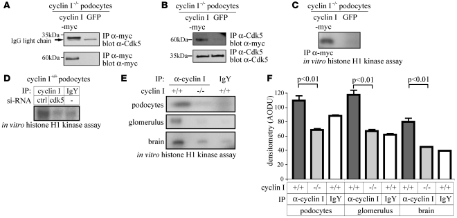
To this end, cultured podocytes were isolated from cyclin I WT and null mice (12), and the latter were stably infected with myc-tagged cyclin I. Reciprocal coimmunoprecipitation studies with antibodies to either myc or Cdk5 revealed that cyclin I bound to and activated endogenous Cdk5, as analyzed by histone H1 phosphorylation (Figure 1, A and B). Moreover, when endogenous protein was immunoprecipitated with α–cyclin I antibody, the histone H1 kinase was active in WT cells, but not in cyclin I–null cells (Figure 1C). Specificity of cyclin I–Cdk5 kinase activity was confirmed by reducing endogenous Cdk5 expression with siRNA (Figure 1D). In addition, we further validated these findings by transducing cells with a kinase-inactive dominant negative Cdk5 mutant. Again, cyclin I–mediated kinase activity was markedly diminished in the presence of a dominant negative Cdk5 mutant (data not shown).
Figure 1. Cyclin I binds and activates constitutive Cdk5 in postmitotic cells.
(A, B) To determine whether cyclin I and Cdk5 coimmunoprecipitate in glomerular podocytes, cyclin I–null (–/–) podocytes were infected with either myc-tagged cyclin I or GFP. Reciprocal coimmunoprecipitation studies with either α-myc (A, lanes 1, 2) or α-Cdk5 (B, lanes 3, 4) antibodies revealed that cyclin I bound to endogenous Cdk5 in podocytes. (C) Histone H1 kinase activity was abundant in cyclin I–null podocytes infected with cyclin I–myc (lane 1). In contrast, kinase activity was not detected in cultured cyclin I–null podocytes infected with GFP (lane 2). (D) To prove that the cyclin I–associated histone H1 kinase activity shown in C was specifically due to the activation of Cdk5, WT (+/+) podocytes were transfected with siRNA. Cyclin I–associated histone H1 kinase activity was present in podocytes transfected with control siRNA (lane 1). In contrast, cyclin I–associated kinase activity was not detected in cells transfected with siRNA targeting Cdk5 (lane 2). As expected, an IP with a control preimmune IgY antibody showed no kinase activity (lane 3). Ctrl, control. (E and F) To determine whether cyclin I–Cdk5 was also active in tissues in vivo, kinase activity was measured in protein extracts from kidney glomeruli and brain of WT and cyclin I–null mice. Cdk5 was active in WT cultured podocytes, glomeruli, and brain tissue. In contrast, histone H1 kinase activity was not detected in corresponding tissues from cyclin I–null mice. Densitometric analysis (n = 3). Data shown represent mean + SD.
In order to extend these findings to cells in vivo, kidney glomeruli and brain were isolated from cyclin I WT and null mice. Figure 1E shows that when protein lysates from isolated glomeruli and brain were immunoprecipitated with a polyclonal–α–cyclin I antibody, histone H1 kinase activity was abundant when quantitated by densitometry (Figure 1F). As expected, histone H1 kinase activity was absent in organs from cyclin I–null mice. Cyclin I–Cdk5 kinase activity was not detected in nonterminally differentiated kidney cells such as kidney tubules (data not shown). Taken together, these combined methods show that the cyclin I–Cdk5 complex is active specifically in neurons and podocytes in vitro and in vivo.
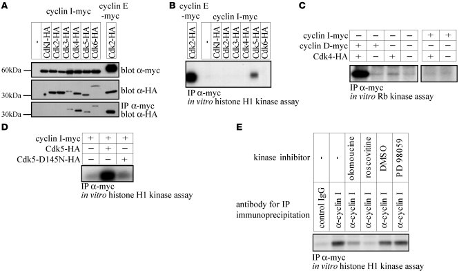
To further validate these findings by different methodology, HEK293 cells, which lack significant levels of cyclin I and Cdk5, were cotransfected with plasmids encoding myc-tagged cyclin I and HA-tagged Cdks 1 through 6. Figure 2A shows that in addition to Cdk5, Cdk3, -4, and -6 coimmunoprecipitated from total cellular lysates with an antibody to the myc epitope tag on cyclin I. Histone H1 and retinoblastoma protein (Rb) were used as kinase substrates to determine whether any of these cyclin I–Cdk complexes were active. Figure 2, B and C, shows that in transfected HEK293 cells, the cyclin I–Cdk5 complex alone was able to phosphorylate histone H1. In contrast, despite proven protein-protein interactions by coimmunoprecipitation, there was no kinase activity detected in the cyclin I–Cdk3, –Cdk4, or –Cdk6 complexes toward either histone H1 or Rb (Figure 2, B and C). Myc-tagged cyclin E and HA-tagged Cdk2 and cyclin D–Cdk4 served as positive controls for histone H1 and Rb phosphorylation assays, respectively.
Figure 2. Cyclin I is a specific binding partner and activator of Cdk5 in HEK293 cells.
(A) Following cotransfection of HEK293 cells with myc-tagged cyclin I and HA-tagged Cdks1–6, cyclin I coimmunoprecipitated with Cdk 3–6. (B, C) To assess which Cdk was activated by cyclin I, immunoprecipitations were performed followed by histone H1 (B) or Rb (C) kinase assays. Kinase activity was only detected in cells cotransfected with cyclin I and Cdk5. HA-Cdk2 and myc–cyclin E (B, histone H1 assay) and HA-Cdk4 and myc–cyclin D (C, Rb assay) served as positive controls. These results show that cyclin I specifically activates Cdk5, but not Cdk1, -2, -3, -4, or -6. (D) To prove specificity, HEK293 cells were cotransfected with cyclin I alone, cyclin I and Cdk5, or cyclin I and a dominant negative Cdk5 (D145N-Cdk5). No kinase activity was detected when cells were transfected with cyclin I alone (lane 1). Kinase activity was detected in cells transfected with cyclin I and Cdk5 (lane 2), but not in cells with cyclin I and the kinase-inactive mutant (lane 3). (E) No histone H1 kinase activity was detected when control IgG was used for immunoprecipitation (lane 1). Following immunoprecipitation with an antibody to myc, kinase activity was present (lane 2). In the presence of the Cdk inhibitors olomoucine (10 μM; lane 3) and roscovitine (50 μM, lane 4), kinase activity was reduced. The controls DMSO and PD98059 (an ERK5 inhibitor) had no effect on cyclin I kinase activity.
To address whether the kinase activity was due to Cdk5 itself or an associated protein, HEK293 cells were also transfected with an HA-tagged kinase-inactive Cdk5 mutant. As shown in Figure 2D, phosphorylation of histone H1 was only detected when cyclin I was cotransfected with WT Cdk5 but not with the kinase-inactive Cdk5 mutant. Furthermore, the kinase activity associated with transfected cyclin I was inhibited by pharmacological agents known to inhibit Cdks, olomoucine, and roscovitine (Figure 2E). PD 98059, an ERK5 inhibitor, showed no effect on cyclin I–associated kinase activity. Taken together, these results confirmed the results that cyclin I is the first known cyclin activator of Cdk5 and that this occurs in terminally differentiated cells (podocytes and neurons).
Activation of Cdk5 by cyclin I is distinct from p35.
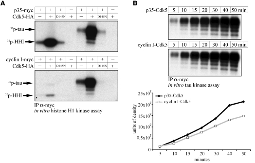
Previously, the noncyclin proteins p35, p39 (reviewed in ref. 17), and p67 (18) were shown to be activators of Cdk5 in podocytes and neurons (16). We therefore aimed to compare cyclin I–Cdk5 and p35-Cdk5 activity and questioned whether the substrate specificity of Cdk5 depends on its activating partner proteins, cyclin I and p35. The experiments represented in Figure 3, A and B, directly compared cyclin I–Cdk5 and p35-Cdk5.
Figure 3. Cyclin I–Cdk5 preferentially phosphorylates tau.
(A) HEK293 cells were transfected with HA-Cdk5, then cotransfected with either myc-p35 or myc–cyclin I. Following an IP to the myc epitope tag on either p35 or cyclin I, in vitro kinase assays were performed using either histone H1 (HH1) or tau as substrates. Histone H1 and tau (lanes 2, 6) were strongly phosphorylated by p35-Cdk5. In contrast, cyclin I–Cdk5 preferentially phosphorylated tau (lane 6) rather than histone H1 (lane 2). No kinase activity was detected in the presence of a kinase-inactive dominant negative Cdk5 mutant (HA-D145N-Cdk5, lane 3, 7) or in the absence of either Cdk5 (lanes 1, 5) or p35/cyclin I (lanes 4, 8). (B) To compare the kinase kinetics by which cyclin I–Cdk5 phosphorylates tau in comparison with p35-Cdk5, a time-course experiment was performed in HEK293 cells cotransfected with myc-p35 and HA-Cdk5 or myc–cyclin I and HA-Cdk5. Following IP for the myc tag, phosphorylation of tau was assessed by incorporation of 32P-ATP quantified on a phosphoimager system. Cyclin I–Cdk5 showed phosphorylation kinetics similar to those of p35-Cdk5. Phosphorylation of tau was already evident after 5 minutes and reached a plateau after 40–50 minutes.
Histone H1 and tau were strongly phosphorylated by p35-Cdk5. In contrast, cyclin I–Cdk5 preferentially phosphorylated tau rather than histone H1. Cyclin I–Cdk5 and p35-Cdk5 were quantitatively similar in their abilities to phosphorylate tau (Figure 3B). These data suggest the substrate specificity of Cdk5, like that of other Cdks, may depend in part on its particular activating subunit, i.e., cyclin I or p35.
Cyclin I–Cdk5 kinase activity is required to protect podocytes from apoptosis.
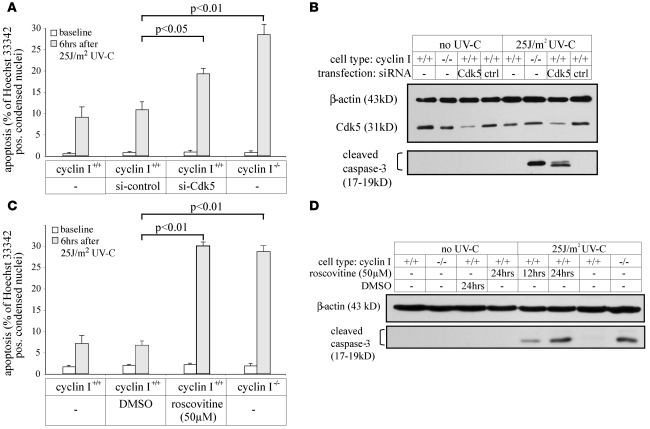
To explore the biological mechanisms for the role of cyclin I in terminally differentiated cells, we began by asking whether Cdk5 had a biological role in enhancing podocyte survival similar to what has been reported in neurons and whether this effect was governed distinctly by cyclin I and/or p35. Griffin et al. (12) first described cyclin I–null podocytes as more susceptible to apoptosis and restoration of cyclin I expression to normal levels as preventative of this. To test the hypothesis that activation of Cdk5 by cyclin I is necessary for survival, we reduced Cdk5 levels and activity in WT podocytes as follows: first, lowering Cdk5 expression with siRNA or inhibiting Cdk activity with roscovitine was not sufficient to induce apoptosis (measured by Hoechst staining and caspase-3 cleavage product) in the absence of an injury such as that caused by ultraviolet light C (UV-C) (Figure 4, A–D) or TGF-β (data not shown). Second, lowering Cdk5 with siRNA (Figure 4, A and B) or roscovitine (Figure 4, C and D) substantially augmented UV-C–induced apoptosis in WT cells. These data show that active Cdk5 is required to reduce injury-induced apoptosis but is not necessary to protect cells under stress-free conditions.
Figure 4. Cyclin I–Cdk5 kinase activity is required to protect podocytes from apoptosis.
(A) Apoptosis was quantitated by Hoechst 33342 staining in podocytes under nonstressed conditions (baseline) and 6 hours after UV-C irradiation (25 J/m2). UV-C–induced apoptosis was increased 2- to 3-fold in WT podocytes transfected with siRNA-targeting Cdk5 (lane 3) compared with cells transfected with control siRNA (lane 2). UV-C induced marked apoptosis in cyclin I–null podocytes (lane 4). Experiments were performed in triplicate, and a minimum of 400 cells were counted per experiment. pos, positive. (B) In the absence of UV-C, Cdk5 protein levels measured by Western blot analysis were similar in nontransfected cyclin I WT and null podocytes (lanes 1, 2). siRNA reduced Cdk5 protein levels in WT cells (middle panel), which resulted in increased caspase-3 cleavage following UV-C irradiation (lower panel); caspase-3 cleavage was not increased in control siRNA–transfected cells exposed to this dose of UV-C (lanes 6, 7). β-actin served as loading control. (C) UV-C irradiation increased apoptosis (Hoechst 33342 staining) 3- to 4-fold in cyclin I WT podocytes when Cdk5 activity was inhibited by roscovitine (lane 3) compared with control cells exposed to DMSO (lane 2). (D) Roscovitine induced a progressive increase in cleaved caspase-3 (lanes 5, 6) following exposure to UV-C. β-actin served as a loading control. Data shown represent mean + SD.
Next, to determine whether the maximal survival benefit for Cdk5 was conferred by its activation by p35 and/or cyclin I, cultured podocytes were isolated and characterized from cyclin I–null, p35–null, and WT mice by standard methods. As expected, UV-C–induced apoptosis was significantly increased in cyclin I–null and p35-null podocytes. To determine the relative contribution of cyclin I and p35, p35 protein levels were reduced in cyclin I–null podocytes by siRNA, and the results are shown in Figure 5. In the absence of cell injury, p35 siRNA transfection was not sufficient to induce apoptosis in cyclin I–null podocytes (data not shown). Thus, cyclin I and p35 are not required for survival under normal nonstressed conditions. In contrast, UV-C irradiation significantly increased caspase-3 cleavage in cyclin I–null cells transfected with p35 siRNA compared with cyclin I–null cells transfected with control siRNA. Similar results were obtained in cyclin I–null cells treated with siRNA targeting Cdk5 or incubated with the Cdk inhibitor roscovitine. Taken together, these data indicate that both cyclin I–Cdk5 and p35-Cdk5 have nonredundant roles in promoting cell survival following injury.
Figure 5. Reducing p35 or Cdk5 activity augments caspase-3 cleavage in cyclin I–null podocytes.
To determine the relative roles of cyclin I, p35, and Cdk5 on apoptosis measured by caspase-3 cleavage, cyclin I–null podocytes were transfected (tx) with siRNA targeting either Cdk5 or p35, and in separate studies, Cdk5 activity was reduced in cyclin I–null cells with roscovitine. Reducing Cdk5 protein levels augmented caspase-3 cleavage in cyclin I–null cells following UV-C irradiation compared with control siRNA–transfected cells (lanes 1, 2). Lowering p35 protein levels increased caspase-3 cleavage following UV-C irradiation in cyclin I–null cells compared with nontransfected and control siRNA–transfected cells (lanes 3–5). Roscovitine increased caspase-3 cleavage (lane 6) following exposure to UV-C to the levels seen in cyclin I–null cells transfected with siRNA that targets p35.
Cyclin I–Cdk5 activates MEK1/2 and ERK1/2.
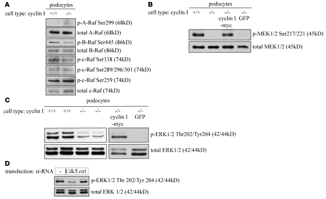
Having shown that Cdk5 can be activated by cyclin I and/or p35, we asked whether the regulation of apoptosis by cyclin I–Cdk5 and p35-Cdk5 were governed by distinct or common pathways. Having recently reported that ERK1/2 is required for podocyte survival (19), we asked whether cyclin I–Cdk5 was an effector of the Raf-MEK1/2-ERK1/2 pathway. Phospho-specific antibodies did not show any substantial differences in Raf activation between WT and cyclin I–null cells (Figure 6A). However, there was a substantial decrease in phosphorylated MEK1/2 and phosphorylated ERK1/2 in 2 different clones of cyclin I–null podocytes compared with WT podocyte clones (Figure 6, B and C). Proof that these differences were indeed due to cyclin I was confirmed by infecting cyclin I–null cells with myc-tagged cyclin I. Restoring cyclin I levels in null cells restored the levels of MEK1/2 and ERK1/2 phosphorylation to that of WT cells. In addition, reducing Cdk5 expression with siRNA also decreased the phosphorylation of ERK1/2 (Figure 6D). Given that phospho-Raf levels were unchanged, we asked whether cyclin I exerts its effects on the MAPK pathway at the level of MEK1. The results showed that when constitutively active MEK1 was transduced in cyclin I–null cells, the levels of phospho-ERK normalized. These in vitro data are consistent with the regulation of the MAPK pathway by cyclin I at the level of MEK1/2. There were no quantitative differences in levels of AKT or p38 or JNK between cyclin I–null and WT cells (data shown in Supplemental Figure 3; supplemental material available online with this article; doi:10.1172/JCI37978DS1).
Figure 6. Cyclin I–Cdk5 regulates apoptosis by activating MEK1/2–ERK1/2.
(A) There were no substantial differences in the phosphorylation status of A-, B- and c-Raf in cyclin I–null (–/–) and WT (+/+) podocytes under nonstressed conditions using several phosphospecific antibodies. (B) Phosphorylation of MEK1/2 on residues Ser217/221 was substantially reduced in cyclin I–null podocytes (lane 2) compared with WT podocytes (lane 1) under physiological, nonstressed conditions. Restoring cyclin I levels in null cells by retroviral infection normalized MEK1/2 phosphorylation (lane 3) to levels comparable to those of WT podocytes. GFP transfection had no effect (lane 4). Total MEK served as a loading control. These results show that MEK1/2 Ser217/221 phosphorylation is cyclin I dependent. (C) Phosphorylation of ERK1/2 on residues Thr202/Tyr204 was reduced in 2 different clones of cyclin I–null podocytes (lanes 3, 4) compared with 2 different WT podocyte clones (lanes 1, 2). Restoring cyclin I levels in null cells by retroviral infection normalized ERK1/2 phosphorylation (lane 5) to levels comparable to those of WT podocytes; GFP infection had no effect. Total ERK1/2 served as loading control. These results show that the cyclin I–dependent activation of MEK1/2 was reflected by an increased phosphorylation of ERK1/2. (D) Reducing Cdk5 expression in cyclin I WT podocytes with siRNA decreased ERK1/2 phosphorylation (lane 2) compared with nontransfected cells. Scrambled siRNA had no effect on ERK1/2 phosphorylation.
Phospho-ERK1/2 staining is altered in cyclin I–null mice with experimental glomerulonephritis.
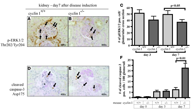
To further validate the in vitro findings, we performed additional in vivo studies by performing immunohistochemistry stainings in mice with experimental glomerulonephritis induced by injection of a sheep anti-glomerulus antibody as previously published (12, 20). Caspase-3 cleavage staining was not detected in normal cyclin I–null or WT animals prior to disease induction. However, the data in Figure 7 show that this model is associated with increased caspase-3 staining, which correlates with our reports of podocyte apoptosis. Thus, like the in vitro models described earlier, the in vivo model was also associated with caspase-3 cleavage. Moreover, there was a substantial increase in caspase-3 cleavage in diseased cyclin I mice (day 7, P < 0.01 compared with diseased WT mice; Figure 7). The increase in caspase-3 cleavage was associated with reduced podocyte number and worsened renal function (data not shown).
Figure 7. Decreased ERK1/2 activation and increased caspase-3 cleavage in cyclin I–null mice with experimental glomerulonephritis.
Experimental glomerulonephritis was induced in 10- to 12-week-old WT and cyclin I–null mice by administration of anti-glomerular antibody. (A–C) Activation of ERK1/2 was assessed by immunostaining for p-ERK1/2 Thr202/Tyr204. There was a significant decrease in glomerular pERK1/2 staining at day 7 of nephritis in cyclin I–null mice (P < 0.05, ANOVA). (D–F) Apoptosis was quantified by immunostaining for caspase-3 cleavage adjacent to Asp175. Both WT and cyclin I–null mice showed increased caspase-3 cleavage in podocytes following disease induction. However, there was significantly more caspase-3 cleavage in cyclin I–null mice at day 7 compared with WT mice (P < 0.01, ANOVA). Depicted are representative glomeruli. Data shown represent mean + SD.
Because of the cell-culture findings described earlier, ERK1/2 activation was examined. At day 7 after disease induction, there was increased staining for phospho-ERK1/2 in glomeruli of diseased WT mice. Quantitation of glomerular active ERK1/2 staining showed a significant decrease in diseased cyclin I–null mice vs. diseased WT mice (P < 0.05) (Figure 7). Similarly to what was found in the cell-culture studies, decreased ERK1/2 activation was associated with increased podocyte apoptosis in cyclin I–null mice compared with WT controls. Importantly, these in vivo studies validate the elements described in the cell-culture studies performed in which more mechanistic approaches can be employed.
Cyclin I requires an intact MEK/ERK pathway to promote cell survival.
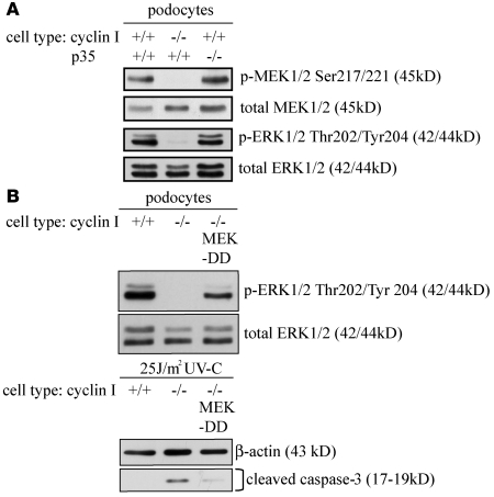
In contrast to cyclin I–null cells, repeated studies at different passages showed no differences in the levels of phospho-MEK1/2 and phospho-ERK1/2 in p35-null podocytes compared with WT cells (Figure 8A). Thus, the signaling pathway downstream of cyclin I–Cdk5 was distinct from that of p35-Cdk5. In order to address the biological significance of reduced phospho-MEK1/2 (and thus phospho-ERK1/2), cyclin I–null cells were stably infected with constitutively active MEK1 (MEK-DD) and exposed to injury. Figure 8B shows that this restored normal levels of phospho-ERK1/2, and UV-C–induced apoptosis was substantially reduced. These results show that despite cyclin I and p35 both activating Cdk5, they promote cell survival by different pathways. Cyclin I, but not p35, is required for normal phosphorylation, and thus activity, of MEK1/2 and ERK1/2. In the absence of cyclin I, decreased phospho-MEK1/2 and phospho-ERK1/2 lower the threshold to injury-induced apoptosis.
Figure 8. The signaling pathway downstream of cyclin I–Cdk5 is distinct from siRNA that targets p35-Cdk5.
(A) There were no differences in MEK1/2 and ERK1/2 phosphorylation in the absence of p35 in podocytes compared with WT podocytes. (B) To prove that the specific decrease in MEK1 was central to the increases in apoptosis in the absence of cyclin I, cyclin I–null podocytes were transfected with constitutively active MEK1 (MEK-DD) mutants. Restoring active MEK1 increased ERK 1/2 phosphorylation (lane 3) similar to that of WT cells (lane 1). Restoring MEK1 reduced UV-C–induced apoptosis measured by caspase-3 cleavage products. These results show that cyclin I is required to phosphorylate MEK1 (and thus ERK1/2), which is necessary to reduce apoptosis.
Cyclin I regulates specific Bcl-2 family proteins.
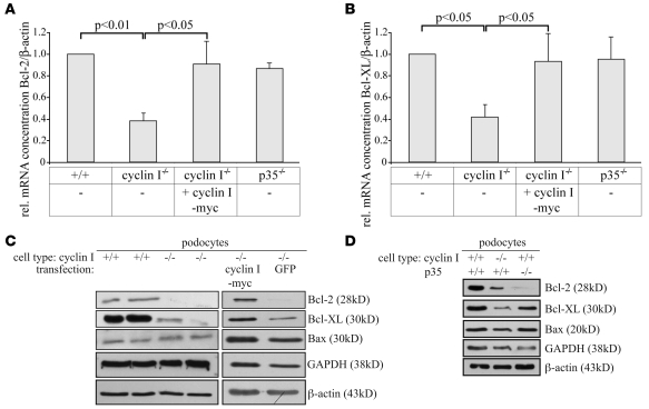
Apoptosis in podocytes in these studies are caspase-3 dependent. Studies have shown that specific Bcl-2 family proteins regulate caspase activation. Having shown that cyclin I–Cdk5 and p35-Cdk5 are necessary for survival but have different effects on the MAPK pathway, we next tested to determine whether cyclin I and p35 had differential effects on Bcl-2 family proteins in podocytes under physiological, nonstressed conditions. Quantitative PCR showed a marked decrease in the constitutive mRNA levels for the prosurvival proteins Bcl-2 and Bcl-XL in cyclin I–null podocytes compared with control WT cells (Figure 9, A and B). In contrast, the absence of p35 in p35-null cells had no effect on mRNA levels for Bcl-2 and Bcl-XL. In addition to changes in mRNA, Figure 9, C and D, shows that the protein levels for Bcl-2 and Bcl-XL were also markedly reduced in cyclin I–null podocytes. In contrast, there were no differences in the levels of Bax. Importantly, restoration of cyclin I expression in cyclin I–null podocytes by retroviral infection returned the protein levels of Bcl-2 and Bcl-XL to that of WT cells (Figure 9, A–D), demonstrating that these changes were the direct effect of cyclin I. By comparison, in p35-null podocytes, only a decrease in Bcl-2 protein was observed while Bcl-XL and Bax remained unchanged.
Figure 9. Cyclin I, but not p35, differentially regulates specific Bcl-2 family proteins.
(A, B) The mRNA levels for Bcl-2 (A) and Bcl-XL (B) were measured by quantitative PCR, and the relative concentrations are shown as ratio normalized to β-actin. Samples were run in triplicate, and mRNA from 3 independent experiments was included. Relative gene expression was analyzed using the 2–standard curve method. Compared with WT cells (lanes 1), the mRNA levels for Bcl-2 and Bcl-XL (lanes 2) were significantly reduced in cyclin I–null podocytes. Restoring cyclin I in null cells by stable infection normalized transcripts for Bcl-2 and Bcl-XL (lanes 3). In contrast, the absence of p35 in p35-null cells had no effect on Bcl-2 and Bcl-XL mRNA levels (lane 4). (C, D) To determine whether cyclin I or p35 altered the protein levels for certain Bcl-2 family proteins, Western blot analyses were performed in WT, cyclin I–null, and p35-null podocytes grown under physiological, nonstressed conditions. GAPDH and β-actin served as loading controls. Compared with WT cells (C, lanes 1, 2), Bcl-2 and Bcl-XL, but not Bax, protein levels were reduced in cyclin I–null podocytes (C, lanes 3, 4). Levels for Bcl-2 and Bcl-XL were normalized upon infection with cyclin I (lane 5), but not GFP (lane 6). In contrast, in the absence of p35, only Bcl-2 protein expression was strongly reduced compared with WT podocytes (D). No effects on Bcl-XL and Bax protein levels were observed. Data shown represent mean + SD.
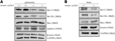
The effect of cyclin I on specific Bcl-2 protein levels was also measured in vivo in normal mouse glomeruli and brains. Protein isolated from the glomeruli of nonstressed cyclin I–null mice had markedly reduced levels of Bcl-2 and Bcl-XL proteins compared with WT animals (Figure 10A). There were no differences in Bax levels in glomerular protein from cyclin I–null and WT mice. Similarly, we found that the levels of Bcl-2 and Bcl-XL were decreased in neuronal tissue from cyclin I–null mice compared with WT mice by Western blot analysis (Figure 10B) and immunostaining (Supplemental Figure 1).
Figure 10. Cyclin I regulates specific Bcl-2 family proteins in vivo.
(A) Glomeruli were isolated and pooled from the kidneys of 6 animals from each strain, divided into 2 samples, and loaded separately on the gel. Compared with WT glomeruli (lanes 1, 2), levels of Bcl-2 and Bcl-XL were reduced in cyclin null glomeruli (lanes 3, 4). Nephrin, a protein expressed selectively and constitutively in podocytes, was included as a loading control in addition to β-actin and GAPDH to rule out any potential loading differences in protein from podocytes. (B) Compared with brain protein lysates harvested from cyclin I WT mice (lane 1) under physiological, nonstressed conditions, the protein levels of Bcl-2 and Bcl-XL were decreased in cyclin I–null mice (lane 2). Bax levels remain unchanged; β-actin was used as loading control. Taken together, these data show that similar to the cultured cells, the absence of cyclin I also regulates Bcl-2 and Bcl-XL in postmitotic organs in vivo.
Cyclin I regulates expression of Bcl-2 family proteins by activating Cdk5.
Having shown that cyclin I regulates the protein levels of specific Bcl-2 family proteins, we next addressed the question of whether Cdk5 activity underlies this effect. To this end, Cdk5 expression and activity were reduced by transient transfection with siRNA against Cdk5 in podocytes. As demonstrated in Figure 11A, reducing Cdk5 levels and activity decreased the levels of Bcl-2 and Bcl-XL. In contrast, the proapoptotic protein Bax was not affected. Similarly, protein levels of Bcl-2 and Bcl-XL, but not Bax, decreased when WT podocytes were incubated with the Cdk inhibitor roscovitine (Figure 11B). Thus, a decrease in Cdk5 or its activity in WT cells recapitulated what we observed in cyclin I–null podocytes.
Figure 11. Cyclin I, but not p35, differentially regulates specific Bcl-2 family proteins.
(A) Reducing Cdk5 expression by siRNA decreased the protein levels of Bcl-2 and Bcl-XL. No effect was observed in control podocytes transfected with control siRNA. (B) Inhibiting Cdk5 activity by roscovitine (50 μM) reduced the protein expression of Bcl-2 and Bcl-XL compared with vehicle (DMSO). GAPDH and β-actin served as loading controls. (C) To prove that regulation of Bcl-2 and Bcl-XL underlies the prosurvival effect of cyclin I, protein expression of Bcl-2 or Bcl-XL was restored in cyclin I–null podocytes by retroviral infection. There was a 3- to 4-fold increase in apoptosis following UV-C irradiation in cyclin I–null cultured podocytes infected with GFP compared with injured cyclin I WT cells. UV-C–induced apoptosis was markedly reduced in cyclin I–null podocytes stably infected with Bcl-2 or Bcl-XL. (D) Accordingly, caspase-3 cleavage products were also decreased in cyclin I–null podocytes infected with Bcl-2 or Bcl-XL 6 hours after UV-C irradiation compared with GFP-infected podocytes. (E) Infecting cyclin I–null podocytes with constitutively active MEK1 (MEK-DD) mutants also increased the expression of Bcl-2 and Bcl-XL linking regulation of the MEK-ERK pathway by cyclin I–Cdk5 to the observed regulation of Bcl-2 and Bcl-XL expression. Data shown represent mean + SD.
We next tested the hypothesis that increased caspase-3 cleavage in cyclin I–null podocytes was indeed due to the changes observed in Bcl-2 and/or Bcl-XL. Cyclin I–null podocytes were stably infected with Bcl-2 or Bcl-XL to levels comparable to those of WT cells (Supplemental Figure 2) and then exposed to UV-C (25 J/m2) to induce apoptosis. The level of apoptosis in cyclin I–null podocytes with restored levels of (exogenous) Bcl-2 or Bcl-XL was similar to the levels of apoptosis in cyclin I WT podocytes (Figure 11C). Restoring Bcl-2 and Bcl-XL levels in cyclin I–null podocytes also reduced caspase-3 cleavage (Figure 11D). These experiments showed that the decreased expression of Bcl-2 and Bcl-XL in cyclin I–null podocytes was causally related to their decreased survival after UV-C damage.
Finally, to determine whether the decrease in phospho-ERK1/2 activation in cyclin I–null podocytes was related to the decrease in Bcl-2 and Bcl-XL protein levels, cyclin I–null cells were stably infected with constitutively active MEK1 (MEK-DD). Active MEK1 increased expression of Bcl-2 and Bcl-XL (Figure 11E). In summary, these results suggest that cyclin I and Cdk5 regulate the phosphorylation and activation of ERK1/2, which in turn modulates cell survival via its effects on the expression of Bcl-2 and Bcl-XL at the mRNA and protein levels.
Discussion
In terminally differentiated and highly specialized cells that do not readily proliferate, such as the kidney and brain, apoptosis is a major cause of reduced cell number, ultimately leading to organ dysfunction. In diseases of podocytes, such as occurs in diabetic and nondiabetic renal disease (4–6), the resultant reduced cell number underlies the development of proteinuria and also progressive kidney scarring. Similarly, neuronal apoptosis in stroke or neurodegenerative diseases (2, 3) can be associated with severe and irreversible pathology. Several different mechanisms are therefore required to maintain survival of terminally differentiated cells. Our studies focused on Cdk5 as a critical survival factor in terminally differentiated cells and its regulation by 2 distinct activators, cyclin I and p35.
Cyclin I is a novel regulator of Cdk5.
Cdk5 reduces apoptosis in neurons (reviewed in ref. 11). Unlike other members of the cyclin-dependent kinase family, Cdk5 is activated by the noncyclins p35, p39 (reviewed in ref. 17), and p67 (18). However, identification of a partner cyclin for Cdk5 has been elusive. We focused on cyclin I as a novel partner for Cdk5 because cyclin I staining colocalizes with Cdk5 in kidney podocytes and brain. Despite structural similarities with other cyclins, several lines of evidence suggest that cyclin I differs from known “classical” cyclins. Cyclin I is most abundant in terminally differentiated (postmitotic) cells and is not involved in the regulation of proliferation; the mRNA and protein levels do not fluctuate during cell cycle, and cyclin I–null mice do not have any developmental abnormalities (12, 14). Moreover, a partner cyclin-dependent kinase has not yet been identified for cyclin I.
The following lines of evidence support the first major finding in these studies, that cyclin I binds to and activates Cdk5. First, cyclin I and Cdk5 coimmunoprecipitate in protein lysates extracted from kidney glomeruli, cultured podocytes, and brain tissue, and the complex is an active kinase. Second, cotransfection studies in HEK293 cells confirmed cyclin I and Cdk5 coimmunoprecipitation and activation. Third, Cdk5 activity was substantially diminished in vivo in glomeruli and neurons in cyclin I–null mice but was restored to WT levels when cyclin I was reconstituted by ectopic expression. Fourth, the specificity of this interaction was confirmed using dominant negative mutants, siRNA, and chemical inhibitors of Cdk activity. We believe these results are the first demonstration that cyclin I has a specific partner Cdk and the first to show a cyclin activator for Cdk5.
Cyclin I–Cdk5 confers a survival benefit independent of p35.
Recently, we showed that in the kidneys of cyclin I–null mice, the terminally differentiated glomerular epithelial podocytes are significantly more susceptible to apoptosis following injury in vitro and in experimental models in vivo (12). This resulted in markedly increased proteinuria and progressive glomerular scarring. In contrast, the absence of cyclin I did not affect survival/apoptosis in cells with a high proliferative capacity, such as kidney mesangial cells (Habu snake venom–induced mesangial proliferative glomerulonephritis; data not shown) and tubular epithelial cells (unilateral ureteral obstruction model of acute tubular apoptosis) (12) in vivo. This suggested that the biological role of cyclin I was confined to postmitotic cells, which are usually terminally differentiated and do not re-enter the cell cycle and proliferate to replenish cell loss.
Given that cyclin I confers a survival benefit in kidney podocytes, we next determined whether the activation of Cdk5 by cyclin I serves a survival role similar to that seen in previous reports of p35-Cdk5 in neurons.
Our data show that reducing cyclin I–Cdk5 activity in the absence of injury was not sufficient to induce apoptosis. In contrast, when cyclin I–Cdk5 activity was reduced, the onset of apoptosis was earlier, and the magnitude substantially increased following apoptotic injuries such as UV-C irradiation or exposure to the proapoptotic cytokine TGF-β (results not shown). To further validate the biological relevance of the increase in caspase-3 activation in the injured cells in culture used in these studies, additional in vivo experiments were performed in WT and in cyclin I–null mice with experimental glomerulonephritis induced by the administration of anti-glomerular antibody as we have previously reported (12, 20). Glomerulonephritis was associated with increased caspase-3 cleavage staining in diseased WT mice, in a podocyte distribution. Moreover, caspase-3 cleavage was augmented in experimental glomerulonephritis in cyclin I–null mice compared with diseased WT mice. These in vivo studies validate the cell-culture studies, as both “models” are characterized by caspase-3–associated apoptosis. Taken together, these data are consistent with the notion that active cyclin I–Cdk5 sets an apoptotic threshold to injury.
We previously reported that cyclin I–null podocytes are more vulnerable to apoptosis induced by several forms of apoptotic injury, and others have shown that p35-Cdk5 also regulates apoptosis in postmitotic cells. Accordingly, 2 strategies were used to determine whether the presence of both p35 and cyclin I are necessary for the maximal survival benefit by Cdk5. First, reducing p35 levels with siRNA further increased the rates of apoptosis in cyclin I–null podocytes. Second, apoptosis was also augmented when Cdk5 activity was reduced in cyclin I–null cells. These data support the notion that the effects of cyclin I–Cdk5 on apoptosis are indeed independent from and nonredundant with that of p35-Cdk5.
MAPK signaling by cyclin I is distinct from p35.
Because Cdk5 can be activated by cyclin I and p35 and their roles on apoptosis are distinct, we next sought to determine whether cyclin I and p35 govern apoptosis through different or common downstream pathways. Accordingly, AKT and several MAPK pathways involved in the regulation of cellular survival processes were measured. No differences in p38, JNK/SAPK, or AKT pathways were observed between null and WT cells (Supplemental Figure 3). We thus focused on the Raf/MEK/ERK pathway because published reports in neurons describe regulation of the MAPK pathway by Cdk5 (21–24).
The current data showed no difference in the levels of total Raf and phospho-Raf in cyclin I–null and WT cells. However, there was a striking decrease in the phosphorylated (active) forms of MEK1/2 (Ser217/221) and ERK1/2 (Thr202/Tyr204) in cyclin I–null podocytes compared with WT cells when grown under nonstressed conditions. Ectopic expression of cyclin I in cyclin I–null cells normalized MEK1/2 and ERK1/2 phosphorylation, thus validating that these changes were indeed dependent on cyclin I. Furthermore, retroviral infection with constitutive active MEK1 normalized phospho-ERK1/2 levels in null cells without the need to restore cyclin I levels. These results support the notion that phosphorylation of MEK1/2 on Ser217/221 and the subsequent phosphorylation of ERK1/2 requires the presence of cyclin I. In order to determine whether MEK1/2 Ser217/221 was a direct substrate for cyclin I–Cdk5, recombinant human MEK1 was used as a substrate in a cyclin I–Cdk5 phosphorylation assay. The results (data not shown) were inconclusive. This raises the question of whether unidentified intermediate kinases may be involved in the regulation of MEK1/2 phosphorylation by cyclin I–Cdk5.
We next asked whether the effects of cyclin I on MAPK differed from the effects of p35 on this signaling pathway. The results of the current study show that both MEK1/2 and ERK1/2 phosphorylation are normal in nonstressed p35-null podocytes, with levels similar to those of WT cells. In contrast, Zheng characterized p35-Cdk5 as the “molecular switch” that modulates the duration of ERK1/2 phosphorylation in PC12 cells and rat cortical neurons (24). In the absence of p35-Cdk5, ERK1/2 was hyperphosphorylated, and hyperphosphorylated ERK1/2 was proapoptotic (22, 24, 25). In contrast with these published results, Wang et al. reported that retinoid acid and brain-derived neurotrophic factor phosphorylate ERK1/2 through active p35-Cdk5 in SH-SY5Y cells and in primary neuronal cultures. Inhibiting p35-Cdk5 or ERK1/2 abrogated the prosurvival effects of p35-Cdk5 and was associated with increased neuronal apoptosis following injury (23). One might conclude that the biological role of p35 on Raf/MEK/ERK phosphorylation differs depending on the conditions of the experiments, the growth factors studied, and the cell types used. Most of the studies on p35-Cdk5 focused principally on neuronal differentiation in primary neuronal cell-culture systems to monitor the role of Cdk5 in differentiation and maturation. The results reported in the current experiments focused on nonproliferating, fully terminally differentiated cells.
Finally, the biological relevance of cyclin I–dependent differences in ERK1/2 phosphorylation was considered. Indeed, this regulation was very significant, as normalizing phospho-ERK1/2 augmented survival of cells compared with those in which ERK1/2 was not phosphorylated. Moreover, silencing Cdk5 expression by siRNA in WT podocytes also decreased ERK1/2 phosphorylation, thus validating the hypothesis that the effects of cyclin I on the ERK pathway are indeed mediated by active Cdk5.
Differential effect of cyclin I and p35 on Bcl-2 family proteins.
Caspase-3–dependent mechanisms characterize many forms of kidney and neuronal apoptosis. The data in the current study showed that caspase-3 cleavage is increased when cyclin I–Cdk5 activity is lowered. While several pathways regulate caspase cleavage, Bcl-2 and Bcl-XL inhibit caspase activation by governing mitochondrial permeability and the release of certain factors that trigger caspase activation. Bax and others counteract the effects of Bcl-2 and Bcl-XL, thereby promoting caspase activation. These data show that both the mRNA and protein levels of Bcl-2 and Bcl-XL were substantially decreased in cyclin I–null podocytes and that the protein levels were also decreased in kidney and brain of cyclin I–null mice. Restoring cyclin I levels normalized Bcl-2 and Bcl-XL levels. The biological significance of these events was shown when apoptosis was decreased by stably infecting cyclin I–null cells with Bcl-2 and Bcl-XL. The absence of cyclin I had no effect on Bax levels.
The effects of cyclin I–Cdk5 on Bcl-2 family proteins described here differs from what has been previously reported for p35-Cdk5. Wang et al. (23) reported that Cdk5 inhibition was accompanied by decreased Bcl-2 protein levels, and this correlated with increased cell death in response to an apoptotic trigger. They did not observe an effect of p35 inhibition on Bcl-XL protein levels, and we confirm that finding here. It has also been shown that Bcl-2 family members can be phosphorylated, thereby regulating their function posttranslationally (26–29). Indeed human Bcl-2 can be phosphorylated on Ser70 by Cdk5, and this promotes survival of neurons (30). These results are consistent with the hypothesis that the effect of p35 on Bcl-2 is posttranscriptional.
In addition to showing that the restoration of phospho-ERK1/2 increased Bcl-2 and Bcl-XL in cyclin I–null cells, additional experiments (data not shown) using a chemical inhibitor for Raf1 (GW 5074) did not show any effect on Bcl-2 family proteins. However, the MEK1/2 inhibitor U0126 reduced levels of Bcl-2 and Bcl-XL, further validating the importance of MEK1/2 and subsequent ERK1/2 activation in the regulation of specific Bcl-2 family proteins independent of Raf.
Taken together, these results support the hypothesis that reduced transcription and translation of Bcl-2 and Bcl-XL secondary to reduced cyclin I–Cdk5 made cells more vulnerable to apoptosis.
The results of these studies provide several lines of evidence that cyclin I–Cdk5 is distinct from p35-Cdk5 in the regulation of Bcl-2 proteins as follows: (i) the mRNA and protein regulation of Bcl-2 and Bcl-XL by cyclin I; the absence of p35 is associated with decreased protein levels of Bcl-2 only (23); (ii) a significant decrease in MEK1/2 and ERK1/2 phosphorylation in cyclin I–null cells but not in p35-null cells; and (iii) retroviral infection with constitutive active MEK1 or cRaf had no effect on Bcl-2 protein levels or podocyte survival in p35-null podocytes (data not shown).
These findings are in line with numerous reports in podocytes and neurons that clearly demonstrate specific mechanisms are required to protect these highly specialized cells from apoptosis and to maintain survival (31–34). Here, we describe what we believe is a novel pathway that regulates apoptosis through the prosurvival effects of cyclin I–Cdk5.
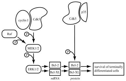
We conclude by proposing that cyclin I–Cdk5 regulates Bcl-2 and Bcl-XL by activating the MEK/ERK pathway and increased transcriptional activity while the effects of p35-Cdk5 occur at the posttranslational level (Figure 12). These results further emphasize the differential role of Cdk5 in protecting postmitotic, terminally differentiated cells against apoptosis.
Figure 12. Proposed model showing the effects of Cdk5 upon activation by cyclin I and p35.
Cdk5 is activated by cyclin I and p35. The pathways by which Cdk5 confers a prosurvival function depend on the activator. Cyclin I–Cdk5 leads to phosphorylation of MEK1/2, and subsequently, ERK1/2, leading to increased mRNA and protein levels for the prosurvival proteins Bcl-2 and Bcl-XL. In contrast, p35-Cdk5 increases Bcl-2 protein levels by posttranslational modification (30). p35-Cdk5 has no effect on Bcl-XL levels. The dual activation of Cdk5 by cyclin I and p35 ensures that maximal survival pathways are operative in terminally differentiated cells.
Methods
Cell culture.
HEK293 cells (ATCC) were cultured under standard conditions at 37°C in DMEM media (Invitrogen). Conditionally immortalized podocytes were generated as previously described (12, 35). Quiescence and differentiation were induced by culturing the cells at 37°C for 13 to 15 days on Primaria plastic plates (BD Biosciences) in the absence of IFN-γ.
Plasmid transfection.
pCS2+ vectors encoding myc-tagged cyclin I or cyclin E and HA-tagged Cdks 1–6 as well as pCMV-myc–tagged p35 (a gift from Li-Huei Tsai, MIT, Boston, Massachusetts, USA), pCMV-HA–tagged Cdk5 (36), and HA-tagged dominant negative (D145N) Cdk5 (36) (both from Addgene) were used to transfect HEK cells using CaCl2 DNA precipitation.
cMMP-IRES-GFP vectors encoding human Bcl-2 or Bcl-XL (37) and pBabe-puro vectors (38) (Addgene) encoding myc-tagged cyclin I, GFP, or MEK-DD (39) (Addgene) were transfected into Phoenix Eco packaging cells (Gary Nolan, The Baxter Laboratory of Genetic Pharmacology, Stanford University School of Medicine, Stanford, California, USA) to generate retrovirus. Selection of infected cells was achieved either by 48-hour selection with puromycin (2.5 μg/ml; Sigma-Aldrich) for cyclin I–myc–, GFP-, and MEK-DD–infected cells or by FACS sorting (cMMP-IRES-GFP–Bcl-2/Bcl-XL).
Recombinant herpes simplex virus 1 encoding dominant negative Cdk5-D145N-IRES-GFP or HSV-p1005-IRES-GFP (gift from Li-Huei Tsai and Rachael Neve, Harvard Medical School, McLean Hospital, Belmont, Massachusetts, USA) was used to transduce differentiated podocytes at day 10 of growth restriction. Transduction efficiency was greater than 90% as assessed by FACS.
siRNA transfection.
On day 10 of growth arrest, podocytes were transfected with 2 μg pooled siRNA-targeting Cdk5 or p35 using electroporation (175 volt, 200 μsec, 5 pulses) according to the manufacturer’s recommendation (Ambion). Nonspecific siRNA (Ambion) was included as transfection control. Gene silencing was confirmed by quantitative PCR. The efficiency of the Cdk5 and p35 siRNA in reducing Cdk5 or p35 protein expression respectively ranged from 70% to 90% as assessed by densitometric analysis (NIH ImageJ software, http://rsbweb.nih.gov/ij/; n = 3). The protein knockdown effect was found to be maximal 72 to 96 hours after siRNA transfection.
Induction and detection of apoptosis.
Apoptosis was induced in 80%–90% confluent, fully differentiated podocytes. Each experiment was performed in triplicate. To induce apoptosis, cells were irradiated with UV-C (25 J/m2), and apoptosis was assessed 6 hours later by staining for nuclear condensation with Hoechst 33342 (10 mM; Sigma-Aldrich) and by the detection of caspase-3 cleavage products by Western blot analysis as previously reported (40).
Immunoprecipitation studies.
In order to determine protein-protein interactions, immunoprecipitation studies were carried out. The ProFound c-Myc tag IP/Co-IP kit (Pierce) was used for immunoprecipitation studies to reduce IgG light- and heavy-chain contamination according to the manufacturer’s instructions.
Isolation of glomeruli.
Kidney glomeruli were isolated from cyclin I WT and null mice as previously reported (41). In brief, mice were perfused through the heart with magnetic Dynabeads (Invitrogen). Kidneys were minced into small pieces, digested with collagenase A (Roche), filtered, and collected using a magnet. To increase protein yield, glomeruli from several animals were pooled and resuspended in protein lysis buffer.
Kinase assay.
To determine the activity of cyclin-Cdk complexes, coimmunoprecipitation studies were performed, followed by histone H1 or Rb kinase assay. Following immunoprecipitation, the kinase reaction was performed at 37°C for 30–60 minutes in a kinase reaction buffer containing 5 μmol/l ATP, 2 μCi [32P]-γATP (NEN Life Science Products), and 0.6 μg histone H1 (Millipore). The reactions were stopped with 2× SDS sample buffer, then separated on a 12% SDS-PAGE under reducing conditions and visualized by exposing to autoradiographic film (GE Healthcare) or exposed to a phosphoimager screen and quantified on a phosphoimager system (Molecular Dynamics).
Western blot analysis.
Protein levels were measured by Western blot analysis. The samples were separated by SDS-PAGE and blotted on a PVDF membrane (Sigma-Aldrich). The membrane was incubated in 5% nonfat milk powder in TBS solution (10 mM Tris-HCl, pH 8.0, 150 mM NaCl), and the blot was incubated overnight at 4°C with primary antibodies followed by incubation with appropriate horseradish peroxidase secondary antibodies (Sigma-Aldrich). Proteins were visualized using enhanced chemiluminescence technology (GE Healthcare). In some cases, membranes were incubated in stripping buffer (100 mM glycine, 1% SDS, pH 2.5) and restained as stated.
Quantitative real-time PCR.
RNA was isolated using TRIzol reagent (Invitrogen) according to the manufacturer’s directions. 1 μg RNA was used for cDNA synthesis using the Fermentas First Strand cDNA Synthesis Kit (Fermentas). Quantitative real-time PCR was carried out on a Rotor-Gene 6000 (Corbett Research) using 6-FAM-phosphoramidite–labeled primer probes (all from Applied Biosciences) and Quantace SensiMix dT reagent (Quantace). The amount of product was determined at the end of each cycle by the Rotor-Gene software (Rotor-Gene 6000 series software 1.7). Relative gene expression was analyzed with a normalizing gene using the 2-standard curve method.
Animal models.
Caspase-3 cleavage and ERK1/2 activation were also studied in vivo in WT and cyclin I–null C57BL/6 mice. Crescentic glomerulonephritis was induced in 10- to 12-week-old male WT and cyclin I–null mice by intraperitoneal injection of sheep anti-rabbit glomerular antibody (20 mg/g body weight) on 2 consecutive days as described previously (12, 20).
Mice were sacrificed on days 3 and 7 after the second injection of anti-glomerular antibody (n = 6/group/time point). Renal tissue was embedded in 10% neutral buffered formalin for immunostaining.
Mice were housed according to the standardized specific pathogen–free conditions in the University of Washington animal facility. The Animal Care Committee of the University of Washington reviewed and approved the experimental protocol.
Statistics.
All results are expressed as mean ± SD. Statistical significance was evaluated using GraphPad Prism version 4.00c for Macintosh (GraphPad Software). ANOVA with Tukey-Kramer adjustment for multiple comparisons was applied. P < 0.05 was considered significant.
Supplementary Material
Supplemental data
Acknowledgments
The authors thank and acknowledge the following individuals for providing research material included in this study: Sander van den Heuvel (pCMV-HA-Cdk5/HA-dn-Cdk5), Li-Huei Tsai (pCMV-myc-p35, HSV Cdk5–D145N-IRES-GFP), Rachael Neve (HSV preparation), Richard C. Mulligan (cMMP-backbone), Bob Weinberg (pBabe-puro), and Garry Nolan (Phoenix Eco cells). This work was supported by NIH grants (DK60525, DK56799, and DK51096 to S.J. Shankland), and the German Research Foundation (to P.T. Brinkkoetter) S.J. Shankland is also an established investigator of the American Heart Association.
Footnotes
Conflict of interest: The authors have declared that no conflict of interest exists.
Citation for this article: J. Clin. Invest. 119:3089–3101 (2009). doi:10.1172/JCI37978.
Paul T. Brinkkoetter’s present address is: Department of Medicine, Centre for Molecular Medicine and Cologne Excellence Cluster on Cellular Stress Responses in Aging-Associated Diseases, University of Cologne, Cologne, Germany.
References
- 1.Kobayashi N., et al. Process formation of the renal glomerular podocyte: is there common molecular machinery for processes of podocytes and neurons? Anat. Sci. Int. 2004;79:1–10. doi: 10.1111/j.1447-073x.2004.00066.x. [DOI] [PubMed] [Google Scholar]
- 2.Bredesen D.E., Rao R.V., Mehlen P. Cell death in the nervous system. Nature. 2006;443:796–802. doi: 10.1038/nature05293. [DOI] [PMC free article] [PubMed] [Google Scholar]
- 3.Cho B.B., Toledo-Pereyra L.H. Caspase-independent programmed cell death following ischemic stroke. J. Invest. Surg. 2008;21:141–147. doi: 10.1080/08941930802029945. [DOI] [PubMed] [Google Scholar]
- 4.Barisoni L., Kriz W., Mundel P., D’Agati V. The dysregulated podocyte phenotype: a novel concept in the pathogenesis of collapsing idiopathic focal segmental glomerulosclerosis and HIV-associated nephropathy. J. Am. Soc. Nephrol. 1999;10:51–61. doi: 10.1681/ASN.V10151. [DOI] [PubMed] [Google Scholar]
- 5.Pagtalunan M.E., et al. Podocyte loss and progressive glomerular injury in type II diabetes. J. Clin. Invest. 1997;99:342–348. doi: 10.1172/JCI119163. [DOI] [PMC free article] [PubMed] [Google Scholar]
- 6.Shankland S.J., et al. Cyclin kinase inhibitors are increased during experimental membranous nephropathy: potential role in limiting glomerular epithelial cell proliferation in vivo. Kidney Int. 1997;52:404–413. doi: 10.1038/ki.1997.347. [DOI] [PubMed] [Google Scholar]
- 7.Kriz W., Gretz N., Lemley K.V. Progression of glomerular diseases: is the podocyte the culprit? Kidney Int. 1998;54:687–697. doi: 10.1046/j.1523-1755.1998.00044.x. [DOI] [PubMed] [Google Scholar]
- 8.Kriz W., Lemley K.V. The role of the podocyte in glomerulosclerosis. Curr. Opin. Nephrol. Hypertens. 1999;8:489–497. doi: 10.1097/00041552-199907000-00014. [DOI] [PubMed] [Google Scholar]
- 9.Wharram B.L., et al. Podocyte depletion causes glomerulosclerosis: diphtheria toxin-induced podocyte depletion in rats expressing human diphtheria toxin receptor transgene. J. Am. Soc. Nephrol. 2005;16:2941–2952. doi: 10.1681/ASN.2005010055. [DOI] [PubMed] [Google Scholar]
- 10.Besson A., Dowdy S.F., Roberts J.M. CDK inhibitors: cell cycle regulators and beyond. Dev. Cell. 2008;14:159–169. doi: 10.1016/j.devcel.2008.01.013. [DOI] [PubMed] [Google Scholar]
- 11.Weishaupt J.H., Neusch C., Bahr M. Cyclin-dependent kinase 5 (CDK5) and neuronal cell death. Cell Tissue Res. 2003;312:1–8. doi: 10.1007/s00441-003-0703-7. [DOI] [PubMed] [Google Scholar]
- 12.Griffin S.V., Olivier J.P., Pippin J.W., Roberts J.M., Shankland S.J. Cyclin I protects podocytes from apoptosis. J. Biol. Chem. 2006;281:28048–28057. doi: 10.1074/jbc.M513336200. [DOI] [PubMed] [Google Scholar]
- 13.Bates S., Rowan S., Vousden K.H. Characterisation of human cyclin G1 and G2: DNA damage inducible genes. Oncogene. 1996;13:1103–1109. [PubMed] [Google Scholar]
- 14.Jensen M.R., Audolfsson T., Factor V.M., Thorgeirsson S.S. In vivo expression and genomic organization of the mouse cyclin I gene (Ccni). Gene. 2000;256:59–67. doi: 10.1016/S0378-1119(00)00361-9. [DOI] [PubMed] [Google Scholar]
- 15.Nakamura T., et al. Cyclin I: a new cyclin encoded by a gene isolated from human brain. Exp. Cell Res. 1995;221:534–542. doi: 10.1006/excr.1995.1406. [DOI] [PubMed] [Google Scholar]
- 16.Griffin S.V., et al. Cyclin-dependent kinase 5 is a regulator of podocyte differentiation, proliferation, and morphology. Am. J. Pathol. 2004;165:1175–1185. doi: 10.1016/S0002-9440(10)63378-0. [DOI] [PMC free article] [PubMed] [Google Scholar]
- 17.Dhavan R., Tsai L.H. A decade of CDK5. Nat. Rev. Mol. Cell Biol. 2001;2:749–759. doi: 10.1038/35096019. [DOI] [PubMed] [Google Scholar]
- 18.Shetty K.T., et al. Molecular characterization of a neuronal-specific protein that stimulates the activity of Cdk5. J. Neurochem. 1995;64:1988–1995. doi: 10.1046/j.1471-4159.1995.64051988.x. [DOI] [PubMed] [Google Scholar]
- 19.Wada T., Pippin J.W., Nangaku M., Shankland S.J. Dexamethasone’s prosurvival benefits in podocytes require extracellular signal-regulated kinase phosphorylation. Nephron Exp. Nephrol. 2008;109:e8–e19. doi: 10.1159/000131892. [DOI] [PubMed] [Google Scholar]
- 20.Ophascharoensuk V., et al. Role of intrinsic renal cells versus infiltrating cells in glomerular crescent formation. Kidney Int. 1998;54:416–425. doi: 10.1046/j.1523-1755.1998.00003.x. [DOI] [PubMed] [Google Scholar]
- 21.Kesavapany S., et al. p35/cyclin-dependent kinase 5 phosphorylation of ras guanine nucleotide releasing factor 2 (RasGRF2) mediates Rac-dependent Extracellular Signal-regulated kinase 1/2 activity, altering RasGRF2 and microtubule-associated protein 1b distribution in neurons. J. Neurosci. 2004;24:4421–4431. doi: 10.1523/JNEUROSCI.0690-04.2004. [DOI] [PMC free article] [PubMed] [Google Scholar]
- 22.Sharma P., et al. Phosphorylation of MEK1 by cdk5/p35 down-regulates the mitogen-activated protein kinase pathway. J. Biol. Chem. 2002;277:528–534. doi: 10.1074/jbc.M109324200. [DOI] [PubMed] [Google Scholar]
- 23.Wang C.X., et al. Cyclin-dependent kinase-5 prevents neuronal apoptosis through ERK-mediated upregulation of Bcl-2. Cell Death Differ. 2006;13:1203–1212. doi: 10.1038/sj.cdd.4401804. [DOI] [PubMed] [Google Scholar]
- 24.Zheng Y.L., et al. Cdk5 Modulation of mitogen-activated protein kinase signaling regulates neuronal survival. Mol. Biol. Cell. 2007;18:404–413. doi: 10.1091/mbc.E06-09-0851. [DOI] [PMC free article] [PubMed] [Google Scholar]
- 25.Harada T., Morooka T., Ogawa S., Nishida E. ERK induces p35, a neuron-specific activator of Cdk5, through induction of Egr1. Nat. Cell Biol. 2001;3:453–459. doi: 10.1038/35074516. [DOI] [PubMed] [Google Scholar]
- 26.Fang G., et al. “Loop” domain is necessary for taxol-induced mobility shift and phosphorylation of Bcl-2 as well as for inhibiting taxol-induced cytosolic accumulation of cytochrome c and apoptosis. Cancer Res. 1998;58:3202–3208. [PubMed] [Google Scholar]
- 27.Haldar S., Jena N., Croce C.M. Inactivation of Bcl-2 by phosphorylation. Proc. Natl. Acad. Sci. U. S. A. 1995;92:4507–4511. doi: 10.1073/pnas.92.10.4507. [DOI] [PMC free article] [PubMed] [Google Scholar]
- 28.May W.S., et al. Interleukin-3 and bryostatin-1 mediate hyperphosphorylation of BCL2 alpha in association with suppression of apoptosis. J. Biol. Chem. 1994;269:26865–26870. [PubMed] [Google Scholar]
- 29.Scatena C.D., et al. Mitotic phosphorylation of Bcl-2 during normal cell cycle progression and Taxol-induced growth arrest. J. Biol. Chem. 1998;273:30777–30784. doi: 10.1074/jbc.273.46.30777. [DOI] [PubMed] [Google Scholar]
- 30.Cheung Z.H., Gong K., Ip N.Y. Cyclin-dependent kinase 5 supports neuronal survival through phosphorylation of Bcl-2. J. Neurosci. 2008;28:4872–4877. doi: 10.1523/JNEUROSCI.0689-08.2008. [DOI] [PMC free article] [PubMed] [Google Scholar]
- 31.Asanuma K., Campbell K.N., Kim K., Faul C., Mundel P. Nuclear relocation of the nephrin and CD2AP-binding protein dendrin promotes apoptosis of podocytes. Proc. Natl. Acad. Sci. U. S. A. 2007;104:10134–10139. doi: 10.1073/pnas.0700917104. [DOI] [PMC free article] [PubMed] [Google Scholar]
- 32.Huber T.B., et al. Nephrin and CD2AP associate with phosphoinositide 3-OH kinase and stimulate AKT-dependent signaling. Mol. Cell. Biol. 2003;23:4917–4928. doi: 10.1128/MCB.23.14.4917-4928.2003. [DOI] [PMC free article] [PubMed] [Google Scholar]
- 33.Schiffer M., et al. Apoptosis in podocytes induced by TGF-beta and Smad7. J. Clin. Invest. 2001;108:807–816. doi: 10.1172/JCI12367. [DOI] [PMC free article] [PubMed] [Google Scholar]
- 34.Schiffer M., Mundel P., Shaw A.S., Bottinger E.P. A novel role for the adaptor molecule CD2-associated protein in transforming growth factor-beta-induced apoptosis. J. Biol. Chem. 2004;279:37004–37012. doi: 10.1074/jbc.M403534200. [DOI] [PubMed] [Google Scholar]
- 35.Shankland S.J., Pippin J.W., Reiser J., Mundel P. Podocytes in culture: past, present, and future. Kidney Int. 2007;72:26–36. doi: 10.1038/sj.ki.5002291. [DOI] [PubMed] [Google Scholar]
- 36.van den Heuvel S., Harlow E. Distinct roles for cyclin-dependent kinases in cell cycle control. Science. 1993;262:2050–2054. doi: 10.1126/science.8266103. [DOI] [PubMed] [Google Scholar]
- 37.Haughn L., et al. BCL-2 and BCL-XL restrict lineage choice during hematopoietic differentiation. J. Biol. Chem. 2003;278:25158–25165. doi: 10.1074/jbc.M212849200. [DOI] [PubMed] [Google Scholar]
- 38.Morgenstern J.P., Land H. Advanced mammalian gene transfer: high titre retroviral vectors with multiple drug selection markers and a complementary helper-free packaging cell line. Nucleic Acids Res. 1990;18:3587–3596. doi: 10.1093/nar/18.12.3587. [DOI] [PMC free article] [PubMed] [Google Scholar]
- 39.Boehm J.S., et al. Integrative genomic approaches identify IKBKE as a breast cancer oncogene. Cell. 2007;129:1065–1079. doi: 10.1016/j.cell.2007.03.052. [DOI] [PubMed] [Google Scholar]
- 40.Logar C.M., Brinkkoetter P.T., Krofft R.D., Pippin J.W., Shankland S.J. Darbepoetin alfa protects podocytes from apoptosis in vitro and in vivo. Kidney Int. 2007;72:489–498. doi: 10.1038/sj.ki.5002362. [DOI] [PubMed] [Google Scholar]
- 41.Takemoto M., et al. A new method for large scale isolation of kidney glomeruli from mice. Am. J. Pathol. 2002;161:799–805. doi: 10.1016/S0002-9440(10)64239-3. [DOI] [PMC free article] [PubMed] [Google Scholar]
Associated Data
This section collects any data citations, data availability statements, or supplementary materials included in this article.
Supplementary Materials
Supplemental data
