Systems genetics of metabolism: the use of the BXD murine reference panel for multiscalar integration of traits - PubMed (original) (raw)
Systems genetics of metabolism: the use of the BXD murine reference panel for multiscalar integration of traits
Pénélope A Andreux et al. Cell. 2012.
Abstract
Metabolic homeostasis is achieved by complex molecular and cellular networks that differ significantly among individuals and are difficult to model with genetically engineered lines of mice optimized to study single gene function. Here, we systematically acquired metabolic phenotypes by using the EUMODIC EMPReSS protocols across a large panel of isogenic but diverse strains of mice (BXD type) to study the genetic control of metabolism. We generated and analyzed 140 classical phenotypes and deposited these in an open-access web service for systems genetics (www.genenetwork.org). Heritability, influence of sex, and genetic modifiers of traits were examined singly and jointly by using quantitative-trait locus (QTL) and expression QTL-mapping methods. Traits and networks were linked to loci encompassing both known variants and novel candidate genes, including alkaline phosphatase (ALPL), here linked to hypophosphatasia. The assembled and curated phenotypes provide key resources and exemplars that can be used to dissect complex metabolic traits and disorders.
Copyright © 2012 Elsevier Inc. All rights reserved.
Figures
Figure 1. Study Design for Identifying Genetic Loci that Control Metabolic Traits
(A) The BXD lines were created by crossing B and D parents. The resulting heterozygous F1 mice were again crossed to generate genetically diverse but nonreproducible F2 animals. These F2 progeny were iteratively inbred until generation F20+, at which point the genome was 99.5+% isogenic and the strains are considered fully inbred and together constitute a GRP. The ~160 BXD strains are numbered 1–183. (B) Top: power depends mainly on strain number instead on number of mice/strain. Calculations are for a trait variance fixed at 33%. Bottom: the power to detect a given fraction of variance with a single QTL. Power was calculated with cohort size fixed at four. Both calculations were done for a trait with heritability of 0.67. (C) All animals underwent the same phenotyping programs as specified in the result section. The age corresponds to the timing of the phenotype experiment. BXD60 was only phenotyped in females—all other female strains overlapped with male strains. (D) Heritability of select traits from each phenotyping group. Traits discussed in-depth in this resource paper are indicated in black. Traits are grouped according to phenotyping test. “Body Composition” represents echocardiography phenotypes, bone size, bone density, and body weight. (E) The analysis flow-chart. The cause-to-consequence effect reads from top to bottom. A SNP induces a change in the expression or function of a candidate gene, which has an impact on a given downstream effect or phenotype. Related to Figure S1.
Figure 2. Strain and Sex Influences Differ Widely between Parameters
(A) Metabolic phenotype variation for all cohorts. (B) Traits with higher variation were more likely to have significant differences between males and females; 41% of traits are significantly different (traits on the x axis lower than rank 22). (C) Example of a sex-independent trait that is mostly determined by environment: the ventricular shortening fraction. (D) Example of a sex-independent trait that is mostly determined by genetics: the mean cell volume of red blood cells. (E) Example of a sex-influenced trait that is explained by a mix of genetic and environmental factors: body weight at 18 weeks. Bar graphs and _X_-Y plots are expressed as mean + SEM.
Figure 3. Analysis of Peak Trait QTLs
(A) The genetic variation attributable to a QTL is calculated by the correlation between the trait values (here, ALPL levels) and the genotype at the peak location. ALPL has only one QTL, which explains 58% of the genetic variation. (B) The genetic variation attributable to each of the three QTLs mapped to oxygen consumption. Each QTL has a much smaller effect, but together account for a large amount of variation. Generalized linear modeling or ANOVA (used here) is necessary to calculate the variation independently attributable to each QTL for traits which map to multiple QTLs. (C) The LOD score, corrected p value, and QTL peak location are given along with the trait for the top 10 distinct phenotypes. Of note, this is not all of the significant or suggestive peaks for each trait; only the most significant peak is listed. One positional candidate is given for traits when a literature analysis of every gene under each peak has yielded a linked result. Candidates not discussed in the text include the link between white blood cells and Irak-M (Xie et al., 2007), Pten and heart rate (Zu et al., 2011), and RER in females and Bckdh (Joshi et al., 2006). Systolic BP LOD score is for analysis as described in (Koutnikova et al., 2009). A list of all 54 traits with suggestive or significant peak QTLs is available in Table S1.
Figure 4. Reducing the ALPL QTL to Its Causative Mutation between DBA and C57 Alleles
(A) Expression variation of serum ALPL activity and mRNA levels from liver transcriptome data. Strains with the D allele (red) have significantly more ALPL protein and Alpl mRNA than strains with the B allele (white) for all comparisons (p < 0.005). Males have significantly less ALPL protein than females, but there is no difference in mRNA levels. (B) Correlation between male and female _Alpl_ mRNA and ALPL protein levels demonstrate high trait heritability (Figure 1D). (C) QTL map for liver _Alpl_ mRNA and blood ALPL levels in males and females. All four QTLs (male and female traits are mapped separately) mapped to the same location and crossed the significance threshold of p < 0.05, indicated by the red horizontal line. The _Alpl_ gene is located below the rectangular peak at right. (D) 3D model of ALPL from Phyre2, based on the crystal structure of the placental form of ALP (ALPP). Enlarged on the right are the structure of the amino acids flanking and including the two missense mutations (noted in green). (E) Forty-four amino acid excerpt of sequence comparison of orthologs of ALPL, the amino acid sequence of the region modeled in (D) is shown here. The full protein sequence is highly conserved in mammals (>90% homology) and moderately from mammals to bacteria (>30% homology). (F) Phylogenetic tree of Alpl. The gene has orthologs in all known species, notably including invertebrates. Bar graphs and _X_-Y plots are expressed as mean+SEM. Related to Figure S2.
Figure 5. Clinical Phenotyping of Mice with Extreme Alkaline Phosphatase Values
(A) ALPL levels in all individuals in a further in-depth study in male and female mice for individuals from the 11 strains described in the text. The effect observed on serum ALPL levels shown in Figure 4A was highly reproducible in this independent study, even given a significant difference in the age of the animals in the two studies (19 weeks versus ~46 weeks). (B) Extracellular PLP levels strongly negatively correlated with ALPL levels in males and females. No correlations were observed between ALPL and extracellular PL, but the ratio of extracellular PLP/PL was also strongly negatively correlated with ALPL. (C) Bone volume and surface area were significantly decreased in animals with the B allele of Alpl, consistent with the hypothesis of hypophosphatasia in the strains with the B allele. (D) Scheme summarizing the multiple roles of ALPL in metabolism. Before entering tissues, pyridoxal-5′-phosphate (PLP) in the plasma must be dephosphorylated by ALPL into pyridoxal (PL), which can traverse cell membranes. PL is converted back into PLP by the pyridoxine kinase (PDXK) in the target tissues, such as liver or brain. PLP is a cofactor in many enzymatic reactions. Circulating ALPL also converts pyrophosphate (PPi) into inorganic phosphate (Pi). Although PPi inhibits bone mineralization, Pi and calcium (Ca2+) stimulate it. Low levels of ALPL activity disrupt proper bone development and remodeling via this mechanism. Related to Figure S3.
Figure 6. Analysis of Response to an Intraperitoneal Glucose Tolerance Test
(A) Variation of the AUC of the glucose levels from 0 to 120 min. (B) Network of glucose tolerance response determinants in males and females. Full details are given in Table S2. (C) Multiple and single QTL heat map for glucose levels at each time point. Loci switch according to the feeding status of the mice. Fasting glucose (t = 0 min) maps appear only on Chr7. The early response to glucose injection (t = 15 min) maps on Chr2 only, whereas the rest of the time points also map on Chr1 and Chr9. Candidates under each QTL were selected according to existing literature showing their link with type 2 or type 1 diabetes. Further details are given in Figure S4. (D) Network built around overall glucose response by using gene expression in the liver. Among the top 500 liver mRNA correlates with AUC, two were located on Chr1 QTL (Arpc5 and BC003331) and one on Chr9 QTL (Rab6b) (yellow boxes). One gene had a transQTL on the Chr1 QTL (Ppp1r3b), four had transQTLs on the Chr2 QTL (Sfrs6, Clic4, Slco4a1 and Mtf2), and one had a transQTL on the Chr9 QTL (Gip) (yellow boxes). Light blue boxes are attributed to the five genes that best correlated with AUC (Wapal, Rin3, Omt2b, Stradb and Myc). Bold dark blue lines represent −1 < r < −0.7, light blue lines −0.7 < r < −0.5, light orange lines 0.5 < r < 0.7 and bold red lines 0.7 < r < 1. Related to Figure S4 and Table S2.
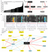
Figure 7. Regulatory Network Underlying Differences in Respiration
(A) Variation among male strains of BXD mice in three parameters of respiration: VO2, VCO2, and RER. Variation across and correlation with female strains is given in Figure S3. (B) QTL graphs of the three respiratory parameters, VO2 (blue), VCO2 (red), and RER (green). Significance is shown by the red horizontal line, suggestive by the blue line (LOD > 3.3 and 3.0, respectively). Candidate genes with established links to energy expenditure are listed below the locus (Aox1, Atp5l, Creb3, Ube2r2, Aqp3, Mrpl54, Pias4, Tab1, and Atf4). (C) Network graph showing all positional candidates with mRNA expression that correlates significantly with the phenotypes in whole-eye tissue (Geisert et al., 2009). (D) Expanded network graph using the same data set, including the top ten mRNA correlates of the phenotypes (light blue boxes). Interestingly, RER had the strongest top mRNA correlates (Acad9, Dusp10, and Adar), whereas Kif4 was the only significant correlate of all three parameters, and the only strong (|r| > 0.7) mRNA correlate to VO2 and none were observed for VCO2. Prkca and Mgat4a were shared significantly between two parameters. Despite the strong correlation between VO2 and VCO2, most top correlates were not shared. Bold dark blue lines represent −1 < r < −0.7, light blue lines −0.7 < r < −0.5, light orange lines 0.5 < r < 0.7 and bold red lines 0.7 < r < 1. Bar graphs are expressed as mean + SEM. Related to Figure S5.
Similar articles
- Echocardiography phenotyping in murine genetic reference population of BXD strains reveals significant QTLs associated with cardiac function and morphology.
Orgil BO, Xu F, Munkhsaikhan U, Alberson NR, Bajpai AK, Johnson JN, Sun Y, Towbin JA, Lu L, Purevjav E. Orgil BO, et al. Physiol Genomics. 2023 Feb 1;55(2):51-66. doi: 10.1152/physiolgenomics.00120.2022. Epub 2022 Dec 19. Physiol Genomics. 2023. PMID: 36534598 Free PMC article. - Using gene expression databases for classical trait QTL candidate gene discovery in the BXD recombinant inbred genetic reference population: mouse forebrain weight.
Lu L, Wei L, Peirce JL, Wang X, Zhou J, Homayouni R, Williams RW, Airey DC. Lu L, et al. BMC Genomics. 2008 Sep 25;9:444. doi: 10.1186/1471-2164-9-444. BMC Genomics. 2008. PMID: 18817551 Free PMC article. - Systems Genetics of Liver Fibrosis.
Hall RA, Lammert F. Hall RA, et al. Methods Mol Biol. 2017;1488:455-466. doi: 10.1007/978-1-4939-6427-7_21. Methods Mol Biol. 2017. PMID: 27933538 - Integrating genetic and gene expression data: application to cardiovascular and metabolic traits in mice.
Drake TA, Schadt EE, Lusis AJ. Drake TA, et al. Mamm Genome. 2006 Jun;17(6):466-79. doi: 10.1007/s00335-005-0175-z. Epub 2006 Jun 12. Mamm Genome. 2006. PMID: 16783628 Free PMC article. Review. - Dissecting quantitative traits in mice.
Mott R, Flint J. Mott R, et al. Annu Rev Genomics Hum Genet. 2013;14:421-39. doi: 10.1146/annurev-genom-091212-153419. Epub 2013 Jul 3. Annu Rev Genomics Hum Genet. 2013. PMID: 23834320 Review.
Cited by
- Two Conserved Histone Demethylases Regulate Mitochondrial Stress-Induced Longevity.
Merkwirth C, Jovaisaite V, Durieux J, Matilainen O, Jordan SD, Quiros PM, Steffen KK, Williams EG, Mouchiroud L, Tronnes SU, Murillo V, Wolff SC, Shaw RJ, Auwerx J, Dillin A. Merkwirth C, et al. Cell. 2016 May 19;165(5):1209-1223. doi: 10.1016/j.cell.2016.04.012. Epub 2016 Apr 28. Cell. 2016. PMID: 27133168 Free PMC article. - MPscore: A Novel Predictive and Prognostic Scoring for Progressive Meningioma.
Liu F, Qian J, Ma C. Liu F, et al. Cancers (Basel). 2021 Mar 5;13(5):1113. doi: 10.3390/cancers13051113. Cancers (Basel). 2021. PMID: 33807688 Free PMC article. - Mitonuclear protein imbalance as a conserved longevity mechanism.
Houtkooper RH, Mouchiroud L, Ryu D, Moullan N, Katsyuba E, Knott G, Williams RW, Auwerx J. Houtkooper RH, et al. Nature. 2013 May 23;497(7450):451-7. doi: 10.1038/nature12188. Nature. 2013. PMID: 23698443 Free PMC article. - Mitochondria: Masters of Epigenetics.
Tatar M, Sedivy JM. Tatar M, et al. Cell. 2016 May 19;165(5):1052-1054. doi: 10.1016/j.cell.2016.05.021. Cell. 2016. PMID: 27203109 Free PMC article. - Pleiotropic influence of DNA methylation QTLs on physiological and ageing traits.
Mozhui K, Kim H, Villani F, Haghani A, Sen S, Horvath S. Mozhui K, et al. Epigenetics. 2023 Dec;18(1):2252631. doi: 10.1080/15592294.2023.2252631. Epigenetics. 2023. PMID: 37691384 Free PMC article.
References
- Auwerx J. Improving metabolism by increasing energy expenditure. Nat. Med. 2006;12:44–45. - PubMed
Publication types
MeSH terms
Substances
Grants and funding
- P20-DA 21131/DA/NIDA NIH HHS/United States
- UO1AA13499/AA/NIAAA NIH HHS/United States
- U01 AA016662/AA/NIAAA NIH HHS/United States
- P20 DA021131/DA/NIDA NIH HHS/United States
- U01 AA017590/AA/NIAAA NIH HHS/United States
- 231138/ERC_/European Research Council/International
- U01 AA013499/AA/NIAAA NIH HHS/United States
- U01AA14425/AA/NIAAA NIH HHS/United States
- U01 AA014425/AA/NIAAA NIH HHS/United States
LinkOut - more resources
Full Text Sources
Medical
Molecular Biology Databases
Miscellaneous


