Real-time imaging of apoptotic cell-membrane changes at the single-cell level in the beating murine heart (original) (raw)
- On the Market
- Published: December 2001
- C.P.M. Reutelingsperger1,
- J.F.M. Smits1,
- M.J.A.P. Daemen1,
- P.A.F. Doevendans1,
- H.J.J. Wellens1 &
- …
- L. Hofstra1
Nature Medicine volume 7, pages 1352–1355 (2001)Cite this article
- 1150 Accesses
- 181 Citations
- 8 Altmetric
- Metrics details
An Erratum to this article was published on 01 January 2002
Abstract
We report a novel real-time imaging model to visualize apoptotic membrane changes of single cardiomyocytes in the injured heart of the living mouse, using fluorescent labeled annexin-V. Annexin-V binds to externalized phosphatidylserine (PS) of cells undergoing programmed cell death. With high-magnification (×100–160) real-time imaging, we visualized the binding of annexin-V to single cardiomyocytes. Kinetic studies at the single-cell level revealed that cardiomyocytes started to bind annexin-V within minutes after reperfusion, following an ischemic period of 30 minutes. The amount of bound annexin-V increased rapidly and reached a maximum within 20–25 minutes. Caspase inhibitors decreased the number of annexin-V–positive cardiomyocytes and slowed down the rate of PS exposure of cardiomyocytes that still bound annexin-V. This technology to study cell biology in the natural environment will enhance knowledge of intracellular signaling pathways relevant for cell-death regulation and strategies to manipulate these pathways for therapeutic effect.
This is a preview of subscription content, access via your institution
Relevant articles
Open Access articles citing this article.
Detection of apoptosis by [18F]ML-10 after cardiac ischemia–reperfusion injury in mice
- Maximilian Fischer
- , Mathias J. Zacherl
- … Andrei Todica
Annals of Nuclear Medicine Open Access 28 October 2022
Targeted optical fluorescence imaging: a meta-narrative review and future perspectives
- H. M. Schouw
- , L. A. Huisman
- … S. Kruijff
European Journal of Nuclear Medicine and Molecular Imaging Open Access 11 October 2021
Phosphatidylserine externalized on the colonic capillaries as a novel pharmacological target for IBD therapy
- Xuerui Zhang
- , Lulu Song
- … Zichun Hua
Signal Transduction and Targeted Therapy Open Access 16 June 2021
Access options
Subscribe to this journal
Receive 12 print issues and online access
$259.00 per year
only $21.58 per issue
Buy this article
- Purchase on SpringerLink
- Instant access to the full article PDF.
USD 39.95
Prices may be subject to local taxes which are calculated during checkout
Additional access options:
Figure 1: Experimental setup.
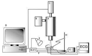
The alternative text for this image may have been generated using AI.
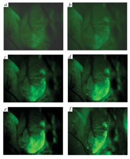
Figure 2: In vivo single-cell imaging.

The alternative text for this image may have been generated using AI.
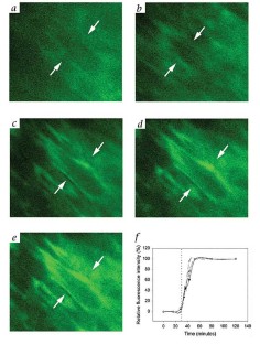
Figure 3: Kinetics of Anx-V-OG binding at the tissue level.
The alternative text for this image may have been generated using AI.
Figure 4: Kinetics of Anx-V-OG binding at the single-cell level.
The alternative text for this image may have been generated using AI.
Figure 5: Caspase inhibition decreases Anx-V-OG binding.
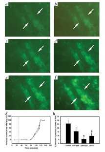
The alternative text for this image may have been generated using AI.
References
- Tempany, C.M. & McNeil, B.J. Advances in biomedical imaging. JAMA 285, 562–567 (2001).
Article CAS Google Scholar - Plymale, D.R., Haskins, J.R. & De La Iglesia, F.A. Monitoring simultaneous subcellular events in vitro by means of coherent multiprobe fluorescence. Nature Med. 5, 351–355 (1999).
Article CAS Google Scholar - Sweeney, T.J. et al. Visualizing the kinetics of tumor-cell clearance in living animals. Proc. Natl. Acad. Sci. USA 96, 12044–12049 (1999).
Article CAS Google Scholar - Yang, M. et al. Whole-body and intravital optical imaging of angiogenesis in orthotopically implanted tumors. Proc. Natl. Acad. Sci. USA 98, 2616–2621 (2001).
Article CAS Google Scholar - Dumont, E.A. et al. Cardiomyocyte death induced by myocardial ischemia and reperfusion: measurement with recombinant human annexin-V in a mouse model. Circulation 102, 1564–1568 (2000).
Article CAS Google Scholar - Koopman, G. et al. Annexin V for flow cytometric detection of phosphatidylserine expression on B cells undergoing apoptosis. Blood 84, 5–20 (1994).
Google Scholar - Martin, S.J. et al. Early redistribution of plasma membrane phosphatidylserine is a general feature of apoptosis regardless of the initiating stimulus: inhibition by overexpression of Bcl-2 and Abl. J. Exp. Med. 182, 552–556 (1995).
Article Google Scholar - Goldstein, J.C., Waterhouse, N.J., Juin, P., Evan, G.I. & Green, D.R. The coordinate release of cytochrome c during apoptosis is rapid, complete and kinetically invariant. Nature Cell Biol. 2, 156–162 (2000).
Article CAS Google Scholar - Xiang, J., Chao, D.T. & Korsmeyer, S.J. Bax-induced cell death may not require interleukin 1β converting enzyme-like proteases. Proc. Natl. Acad. Sci. USA 93, 14559–14563 (1996).
Article CAS Google Scholar - Bell, J.I. Clinical research is dead; long live clinical research. Nature Med. 5, 477–478 (1999).
Article CAS Google Scholar - Contag, P. R., Olomu, I. N., Stevenson, D. K. & Contag, C. H. Bioluminescent indicators in living mammals. Nature Med. 4, 245–247 (1998).
Article CAS Google Scholar
Acknowledgements
We thank N. Steinmetz and B. Armstrong for comments and suggestions. This study was supported from grants from the Dutch Heart Foundation (NHS 98.195 and NHS 2000-D035) and the Wynand-Pon Foundation. L.H. is a clinical research fellow for the Dutch Heart Foundation (NHS 2000-D035)
Author information
Authors and Affiliations
- Cardiovascular Research Institute Maastricht, Universiteitssingel 50, Maastricht, the Netherlands
E.A. Dumont, C.P.M. Reutelingsperger, J.F.M. Smits, M.J.A.P. Daemen, P.A.F. Doevendans, H.J.J. Wellens & L. Hofstra
Authors
- E.A. Dumont
- C.P.M. Reutelingsperger
- J.F.M. Smits
- M.J.A.P. Daemen
- P.A.F. Doevendans
- H.J.J. Wellens
- L. Hofstra
Corresponding author
Correspondence toE.A. Dumont.
Rights and permissions
About this article
Cite this article
Dumont, E., Reutelingsperger, C., Smits, J. et al. Real-time imaging of apoptotic cell-membrane changes at the single-cell level in the beating murine heart.Nat Med 7, 1352–1355 (2001). https://doi.org/10.1038/nm1201-1352
- Issue date: December 2001
- DOI: https://doi.org/10.1038/nm1201-1352
This article is cited by
Detection of apoptosis by [18F]ML-10 after cardiac ischemia–reperfusion injury in mice
- Maximilian Fischer
- Mathias J. Zacherl
- Andrei Todica
Annals of Nuclear Medicine (2023)
Phosphatidylserine externalized on the colonic capillaries as a novel pharmacological target for IBD therapy
- Xuerui Zhang
- Lulu Song
- Zichun Hua
Signal Transduction and Targeted Therapy (2021)
Targeted optical fluorescence imaging: a meta-narrative review and future perspectives
- H. M. Schouw
- L. A. Huisman
- S. Kruijff
European Journal of Nuclear Medicine and Molecular Imaging (2021)
Deep insights: intravital imaging with two-photon microscopy
- Ina Maria Schießl
- Hayo Castrop
Pflügers Archiv - European Journal of Physiology (2016)
Biomarkers in preclinical cancer imaging
- Monique R. Bernsen
- Klazina Kooiman
- Marion de Jong
European Journal of Nuclear Medicine and Molecular Imaging (2015)