Proteome-wide identification of ubiquitin interactions using UbIA-MS (original) (raw)
- Protocol
- Published: 15 February 2018
Nature Protocols volume 13, pages 530–550 (2018)Cite this article
- 11k Accesses
- 825 Citations
- 13 Altmetric
- Metrics details
Subjects
Abstract
Ubiquitin-binding proteins play an important role in eukaryotes by translating differently linked polyubiquitin chains into proper cellular responses. Current knowledge about ubiquitin-binding proteins and ubiquitin linkage-selective interactions is mostly based on case-by-case studies. We have recently reported a method called ubiquitin interactor affinity enrichment–mass spectrometry (UbIA-MS), which enables comprehensive identification of ubiquitin interactors for all ubiquitin linkages from crude cell lysates. One major strength of UbIA-MS is the fact that ubiquitin interactors are enriched from crude cell lysates, in which proteins are present at endogenous levels, contain biologically relevant post-translational modifications (PTMs) and are assembled in native protein complexes. In addition, UbIA-MS uses chemically synthesized nonhydrolyzable diubiquitin, which mimics native diubiquitin and is inert to cleavage by endogenous deubiquitinases (DUBs). Here, we present a detailed protocol for UbIA-MS that proceeds in five stages: (i) chemical synthesis of ubiquitin precursors and click chemistry for the generation of biotinylated nonhydrolyzable diubiquitin baits, (ii) in vitro affinity purification of ubiquitin interactors, (iii) on-bead interactor digestion, (iv) liquid chromatography (LC)–MS/MS analysis and (v) data analysis to identify differentially enriched proteins. The computational analysis tools are freely available as an open-source R software package, including a graphical interface. Typically, UbIA-MS allows the identification of dozens to hundreds of ubiquitin interactors from any type of cell lysate, and can be used to study cell type or stimulus-dependent ubiquitin interactions. The nonhydrolyzable diubiquitin synthesis can be completed in 3 weeks, followed by ubiquitin interactor enrichment and identification, which can be completed within another 2 weeks.
This is a preview of subscription content, access via your institution
Access options
Access Nature and 54 other Nature Portfolio journals
Get Nature+, our best-value online-access subscription
$32.99 / 30 days
cancel any time
Subscribe to this journal
Receive 12 print issues and online access
$259.00 per year
only $21.58 per issue
Buy this article
- Purchase on SpringerLink
- Instant access to the full article PDF.
USD 39.95
Prices may be subject to local taxes which are calculated during checkout
Additional access options:
Figure 1: Schematic overview of UbIA-MS workflow.
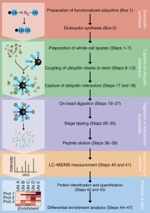
The alternative text for this image may have been generated using AI.
Figure 2: Schematic representation of the DEP analysis workflow.
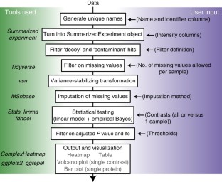
The alternative text for this image may have been generated using AI.
Figure 3: Interactors of different diubiquitin linkages in HeLa cells.
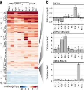
The alternative text for this image may have been generated using AI.
Figure 4: K48 diubiquitin interactors.
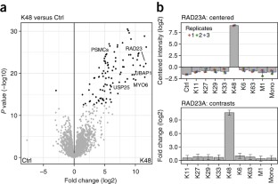
The alternative text for this image may have been generated using AI.
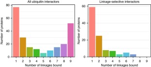
Figure 5: Distribution of the number of diubiquitin linkages bound by each interactor.
The alternative text for this image may have been generated using AI.
References
- Ciechanover, A. The unravelling of the ubiquitin system. Nat. Rev. Mol. Cell Biol. 16, 322–324 (2015).
Article CAS Google Scholar - Husnjak, K. & Dikic, I. Ubiquitin-binding proteins: decoders of ubiquitin-mediated cellular functions. Annu. Rev. Biochem. 81, 291–322 (2012).
Article CAS Google Scholar - Swatek, K.N. & Komander, D. Ubiquitin modifications. Cell Res. 26, 399–422 (2016).
Article CAS Google Scholar - Dikic, I., Wakatsuki, S. & Walters, K.J. Ubiquitin-binding domains — from structures to functions. Nat. Rev. Mol. Cell Biol. 10, 659–671 (2009).
Article CAS Google Scholar - Zhang, X. et al. An interaction landscape of ubiquitin signaling. Mol. Cell 65, 941–955.e8 (2017).
Article CAS Google Scholar - Rösner, D., Schneider, T., Schneider, D. & Scheffner, M. Click chemistry for targeted protein ubiquitylation and ubiquitin chain formation. Nat. Protoc. 10, 1594–1611 (2015).
Article Google Scholar - Chojnacki, M., Mansour, W., Hameed, D.S. & Singh, R.K. Polyubiquitin-photoactivatable crosslinking reagents for mapping ubiquitin interactome identify Rpn1 as a proteasome ubiquitin-associating subunit. Cell Chem. Biol. 24, 443–457.e6 (2017).
Article CAS Google Scholar - Hurley, J.H., Lee, S. & Prag, G. Ubiquitin-binding domains. Biochem. J. 399, 361–372 (2006).
Article CAS Google Scholar - Geiger, T., Wehner, A., Schaab, C., Cox, J. & Mann, M. Comparative proteomic analysis of eleven common cell lines reveals ubiquitous but varying expression of most proteins. Mol. Cell. Proteomics 11, M111.014050 (2012).
Article Google Scholar - Kumar, K.S.A., Spasser, L., Erlich, L.A., Bavikar, S.N. & Brik, A. Total chemical synthesis of di-ubiquitin chains. Angew. Chem. Int. Ed. Engl. 49, 9126–9131 (2010).
Article CAS Google Scholar - Weikart, N.D., Sommer, S. & Mootz, H.D. Click synthesis of ubiquitin dimer analogs to interrogate linkage-specific UBA domain binding. Chem. Commun. (Camb.) 48, 296–298 (2012).
Article CAS Google Scholar - El Oualid, F. et al. Chemical synthesis of ubiquitin, ubiquitin-based probes, and diubiquitin. Angew. Chem. Int. Ed. Engl. 49, 10149–10153 (2010).
Article CAS Google Scholar - Eger, S., Scheffner, M., Marx, A. & Rubini, M. Synthesis of defined ubiquitin dimers. J. Am. Chem. Soc. 132, 16337–16339 (2010).
Article CAS Google Scholar - Flierman, D. et al. Non-hydrolyzable diubiquitin probes reveal linkage-specific reactivity of deubiquitylating enzymes mediated by S2 pockets. Cell Chem. Biol. 23, 472–482 (2016).
Article CAS Google Scholar - Kimple, M.E. & Sondek, J. Overview of affinity tags for protein purification. Curr. Protoc. Protein Sci. Chapter 9 Unit 9.9 (2004).
- Nielsen, M.L. et al. Iodoacetamide-induced artifact mimics ubiquitination in mass spectrometry. Nat. Methods 5, 459–460 (2008).
Article CAS Google Scholar - Smits, A.H. & Vermeulen, M. Characterizing protein-protein interactions using mass spectrometry: challenges and opportunities. Trends Biotechnol. 34, 825–834 (2016).
Article CAS Google Scholar - Cox, J. et al. Accurate proteome-wide label-free quantification by delayed normalization and maximal peptide ratio extraction, termed MaxLFQ. Mol. Cell. Proteomics 13, 2513–2526 (2014).
Article CAS Google Scholar - Cox, J. et al. A practical guide to the MaxQuant computational platform for SILAC-based quantitative proteomics. Nat. Protoc. 4, 698–705 (2009).
Article CAS Google Scholar - Tyanova, S., Temu, T. & Cox, J. The MaxQuant computational platform for mass spectrometry-based shotgun proteomics. Nat. Protoc. 11, 2301–2319 (2016).
Article CAS Google Scholar - Huber, W. et al. Orchestrating high-throughput genomic analysis with Bioconductor. Nat. Methods 12, 115–121 (2015).
Article CAS Google Scholar - Huber, W., Von Heydebreck, A. & Sültmann, H. Variance stabilization applied to microarray data calibration and to the quantification of differential expression. Bioinformatics 18, S96–104 (2002).
Article Google Scholar - Gatto, L. & Lilley, K.S. MSnbase-an R/Bioconductor package for isobaric tagged mass spectrometry data visualization, processing and quantitation. Bioinformatics 28, 288–289 (2012).
Article CAS Google Scholar - Smyth, G.K. Linear models and empirical Bayes methods for assessing differential expression in microarray experiments. Stat. Appl. Genet. Mol. Biol. 3, 3 (2004).
Article Google Scholar - Ritchie, M.E. et al. limma powers differential expression analyses for RNA-sequencing and microarray studies. Nucleic Acids Res. 43, e47 (2015).
Article Google Scholar - Rappsilber, J., Mann, M. & Ishihama, Y. Protocol for micro-purification, enrichment, pre-fractionation and storage of peptides for proteomics using StageTips. Nat. Protoc. 2, 1896–1906 (2007).
Article CAS Google Scholar - Tammsalu, T. et al. Proteome-wide identification of SUMO modification sites by mass spectrometry. Nat. Protoc. 10, 1374–1388 (2015).
Article CAS Google Scholar - Oualid, F.E., Hameed, D.S. & Atmioui, D.E. Synthesis of atypical diubiquitin chains. Methods Mol. Biol. 832, 597–609 (2012).
Article Google Scholar - Valverde, I.E., Bauman, A. & Kluba, C.A. 1, 2, 3-Triazoles as amide bond mimics: triazole scan yields protease-resistant peptidomimetics for tumor targeting. Angew. Chem. Int. Ed. Engl. 52, 8957–8960 (2013).
Article CAS Google Scholar - Zhou, Z. & Fahrni, C.J. A fluorogenic probe for the copper(I)-catalyzed azide-alkyne ligation reaction: modulation of the fluorescence emission via 3(n,pi)-1(pi,pi) inversion. J. Am. Chem. Soc. 126, 8862–8863 (2004).
Article CAS Google Scholar
Acknowledgements
We thank B. Klaus for statistical advice and members of the Vermeulen and Ovaa labs for fruitful discussions. Work in the Zhang lab was supported by the “Hundred Talents Program C” of the Chinese Academy of Sciences (no. 2017-045) and by Guangdong Science and Technology Projects (2014B050504008, 2014B050502012, 2014B020225002 and 2014B030301058). Work in the Vermeulen lab was supported by the NWO Gravitation program CGC.nl. Work in the Ovaa lab was supported by the ERC grant Ubicode (no. 281699). A.H.S. was supported by a fellowship from the EMBL Interdisciplinary Postdoc (EIPOD) Programme under a grant from the Marie Sklodowska-Curie Actions COFUND (no. 664726).
Author information
Author notes
- Xiaofei Zhang, Arne H Smits and Gabrielle BA van Tilburg: These authors contributed equally to this work.
Authors and Affiliations
- CAS Key Laboratory of Regenerative Biology, Guangdong Provincial Key Laboratory of Stem Cells and Regenerative Medicine, South China Institute for Stem Cell Biology and Regenerative Medicine, Guangzhou Institutes of Biomedicine and Health, Chinese Academy of Sciences, Guangzhou, China
Xiaofei Zhang - Department of Molecular Biology, Faculty of Science, Radboud Institute for Molecular Life Sciences, Radboud University Nijmegen, Nijmegen, the Netherlands
Xiaofei Zhang & Michiel Vermeulen - Genome Biology Unit, European Molecular Biology Laboratory, Heidelberg, Germany
Arne H Smits & Wolfgang Huber - Department of Chemical Immunology, Leiden University Medical Center, Leiden, the Netherlands
Gabrielle BA van Tilburg & Huib Ovaa
Authors
- Xiaofei Zhang
- Arne H Smits
- Gabrielle BA van Tilburg
- Huib Ovaa
- Wolfgang Huber
- Michiel Vermeulen
Contributions
X.Z. and M.V. designed the method with input from A.H.S., G.B.A.v.T., W.H. and H.O. A.H.S. prepared figures, and developed and maintained the R package. X.Z., A.H.S., G.B.A.v.T. and M.V. wrote the manuscript with input from H.O. and W.H.
Corresponding authors
Correspondence toXiaofei Zhang, Arne H Smits or Michiel Vermeulen.
Ethics declarations
Competing interests
H.O. is a shareholder in the biotechnology company UbiQ. The other authors declare no competing financial interests.
Integrated supplementary information
Supplementary Figure 1 Quality-control plots.
(a) The number of proteins identified in every sample. Colors indicate the conditions and the solid line indicates the number of proteins identified in all samples (b) Stacked barplot to visualize the total number of proteins classified by the number of samples they are identified in (stacks and colors). (c) Barplot to visualize the number of proteins identified classified by the number of samples they are identified in (colors). (d) Boxplots of protein intensity distributions before (bottom) and after (top) normalization. (e) Heatmap of the proteins with missing values. Colors indicate whether the values are missing (white) or are valid (black).
Supplementary information
Rights and permissions
About this article
Cite this article
Zhang, X., Smits, A., van Tilburg, G. et al. Proteome-wide identification of ubiquitin interactions using UbIA-MS.Nat Protoc 13, 530–550 (2018). https://doi.org/10.1038/nprot.2017.147
- Published: 15 February 2018
- Version of record: 15 February 2018
- Issue date: March 2018
- DOI: https://doi.org/10.1038/nprot.2017.147
This article is cited by
Comments
Commenting on this article is now closed.
- Platform Support 20 February 2018, 16:47
Hi