Central role of E3 ubiquitin ligase MG53 in insulin resistance and metabolic disorders (original) (raw)
- Letter
- Published: 27 January 2013
- Wei Peng1 na1,
- Yan Zhang1 na1,
- Fengxiang Lv1,
- Hong-Kun Wu1,
- Jiaojiao Guo1,
- Yongxing Cao3,
- Yanbin Pi3,
- Xin Zhang3,
- Li Jin1,
- Mao Zhang1,
- Peng Jiang1,
- Fenghua Liu1,
- Shaoshuai Meng1,
- Xiuqin Zhang1,
- Ping Jiang1,
- Chun-Mei Cao1 &
- …
- Rui-Ping Xiao1,4
Nature volume 494, pages 375–379 (2013) Cite this article
- 31k Accesses
- 285 Citations
- 29 Altmetric
- Metrics details
Subjects
Abstract
Insulin resistance is a fundamental pathogenic factor present in various metabolic disorders including obesity and type 2 diabetes1. Although skeletal muscle accounts for 70–90% of insulin-stimulated glucose disposal2,3, the mechanism underlying muscle insulin resistance is poorly understood. Here we show in mice that muscle-specific mitsugumin 53 (MG53; also called TRIM72) mediates the degradation of the insulin receptor and insulin receptor substrate 1 (IRS1), and when upregulated, causes metabolic syndrome featuring insulin resistance, obesity, hypertension and dyslipidaemia. MG53 expression is markedly elevated in models of insulin resistance, and MG53 overexpression suffices to trigger muscle insulin resistance and metabolic syndrome sequentially. Conversely, ablation of MG53 prevents diet-induced metabolic syndrome by preserving the insulin receptor, IRS1 and insulin signalling integrity. Mechanistically, MG53 acts as an E3 ligase targeting the insulin receptor and IRS1 for ubiquitin-dependent degradation, comprising a central mechanism controlling insulin signal strength in skeletal muscle. These findings define MG53 as a novel therapeutic target for treating metabolic disorders and associated cardiovascular complications.
This is a preview of subscription content, access via your institution
Access options
Subscribe to this journal
Receive 52 print issues and online access
$199.00 per year
only $3.83 per issue
Buy this article
- Purchase on SpringerLink
- Instant access to the full article PDF.
USD 39.95
Prices may be subject to local taxes which are calculated during checkout
Additional access options:
Figure 1: MG53 ablation protects mice against diet-induced metabolic syndrome.

The alternative text for this image may have been generated using AI.
Figure 2: MG53 ablation blocks diet-induced systemic insulin resistance.

The alternative text for this image may have been generated using AI.
Figure 3: Overexpression of MG53 triggers systemic insulin resistance and metabolic syndrome.
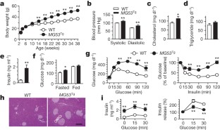
The alternative text for this image may have been generated using AI.
Figure 4: Regulation of muscle insulin signalling by MG53.

The alternative text for this image may have been generated using AI.
Figure 5: MG53 E3 ligase activates ubiquitination of the insulin receptor and IRS1.
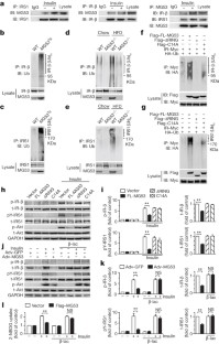
The alternative text for this image may have been generated using AI.
References
- Eckel, R. H., Grundy, S. M. & Zimmet, P. Z. The metabolic syndrome. Lancet 365, 1415–1428 (2005)
Article CAS Google Scholar - DeFronzo, R. A. et al. The effect of insulin on the disposal of intravenous glucose. Results from indirect calorimetry and hepatic and femoral venous catheterization. Diabetes 30, 1000–1007 (1981)
Article CAS Google Scholar - Shulman, G. I. et al. Quantitation of muscle glycogen synthesis in normal subjects and subjects with non-insulin-dependent diabetes by 13C nuclear magnetic resonance spectroscopy. N. Engl. J. Med. 322, 223–228 (1990)
Article CAS Google Scholar - McMillen, I. C. & Robinson, J. S. Developmental origins of the metabolic syndrome: prediction, plasticity, and programming. Physiol. Rev. 85, 571–633 (2005)
Article CAS Google Scholar - Grundy, S. M. Metabolic syndrome: connecting and reconciling cardiovascular and diabetes worlds. J. Am. Coll. Cardiol. 47, 1093–1100 (2006)
Article CAS Google Scholar - DeFronzo, R. A. & Tripathy, D. Skeletal muscle insulin resistance is the primary defect in type 2 diabetes. Diabetes Care 32 (Suppl. 2). S157–S163 (2009)
Article CAS Google Scholar - Lillioja, S. et al. Insulin resistance and insulin secretory dysfunction as precursors of non-insulin-dependent diabetes mellitus. Prospective studies of Pima Indians. N. Engl. J. Med. 329, 1988–1992 (1993)
Article CAS Google Scholar - Martin, B. C. et al. Role of glucose and insulin resistance in development of type 2 diabetes mellitus: results of a 25-year follow-up study. Lancet 340, 925–929 (1992)
Article CAS Google Scholar - Cai, C. et al. MG53 nucleates assembly of cell membrane repair machinery. Nature Cell Biol. 11, 56–64 (2009)
Article CAS Google Scholar - Cao, C. M. et al. MG53 constitutes a primary determinant of cardiac ischemic preconditioning. Circulation 121, 2565–2574 (2010)
Article CAS Google Scholar - Fu, J. et al. Oleylethanolamide regulates feeding and body weight through activation of the nuclear receptor PPAR-α. Nature 425, 90–93 (2003)
Article ADS CAS Google Scholar - Rao, R. H. Insulin resistance in spontaneously hypertensive rats. Difference in interpretation based on insulin infusion rate or on plasma insulin in glucose clamp studies. Diabetes 42, 1364–1371 (1993)
Article CAS Google Scholar - Zhang, X. et al. Rhesus macaques develop metabolic syndrome with reversible vascular dysfunction responsive to pioglitazone. Circulation 124, 77–86 (2011)
Article CAS Google Scholar - Coleman, D. L. Obese and diabetes: two mutant genes causing diabetes-obesity syndromes in mice. Diabetologia 14, 141–148 (1978)
Article CAS Google Scholar - Saltiel, A. R. & Kahn, C. R. Insulin signalling and the regulation of glucose and lipid metabolism. Nature 414, 799–806 (2001)
Article ADS CAS Google Scholar - Thirone, A. C., Huang, C. & Klip, A. Tissue-specific roles of IRS proteins in insulin signaling and glucose transport. Trends Endocrinol. Metab. 17, 72–78 (2006)
Article Google Scholar - Kerouz, N. J., Horsch, D., Pons, S. & Kahn, C. R. Differential regulation of insulin receptor substrates-1 and -2 (IRS-1 and IRS-2) and phosphatidylinositol 3-kinase isoforms in liver and muscle of the obese diabetic (ob/ob) mouse. J. Clin. Invest. 100, 3164–3172 (1997)
Article CAS Google Scholar - Goodyear, L. J. et al. Insulin receptor phosphorylation, insulin receptor substrate-1 phosphorylation, and phosphatidylinositol 3-kinase activity are decreased in intact skeletal muscle strips from obese subjects. J. Clin. Invest. 95, 2195–2204 (1995)
Article CAS Google Scholar - Olefsky, J., Bacon, V. C. & Baur, S. Insulin receptors of skeletal muscle: specific insulin binding sites and demonstration of decreased numbers of sites in obese rats. Metabolism 25, 179–191 (1976)
Article CAS Google Scholar - Capozza, F. et al. Caveolin-3 knockout mice show increased adiposity and whole body insulin resistance, with ligand-induced insulin receptor instability in skeletal muscle. Am. J. Physiol. Cell Physiol. 288, C1317–C1331 (2005)
Article CAS Google Scholar - Zisman, A. et al. Targeted disruption of the glucose transporter 4 selectively in muscle causes insulin resistance and glucose intolerance. Nature Med. 6, 924–928 (2000)
Article CAS Google Scholar - Brüning, J. C. et al. A muscle-specific insulin receptor knockout exhibits features of the metabolic syndrome of NIDDM without altering glucose tolerance. Mol. Cell 2, 559–569 (1998)
Article Google Scholar - Laustsen, P. G. et al. Essential role of insulin and insulin-like growth factor 1 receptor signaling in cardiac development and function. Mol. Cell. Biol. 27, 1649–1664 (2007)
Article CAS Google Scholar - Nakao, R. et al. Ubiquitin ligase Cbl-b is a negative regulator for insulin-like growth factor 1 signaling during muscle atrophy caused by unloading. Mol. Cell. Biol. 29, 4798–4811 (2009)
Article CAS Google Scholar - Ramos, F. J., Langlais, P. R., Hu, D., Dong, L. Q. & Liu, F. Grb10 mediates insulin-stimulated degradation of the insulin receptor: a mechanism of negative regulation. Am. J. Physiol. Endocrinol. Metab. 290, E1262–E1266 (2006)
Article CAS Google Scholar - Rui, L., Yuan, M., Frantz, D., Shoelson, S. & White, M. F. SOCS-1 and SOCS-3 block insulin signaling by ubiquitin-mediated degradation of IRS1 and IRS2. J. Biol. Chem. 277, 42394–42398 (2002)
Article CAS Google Scholar - Xu, X. et al. The CUL7 E3 ubiquitin ligase targets insulin receptor substrate 1 for ubiquitin-dependent degradation. Mol. Cell 30, 403–414 (2008)
Article CAS Google Scholar - Shi, J., Luo, L., Eash, J., Ibebunjo, C. & Glass, D. J. The SCF-Fbxo40 complex induces IRS1 ubiquitination in skeletal muscle, limiting IGF1 signaling. Dev. Cell 21, 835–847 (2011)
Article Google Scholar - Joazeiro, C. A. et al. The tyrosine kinase negative regulator c-Cbl as a RING-type, E2-dependent ubiquitin-protein ligase. Science 286, 309–312 (1999)
Article CAS Google Scholar - Huang, H. et al. Profiling of mismatch discrimination in RNAi enabled rational design of allele-specific siRNAs. Nucleic Acids Res. 37, 7560–7569 (2009)
Article CAS Google Scholar - Zhong, L. et al. The histone deacetylase Sirt6 regulates glucose homeostasis via Hif1α. Cell 140, 280–293 (2010)
Article CAS Google Scholar
Acknowledgements
We thank H. P. Cheng, G. Feng, X. Fu and L. P. Wei for discussions, and S. L. Guo, T. Zhang, X. H. Wang, D. Y. Chen, J. Y. Peng, L. Huang, W. Q. Zhang, N. Hou, L. Pan, L. Chen and Y. L. Liu for their technical support. Special thanks to H. Takeshima and J. J. Ma for their support in providing _MG53_−/− mice. This work was supported by the National Basic Research Program of China (2012CB518000, 2013CB531200, 2012CB944501) and the National Natural Science Foundation of China (81070674, 81070116, 3/22/002 and 81130073).
Author information
Author notes
- Ruisheng Song, Wei Peng and Yan Zhang: These authors contributed equally to this work.
Authors and Affiliations
- Institute of Molecular Medicine, State Key Laboratory of Biomembrane and Membrane Biotechnology, Peking University, Beijing 100871, China
Ruisheng Song, Wei Peng, Yan Zhang, Fengxiang Lv, Hong-Kun Wu, Jiaojiao Guo, Li Jin, Mao Zhang, Peng Jiang, Fenghua Liu, Shaoshuai Meng, Xiuqin Zhang, Ping Jiang, Chun-Mei Cao & Rui-Ping Xiao - Institute of Cardiovascular Sciences, Health Science Center, Peking University, Beijing 100083, China
Ruisheng Song - Institute of Sports Medicine, Peking University Third Hospital, Beijing 100191, China
Yongxing Cao, Yanbin Pi & Xin Zhang - Center for Life Sciences, Peking University, Beijing 100871, China
Rui-Ping Xiao
Authors
- Ruisheng Song
- Wei Peng
- Yan Zhang
- Fengxiang Lv
- Hong-Kun Wu
- Jiaojiao Guo
- Yongxing Cao
- Yanbin Pi
- Xin Zhang
- Li Jin
- Mao Zhang
- Peng Jiang
- Fenghua Liu
- Shaoshuai Meng
- Xiuqin Zhang
- Ping Jiang
- Chun-Mei Cao
- Rui-Ping Xiao
Contributions
R.S., W.P. and Y.Z. are equally contributing first authors. R.S. generated the initial idea and conducted key experiments. R.S., W.P., Y.Z., C.-M.C. and R.-P.X. designed the study, analysed the data and wrote the manuscript. C.-M.C. and R.-P.X. interpreted significance of the study. R.S., W.P., Y.Z., F. Lv, H.-K.W., J.G., Y.C., Y.P., Xin Z., L.J., M.Z., Pe.J., F. Liu and S.M. performed the experiments. Pi.J. helped in the generation of MG53 transgenic mice. Xiu.Z. provided the nonhuman primate tissues.
Corresponding authors
Correspondence toChun-Mei Cao or Rui-Ping Xiao.
Ethics declarations
Competing interests
The authors declare no competing financial interests.
Supplementary information
PowerPoint slides
Rights and permissions
About this article
Cite this article
Song, R., Peng, W., Zhang, Y. et al. Central role of E3 ubiquitin ligase MG53 in insulin resistance and metabolic disorders.Nature 494, 375–379 (2013). https://doi.org/10.1038/nature11834
- Received: 19 October 2010
- Accepted: 11 December 2012
- Published: 27 January 2013
- Issue date: 21 February 2013
- DOI: https://doi.org/10.1038/nature11834
This article is cited by
Editorial Summary
Muscle enzyme MG53 as drug target
This paper reports the surprising finding that dysregulation of the muscle-specific E3 ligase mitsugumin (MG53) causes insulin resistance and metabolic disorders in mice. When MG53 is upregulated metabolic syndrome ensues; removal of MG53 leaves insulin signalling intact, and prevents diet-induced metabolic syndrome. This work identifies MG53 as a promising therapeutic target for the treatment of metabolic diseases such as type 2 diabetes and associated cardiovascular complications.