Caspase-9 is activated in a cytochrome c-independent manner early during TNFα-induced apoptosis in murine cells (original) (raw)
Introduction
Apoptosis is a tightly regulated process important for differentiation, for the regulation of cell numbers, and for the removal of aged, damaged and autoreactive cells.1,2 A variety of extracellular and intracellular signals can trigger an apoptotic response, including growth factor deprivation, overexpression of oncogenes and tumor suppressor genes, radiation, chemotherapeutic drugs, and crosslinking of receptors such as the Fas or tumor necrosis factor (TNF) receptor. Apoptotic triggers activate intracellular response pathways that lead to the controlled activation of cysteine proteases known as caspases.3 Two major pathways of caspase activation have been described – one involving the release of multiple polypeptides from mitochondria as a result of its destabilization and the other involving cell surface ‘death’ receptor activation by ligand binding.4
Human cells have been classified as type I or type II based on their responsiveness to activation of the TNF family of death receptors, particularly Fas ligand-mediated death.5 Ligand binding causes trimerization of these receptors on the cell surface and recruitment of cytoplasmic adaptor proteins. These, in turn, recruit procaspase-8 molecules that self-process into their active forms.6,7 Once activated, caspase-8 can trigger two distinct pathways of apoptosis. In type I cells, the release of large amounts of activated caspase-8 from the death-inducing signaling complex (DISC), enables direct activation of downstream caspases such as caspase-3, leading to cell death. In contrast, in type II cells DISC formation is greatly reduced, but the small amounts of active caspase-8 molecules that are generated are sufficient to induce mitochondrial apoptogenic activity. Caspase-8 cleaves the Bcl-2 family protein, Bid, to generate a C-terminal fragment that then translocates to the mitochondria causing its disruption, and triggering the release of cytochrome c (cyt c).8,9,10 A cytoplasmic multiprotein complex, comprising Apaf-1, cyt c and the ‘initiator’ caspase, caspase-9, then activates a series of ‘effector’ caspases beginning with caspase-3 and culminating in the death of the cell.11,12 In cells of type II, such as Jurkat, the proapoptotic effects of activated Bid on the mitochondria can be effectively inhibited in the presence of high levels of Bcl-2 or Bcl-xL proteins.5,13 Murine hepatocytes appear to exhibit properties of type II cells,14 but some hematopoietic cell lines of murine origin have eluded categorization as type I or type II in terms of their responsiveness to Fas and/or TNF-receptor activation. For instance, IL-3-dependent murine pro-B FL5.12 cells recruit small amounts of procaspase-8 to the DISC and clearly utilize Bid cleavage as the primary mechanism of amplification15 (AK, unpublished). Despite this, however, overexpressed Bcl-xL cannot protect FL5.12 cells against TNF-receptor-mediated death. It was of interest, therefore, to understand how amplification of the apoptotic process occurred in these and other murine cell types in response to TNF-receptor crosslinking.
Release of mitochondrial cyt c into the cytoplasm and its subsequent association with the Apaf-1 protein is thought to be an absolute requirement for the activation of caspase-9, the apical caspase in the mitochondrial pathway of apoptosis.12 In the presence of ATP/dATP, cyt c that has been released into the cytosol binds to and triggers the oligomerization of the cytosolic Apaf-1 protein. The resultant complex recruits multiple copies of procaspase-9 leading to its activation.12 Although autoprocessing occurs rapidly, unprocessed forms of caspase-9 are also catalytically active as part of the caspase-9–Apaf-1 holoenzyme complex.16,17 A purified truncated form of the Apaf-1 protein lacking the WD-repeats and cyt c binding site was shown in vitro to be constitutively active in its ability to induce self-processing of procaspase-9.18 However, two recent studies have indicated that caspase-9 can also be activated by mechanisms involving neither cyt c release nor Apaf-1 activation.19,20 One study showed that, in response to endoplasmic reticular (ER) stress, caspase-12 was able to process and activate caspase-9, while another indicated that virus infection could trigger a novel, yet unknown, pathway of caspase-9 cleavage.
The primary objective of the present study was to determine the basis for the inability of overexpressed Bcl-xL to protect FL5.12 pro-B cells from launching a rapid apoptotic response to TNF-α/cycloheximide (CHX) treatment, when the mitochondrial pathway (via Bid cleavage) and cyt c release appeared to be the preferred route to apoptotic death in untransfected and vector-transfected controls.
Results
Bcl-xL does not protect murine pro-B cells from TNF_α_-induced death
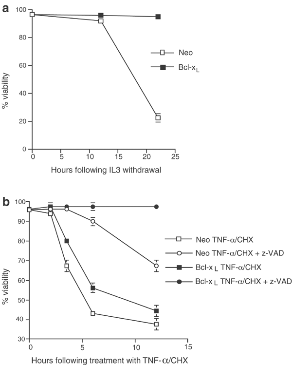
Interleukin-3 (IL-3)-dependent murine FL5.12 pro-B lymphoma cells expressing high levels of transfected Bcl-xL or the empty Neo vector were either subjected to a growth factor withdrawal assay or treated with TNF-α in the presence of CHX (described in Materials and Methods). Figure 1a and b shows that FL5.12 cells overexpressing Bcl-xL are protected against growth factor withdrawal-induced death, as assayed by propidium iodide (PI) exclusion and flow cytometry, for as long as 96 h (22-h experiment shown in Figure 1a), but not against apoptosis caused by treatment with TNF-α and CHX. However, TNF-α/CHX-induced death in both control and Bcl-xL cells could be inhibited in the presence of a broad-spectrum caspase inhibitor, z-VAD-fmk. The inhibition in control cells was manifest as a delayed death response, while the Bcl-xL cells exhibited almost total resistance to apoptosis for at least 24 h (a 12-h experiment is shown in Figure 1b).
Figure 1
Bcl-xL protects murine FL5.12 pro-B cells against growth factor withdrawal, not against death receptor activation. FL5.12 cells transfected with either pSFFV–Bcl-xL or with the empty vector, pSFFV–Neo, were cultured in IL-3-free medium for 22h (a) or incubated with TNF-α (5 ng/ml) and CHX (20 _μ_g/ml) in the presence or absence of 50 _μ_M of the caspase inhibitor z-VAD-fmk for 12 h (b). Viability (mean and standard deviations, _n_=3) of the transfected clones under conditions of IL3 deprivation was determined by PI exclusion and FACS analysis at the indicated time points
Bcl-xL delays the release of mitochondrial cyt c in response to TNF-receptor activation
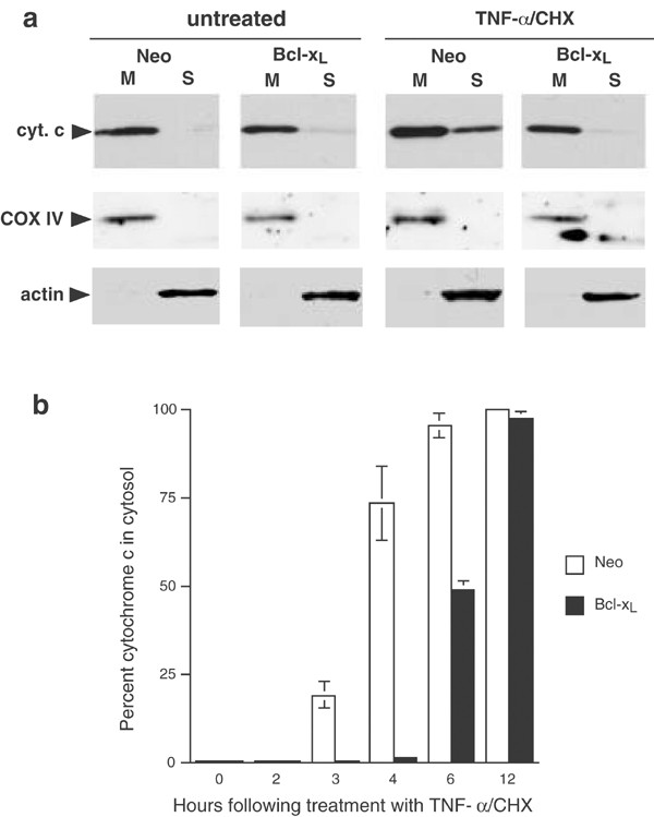
Since Bcl-xL has been shown to be effective at maintaining mitochondrial integrity and preventing cyt c release during growth factor withdrawal,21 we determined the cellular distribution of cyt c in control and Bcl-xL-expressing FL5.12 cells exposed to TNF-α/CHX for a few hours. The Western blot in Figure 2a (top panel) shows that 3 h after induction, when apoptotic cells are easily detectable by flow cytometry (Figure 1b), mitochondria from Bcl-xL-expressing FL5.12 cells still retain cyt c. Shown in the center and bottom panels are cytochrome oxidase subunit IV (COX IV) and actin Western blots, controls for the mitochondrial and cytosolic fractions, respectively. Bcl-xL protein levels, as detected by Western blotting, did not decrease for at least 4 h following treatment with either TNF-α/CHX or CHX alone (not shown). The cyt c release observed in the FL5.12 Neo cells is the result of caspase-8-mediated cleavage and activation of the Bcl-2 family BH3 protein, Bid, in response to TNF-receptor crosslinking.15 We were able to confirm the cleavage of Bid in death receptor-activated control and Bcl-xL-expressing cells by Western blotting using antibodies against both full-length and cleaved mouse Bid protein (not shown). However, while Johnson et al.15 demonstrate no release of cyt c from mitochondria of FL5.12 Bcl-xL cells as late as 6 h after induction of apoptosis, our data consistently show 30–50% loss of the protein from mitochondria in Bcl-xL cells by this time point and almost total release 12 h post-treatment. The data are summarized in Figure 2b. It may be noted that the Neo- and Bcl-xL-expressing lines used in our experiments were clonal isolates. Additionally, the Bcl-xL lines were selected for high levels of expression of the transfected plasmid.22 Under conditions of IL-3 withdrawal, these cell lines show mitochondrial retention of cyt c for at least 72 h postdeprivation (not shown). Since FL5.12 control cells primarily adopt the (type II) cyt _c_-dependent caspase-9 pathway in the amplification of downstream apoptotic cascades in response to TNF-α, it was important to determine the mechanism underlying the early and rapid apoptosis of the Bcl-xL-expressing cells in the absence of cyt c release.
Figure 2
Bcl-xL prevents early mitochondrial cytochrome c release in FL5.12 cells in response to TNF-α/CHX treatment. (a) Western blot of fractionated FL5.12 cells showing the distribution of cyt c. Untreated Neo and Bcl-xL FL5.12 cells, and cells treated with TNF-α/CHX for 3 h were fractionated into mitochondrial (M) and cytoplasmic, S100 (S), fractions, and immunoblotted with a monoclonal antibody against cyt c, COX IV and actin (see Materials and Methods). (b) Bar graph showing the ratio of cytosolic cyt c to total cyt c in Neo and Bcl-xL FL5.12 cells 0, 2, 3, 4, 6 and 12 h following apoptotic induction with TNF-α/CHX (mean and standard deviations, _n_=3). Fractionated cell extracts were immunoblotted with antibody against cyt c and the chemiluminescent cyt c bands, visualized by autoradiography, were later quantified by densitometry using a BioRadGS-363 Molecular Imager
Caspase-9 is processed prior to mitochondrial cyt c release in response to TNF-receptor occupation in Bcl-xL-expressing FL5.12 cells
Next, we examined the processing of caspase-9 and -3, the two major proteases associated with mitochondrial and nonmitochondrial pathways downstream of activated caspase-8, under conditions of TNF-receptor crosslinking. Figure 3 shows lysates of FL5.12 control and Bcl-xL cells at 0, 2, 3, 4 and 6 h after treatment with TNF-α/CHX, immunoblotted with antibodies against caspase-9 (upper panels) or a cleaved (active) form of caspase-3 (lower panels). Caspase-9 is being processed within the first 2 h of induction in both the cell lines. We have consistently observed the early cleavage of caspase-9 in response to TNF-α; the results shown in Figure 3 are representative of six different experiments. A number of groups have already established that caspase-9 requires cyt c release and recruitment into the apoptosome to autoprocess and become activated.12,23 However, data in Figure 2 showing effective inhibition of cyt c release by overexpressed Bcl-xL for three or more hours after induction of apoptosis through the TNF receptor (Figure 2), taken together with results in Figure 3, strongly indicate that caspase-9 is processed prior to mitochondrial cyt c release in the FL5.12 cells. The cleaved caspase-3 (Figure 3, lower panels) detected within the first 3 h after induction probably results from caspase-8 processing activity. It may be noted that levels of this cleaved, presumably active, form increase noticeably at later time points in both vector and Bcl-xL-expressing cells, and this was confirmed using a colorimetric caspase-3 activity detection kit (R&D Systems, not shown). Additionally, it may be noted that the 38 kDa processed product of caspase-9 consistently appeared at least as early after TNF treatment as the 40 kDa cleavage product expected from caspase-3 processing activity (Figure 3, upper panels). It was important to determine whether the observed early processing of caspase-9 was leading to its activation or was merely a consequence of the degradative activity associated with apoptotic progression.
Figure 3
Caspase-9 processing in FL5.12 Bcl-xL cells occurs within 2 h of apoptotic induction with TNF-α/CHX. Autoradiographs of lysates from Neo and Bcl-xL FL5.12 cells treated with TNF-α/CHX for 0, 2, 3, 4, 6 and 12 h immunoblotted with antibody against caspase-9 (top panels) or a cleaved form of caspase-3 (bottom panels)
Early caspase-9 activity contributes to the rapid amplification of apoptosis in Bcl-xL-expressing FL5.12 cells in response to TNF-α
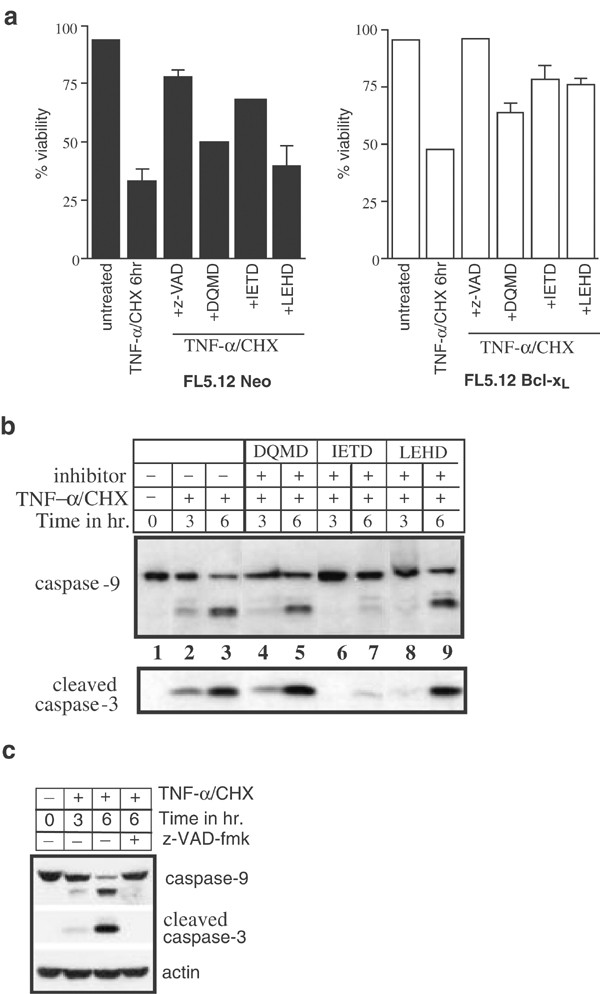
We predicted that if early caspase-9 activation was critical for the rapid and early amplification of this apoptotic pathway, inhibiting caspase-9 activity would delay apoptosis. To test this, FL5.12 cells were treated with TNF-α and CHX in the presence of either the pan-caspase inhibitor z-VAD-fmk, or specific caspase inhibitors. Shown in Figure 4a are bar graphs depicting the viability of FL5.12 cells at 6 h following TNF-α treatment, as assayed by flow cytometric analysis of Annexin V and PI uptake. As expected, z-VAD-fmk blocked apoptosis of the Bcl-xL cells while providing only partial protection to the controls. It has previously been shown that 50 _μ_M z-VAD-fmk is unable to block cyt c release in FL5.12 Neo cells15 (and AK, unpublished). The caspase-3-specific inhibitor, z-DQMD-fmk, imparted 30–50% protection over the TNF-α/CHX controls in both the cell lines. It is known that related effector caspases, such as caspase-7, can effectively replace active caspase-324,25 in processing downstream targets, and this may be the reason for the lower levels of protection observed in the presence of z-DQMD-fmk. The caspase-8 inhibitor, z-IETD-fmk, was more effective in inhibiting apoptosis in both the cell lines. Figure 4a also shows that z-LEHD-fmk, an inhibitor of caspase-9 activity, delayed apoptosis almost as efficiently in the Bcl-xL cells as did z-IETD (right panel), suggesting that caspase-9 proteolytic activity plays an important role in this death pathway even in the absence of cyt c release. The low level of inhibition observed in control FL5.12 cells (Figure 4a, left panel) in the presence of the caspase-9 inhibitor can be attributed to the other proapoptotic compounds, such as Smac/DIABLO, AIF, and even preprocessed caspase-3, that are released from the mitochondrial intermembrane space along with cyt c. Although, IETD and LEHD were able to delay the onset of apoptosis more effectively than the caspase-3 inhibitor, only the broad-spectrum inhibitor z-VAD could protect the Bcl-xL cells against TNF-induced death for over 24 h (not shown). Figure 4b is a Western blot of FL5.12 Bcl-xL cell lysates showing caspase-9 and -3 processing, 3 and 6 h following TNF-α/CHX treatment in the presence or absence of caspase inhibitors. While the caspase-8 inhibitor, IETD, effectively prevented processing of both caspase-9 and -3 (lanes 6 and 7), the caspase-3 inhibitor (lanes 4 and 5) did not affect the processing of either of these caspases. Figure 4c (top panel) represents a caspase-9 immunoblot of lyates from untreated FL5.12 Bcl-xL cells, and cells treated with TNF-α/CHX in the presence of 50 _μ_M z-VAD. The membrane was sequentially stripped and reprobed with antibodies against active caspase-3 and actin (center and bottom panels, respectively). As expected, based on the viability assays shown in Figure 4a (right panel), both caspase-9 and -3 processing were completely inhibited.
Figure 4
Early caspase-9 processing contributes to the rapid amplification of apoptosis observed in Bcl-xL-expressing FL5.12 cells in response to TNF-α/CHX. (a) FL5.12 cells were incubated with 100 _μ_M concentrations of specific cell-permeable caspase inhibitors z-DQMD-fmk (caspase-3), z-IETD-fmk (caspase-8) or z-LEHD-fmk (caspase-9) and 50 _μ_M of the pancaspase inhibitor z-VAD-fmk for 30 min before being treated with TNF-α and CHX. Viability of cells was determined at specific intervals by flow cytometric analysis Annexin V–FITC labeling and PI uptake (see Materials and Methods). Figure represents percent viability of untreated FL5.12 Neo (left panel) and Bcl-xL (right panel) cells, and cells treated with TNF-α/CHX for 6 h in the absence or presence of caspase inhibitors (mean and standard deviations, _n_=3). (b) Western blots of lysates from 0, 3 and 6 h-treated FL5.12 Bcl-xL cells from the experiment in (4a). Top panel shows an autoradiograph of 25 _μ_g total protein immunoblotted with antibody against caspase-9, and bottom panel represents the same Western blot, stripped and reprobed with an antibody against the cleaved (active) form of caspase-3. (c) Caspase-9 immunoblot of z-VAD-fmk-incubated Bcl-xL cell lysates (top panel) from the above experiment, stripped and reprobed with active caspase-3 (center panel) and actin (bottom panel) antibodies
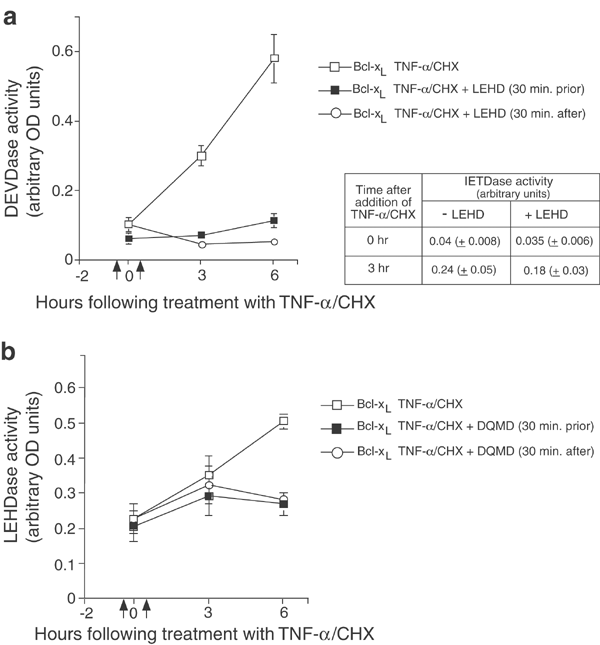
Although caspase-9 inhibition effectively blocked the processing of caspase-3 at early times, by the end of 6 h levels of cleaved caspase-3 appeared to have been restored to those observed in the controls (Figure 4b, lower panel, lanes 8 and 9). To resolve this issue, DEVDase activity was measured in TNF-treated FL5.12 Bcl-xL cells in the presence of LEHD added 30 min before or after the addition of inducer (Figure 5a). Results indicated that caspase-3 activity was effectively blocked at the earliest time point in both the instances. Activity remained low in both samples at the end of 6 h, and consistently showed steady increases in DEVDase levels thereafter (not shown). If caspase-3 was being directly inhibited by LEHD rather than through inhibition of caspase-9, we should also detect caspase-8 inhibition, since caspase-8 and -9 are more closely related in terms of their substrate specificities.26 However, IETDase activity measured at 0 and 3 h in the FL5.12 cells treated with LEHD prior to apoptosis induction, indicated that caspase-8 was not significantly inhibited at the 3 h time point (Table inset, Figure 5a). The 25% reduction observed in IETDase activity is probably responsible for part of the decrease in caspase-3 activity, but we believe that these data indicate that a bulk of the early inhibition of caspase-3 activity results from inhibition of caspase-9 function.
Figure 5
Inhibition of caspase-9 inhibits early DEVDase activity, but inhibition of caspase-3 does not affect early LEHDase activity in FL5.12 Bcl-xL cells treated with TNF-α/CHX. Cells were incubated with 100 _μ_M z-LEHD-fmk (a) or z-DQMD-fmk (b) 30 min prior to, or 30 min. following (shown by arrows) the addition of TNF-α/CHX, and DEVDase (a) or LEHDase (b) activity measured at the indicated time points using colorimetric assays (R&D Systems). Table inset in (a) shows IETDase activity measured at 0 and 3 h in the FL5.12 cells treated with z-LEHD-fmk prior to apoptosis induction
Western blots of caspase-9 in Figure 4b (upper panel, lanes 4 and 5) showed no effect of DQMD, the caspase-3-specific synthetic inhibitor, on caspase-9 processing. Colorimetric caspase-9 activity assays in the presence and absence of DQMD (Figure 5b) showed that caspase-3 inhibition did not block early caspase-9 activation, but inhibited late LEHDase activity. These results confirm that caspase-9 is activated within 3 h following TNF-receptor crosslinking and suggest, furthermore, that active caspase-3 is not the protease responsible for the early activation.
RNA interference-mediated downregulation of endogenous caspase-9 protects FL5.12. Bcl-xL cells from TNF-receptor-activated death
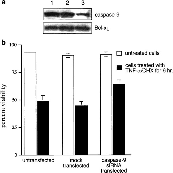
Although data in Figure 4 were suggestive of caspase-9 participation in early amplification of apoptosis in FL5.12 Bcl-xL cells, it was important to confirm this by methods that did not involve the use of synthetic caspase inhibitors. For an alternative approach, FL5.12 Bcl-xL cells were transiently transfected with siRNA directed against nucleotides 289–309 of the murine caspase-9 (mC-9) RNA sequence (see Materials and Methods). Figure 6a shows a caspase-9 immunoblot of lysates 48 h following transfection from parental, mock transfected and siRNA-transfected FL5.12 Bcl-xL cells (upper panel). The membrane was stripped and reprobed with an antibody against Bcl-xL to confirm even loading of lysates (lower panel). The decrease in caspase-9 protein expression was determined by densitometry to be roughly 50%. Figure 6b shows the results of viability assays carried out on control and transfected populations treated with TNF-α and CHX for 6 h. While the mean viability exhibited by the mock-transfected cells did not differ from the 48% viability observed in the untransfected parental population, caspase-9 siRNA-transfected cells were 64% viable at the end of 6 h. The observed delay in apoptosis in the presence of reduced levels of caspase-9 supports earlier results (see Figures 4 and 5) that caspase-9 function is involved in accelerating TNF-activated apoptosis in cells in which the mitochondrial cyt c release pathway is effectively blocked.
Figure 6
Downregulation of caspase-9 levels by RNA interference partially protects FL5.12 Bcl-xL cells against TNF-receptor-activated apoptosis. FL5.12 Bcl-xL cells were either mock transfected or transfected with mC-9 siRNA (100 nM) using siPORT Amine (Ambion) as transfection agent. (a) Western blot of lysates from parental FL5.12 Bcl-xL cells (lane 1), mock-transfected FL5.12 Bcl-xL cells (lane 2) and cells transfected with caspase-9 siRNA (lane 3). Upper panel represents an immunoblot of caspase-9 and lower panel shows the same blot stripped and reprobed with an antibody against the Bcl-xL protein. (b) Untransfected, mock-transfected and caspase-9 siRNA-transfected FL5.12 Bcl-xL cells were treated with TNF-α and CHX and viability of cells was determined by flow cytometric analysis of Annexin V–FITC labeling and PI uptake. Bar graphs represent percent viability of unstimulated and 6 h-stimulated cell populations (_n_=3)
Early caspase-9 processing occurs in cells lacking the Apaf-1 protein
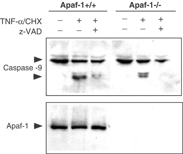
Having determined that the early proteolytic activity of caspase-9 was playing a role in the amplification of apoptosis in the Bcl-xL-transfected cells, we proceeded to determine the source of this early activation. It was unlikely that active caspase-3 molecules resulting from the proteolytic activity of caspase-8 were acting on procaspase-9 to process and activate it (Figures 4 and 5). It was possible that a constitutively active form of Apaf-1 was being generated in response to receptor occupation that could bypass the requirement for cyt c association in order to activate the autoprocessing of procaspase-9 molecules. We surmised that if Apaf-1 was required for the early activation of caspase-9 in the TNF-α pathway, then early processing of caspase-9 should not be detectable in Apaf-1-deficient cells exposed to TNF. Additionally, by using Apaf-1−/− cells, we would ensure that no activation of caspase-9 could occur via the cyt c release pathway, while eliminating the need for overexpressing Bcl-xL to block this pathway. Figure 7 shows Western blots of lysates from Apaf−/− and control mouse embryo fibroblasts (MEFs) that had been treated with TNF-α/CHX in the presence or absence of z-VAD-fmk, the broad-spectrum caspase inhibitor. Upper panel in Figure 7 shows that caspase-9 is processed in both types of MEFs within 3 h following exposure to TNF-α and CHX. Although, z-VAD completely inhibits this processing in Apaf-1−/− cells, control MEFs continue to activate caspase-9 at least partially via the mitochondrial pathway. The Western blot was stripped and reprobed with an antibody against Apaf-1 (Figure 7, lower panel). These data suggest that the early cyt _c_-independent processing of caspase-9 in the TNF pathway in these cells did not result from Apaf-1 activity.
Figure 7
Early caspase-9 processing in murine fibroblasts following exposure to TNF-α/CHX is dependent on caspase activity and not on the Apaf-1 protein. The figure shows autoradiographs of Western blots from lysates of Apaf+/+ and Apaf−/− MEFs that had been treated with TNF-α/CHX for 3 h in the presence or absence of the caspase inhibitor z-VAD-fmk. Upper panel shows caspase-9 processing, and lower panel shows the same Western blot, stripped and reprobed with an antibody against Apaf-1 (see Materials and Methods)
Murine caspase-9 is processed by active caspase-8 in an in vitro cleavage assay
Yet another possibility was that active caspase-8 molecules were themselves directly acting on procaspase-9 to process and activate it. Although caspase-8 had been shown to activate caspase-9 indirectly in type II cells via cleavage of the Bid protein and the subsequent release of mitochondrial cyt c, to date there has been no evidence of direct processing of caspase-9 in vivo by this initiator caspase.
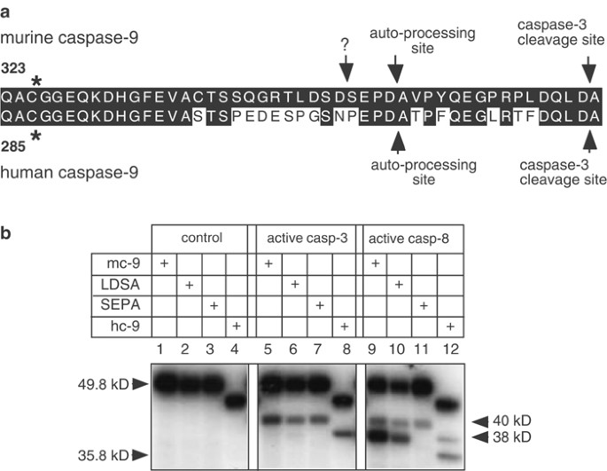
The autoprocessed form of caspase-9 is 38 kDa (see Figure 3, upper panels), but autoprocessing of the latter has only been shown to occur in vivo by ‘induced proximity’ in the context of apoptosome formation in reponse to cyt c release.23,27 The autoprocessing motif, SEPD (residues 350–353), in mC-9 is preceded directly by LDSD (346–349), both putative caspase-8 cleavage consensi26 (see Figure 8a). The LDSD motif is absent in the human caspase-9 (hC-9) protein.
Figure 8
Active recombinant caspase-8 cleaves murine caspase-9 at SEPD, the site of caspase-9 autoprocessing. (a) Representation of the amino-acid sequence between the active site, QACGG (*), and the caspase-3 processing site (marked by arrows) in mC-9 and hC-9. Conserved residues are shaded in black. Additional arrows depict the caspase-9 autoprocessing sites, SEPD (mouse), PEPD (human) and a putative caspase-8 cleavage consensus motif, LDSD (?) present only in the murine protein. (b) In vitro synthesized, radiolabeled mC-9, LDSA, SEPA or hC-9 proteins were synthesized using the TNT transcription/translation system (Promega) and incubated with 15 mU/_μ_l of active human recombinant caspase-3 (lanes 5–8), or 150 mU/_μ_l of active caspase-8 (lanes 9–12) at 37°C for 60 min. Lanes 1–4 show the ‘input’ or corresponding labeled translation products that went into each reaction. Cleavage reactions were resolved by SDS-PAGE, and the gels fixed, dried and autoradiographed
Full-length murine procaspase-9 was cloned from mouse brain cDNA and tagged with a myc epitope at the C-terminus. Two point mutants of murine caspase-9, LDSA and SEPA, were also generated in which residues D349 and D353, respectively, were replaced with alanine. Radiolabeled in vitro translated murine caspase-9, LDSA and SEPA, as well as in vitro translated human wild-type protein were incubated either with active recombinant caspase-3 or -8 at 37°C for 1h. Figure 8b shows an autoradiograph of SDS-PAGE analysis of the reactions. Active caspase-3 (lanes 5–8) processed both the human and murine proteins generating cleavage products of the expected size; 40 kDa for murine caspase-9 and 37 kDa for the human protein. The panel on the right (lanes 9–12) shows the results of active caspase-8 processing activity on caspase-9 and the two point mutants. A major processed fragment of 38 kDa and a minor 40 kDa product resulted from cleavage of wild-type mC-9 as well as the LDSA mutant. The 38 kDa fragment generated by caspase-8 cleavage activity is absent in the cleavage reaction with the SEPA mutant (lane 11) indicating that it was the SEPD, and not the LDSD, motif that served as a cleavage site for active caspase-8. Two cleavage products of approximately 37 and 35 kDa, resulting from processing at the DQLD and PEPD motifs, were also observed in the cleavage reaction with human caspase-9 (lane 12), however, processing at PEPD by active caspase-8 was consistently weaker than at the corresponding (SEPD) motif in the murine protein (lane 9). Srinivasula et al.28 have demonstrated the cleavage of purified recombinant hC-9 by active caspase-8, and their data indicate that although hC-9 is processed at both sites, the DQLD motif is the preferred site of cleavage. Thus, mC-9 serves as a direct processing substrate for active caspase-8, and may be one of the first caspases to be activated in the TNF pathway of apoptosis.
Discussion
Human cells have been classified as cells of type I or type II based on the nature of their response to death receptor activation by ligands such as Fas and TNF-α.5 In cells of type I death receptor ligation leads to vigorous activation of caspase-8, followed by the direct cleavage and activation of procaspase-3.5,7 DISC assembly in type II cells, on the other hand, is both delayed and subdued and the resulting autoprocessed caspase-8 insufficient for the direct activation of caspase-3. Caspase-8 can still cleave the Bcl-2 family protein, Bid, however, to generate a truncated active form of the latter that translocates to the mitochondria and promotes the release of mitochondrial cyt c into the cytosol.8,9,10 Most activated effector caspases have the ability to cleave other caspases, including initiator caspases, but such processing activity is unregulated and primarily associated with the degradative phase of the apoptotic process. Caspase-2, -6 and -9, for instance, are efficiently processed by activated caspase-3 during the rapid cell death observed in type I cells in response to Fas ligand.24,29 In these cells, caspase-9 functions not as an initiator, but as one more target of an effector caspase. Caspase-9 activation follows that of caspase-3 in such a pathway. Survival members of the Bcl-2 family, by virtue of their ability to prevent cyt c release, therefore, impart almost total protection against apoptosis induced via Fas receptor crosslinking to type II cells. The murine FL5.12 cell line in the present study exhibits both type I and type II characteristics. Preventing cyt c release by overexpressing an antiapoptotic Bcl-2 family protein, does not protect FL5.12 cells from death receptor activation-induced apoptosis15 (and Figure 1). Given the close conservation of specific death pathways among humans and mice, as well as the high degree of homology shared by components of different apoptotic pathways among the two species, it was important to investigate further the biochemical basis for this difference in responsiveness.
Apoptosis is discernible by flow cytometry within 2 h of addition of TNF-α/CHX, even as overexpressed Bcl-xL is able to prevent mitochondrial cyt c release for at least 4 h in FL5.12 cells. However, while Johnson et al.15 observed no cyt c release for 6 h in response to TNF-α/CHX, our studies show that Bcl-xL FL5.12 cells release almost 50% of their cyt c into the cytoplasm by the end of 6 h under the same conditions (Figure 2b). The FL5.12 Bcl-xL lines used in the present study have been selected for high-level expression of transfected Bcl-xL22 and have consistently exhibited this delayed release of cyt c in response to TNF-α, while protecting mitochondria for over 72 h following growth factor (IL-3) withdrawal. We believe that the positive feedback of the cytosolic caspase activity on the mitochondria causes the release of cyt c as a late event during the apoptotic process.30 This feedback loop could be manifest in a number of ways, including cleavage of the membrane-inserted survival-promoting proteins themselves.31,32
We show that caspase-9 is processed within 2 h of activation of the death receptor pathway in Bcl-xL-expressing FL5.12 cells. Additionally, using specific inhibitors we observe that caspase-9 activity is essential for the early amplification of apoptosis, under these circumstances. This processing of caspase-9 proceeds in the absence of mitochondrial cyt c release and occurs too early to be the result of a feedback loop involving active caspase-3.24 Furthermore, the Western blots in Figure 3 (lower panels) show relatively low amounts of cleaved/active caspase-3 resulting from caspase-8 processing activity at 2 and 3 h as compared to the cleaved form detected at later time points. Additionally, the first detectable processing intermediate is the 38 kDa product, and not the 40 kDa band expected from caspase-3 cleavage activity. The processing of procaspase-9 by active caspase-3 (at residue D368), known to occur during amplification of caspase cascades, generates a 40 kDa cleavage product in the in vitro cleavage assays (shown in Figure 8b), and confirms this. The inhibitor studies further confirm that active caspase-3 is not the protease responsible for the observed early caspase-9 processing (Figures 4b and 5b). Procaspase-9 has been shown to be cleaved as cells undergo apoptosis; however, such cleavage occurs late during apoptotic progression and is thought to be involved in the turnover of the protease rather than in its enzymatic activation.17,33
Apaf-1 oligomerization in the presence of cyt c and dATP has been shown to be the primary cause of the autoprocessing that results in the activation of caspase-9.12,23,34. Although the full-length Apaf-1 molecule requires cyt c for oligomerization, a truncated form of Apaf-1 lacking the WD-40 repeat region has been shown to be constitutively active in vitro.18 We were able to detect truncated forms of Apaf-1 in vivo in response to TNF-α/CHX in the FL5.12 cells (not shown), but these truncated forms appeared 3–4 h after the initiation of the signal, too late to initiate caspase-9 autoprocessing and were more likely the result of caspase-3 processing activity.35,36 Furthermore, we could demonstrate that caspase-9 cleavage occurs within 3 h of TNF-receptor occupation in Apaf-1-deficient MEFs (Figure 7).
Caspase activity determinations in both FL5.12 Bcl-xL cells and Apaf-1−/− MEFs, using colorimetric substrates, demonstrated that the processed caspase-9 protein had caspase activity (Figure 5b, and not shown). Additionally, FL5.12 Bcl-xL cells expressing half as much caspase-9 protein as the parental controls, showed 30% increased viability when exposed to TNF-α/CHX for 6 h (Figure 6). The partial protection imparted by downregulated caspase-9 is strong indication that the activity of this caspase contributes to the early, rapid apoptosis observed in the parental cells (Figure 1b).
The 38 kDa processing intermediate of caspase-9 appears to be a product of caspase-8 cleavage activity. In vitro cleavage assays, using active recombinant caspase-8, confirm that the 38 kDa band is a major processing product and is a result of cleavage at the SEPD (D353) and not the LDSD motif (D349) on murine caspase-9 (Figure 6). Furthermore, the early appearance of the shorter processing intermediate, in TNF-treated FL5.12 cells, supports the possibility that caspase-9 is a direct processing substrate for active caspase-8 in this pathway of apoptosis. In vitro cleavage data demonstrate that human caspase-9 is also cleaved, albeit less efficiently than its murine counterpart, suggesting that the SEPD motif serves as a better substrate for active caspase-8 than does the PEPD site (Figure 6a). Previously published studies demonstrating the processing of purified recombinant hC-9 by active caspase-828, indicate that DQLD (D330), rather than PEPD (D315), is the preferred site of cleavage activity. This difference in susceptibility between hC-9 and mC-9 to processing by active caspase-8 may help explain the difference in response to death receptor activation between human cells of type I and murine FL5.12 cells. Additional evidence that hC-9 and mC-9 are differently regulated comes from a study showing that the Akt phosphorylation site (located at Serine196) presumed to be involved in the inactivation of hC-9 is absent from its murine counterpart.37,38
In mitochondrial pathways of apoptosis, the zymogen form of caspase-9 is rapidly recruited into the apoptosome following the release of cyt c.39 The active form of caspase-9 is believed to be the Apaf-1-bound holoenzyme. Since procaspase-9 is not normally detected in the apoptosome, it is thought to undergo rapid autoprocessing. However, studies with noncleavable mutants have shown that the unprocessed caspase can also associate with oligomerized Apaf-1 and rapidly recruit and activate caspase-3.16,17 It is unlikely that a holoenzyme is involved in the processing and activation of caspase-9 in response to TNF-α in the FL5.12 cells. Our data suggest, instead, that caspase-9 is functioning not as an initiator, but as an effector caspase and as a direct substrate for active caspase-8 early after apoptotic stimulation. The possibility that the murine caspase-9 cleavage site is modified in healthy, proliferating cells in a manner similar to the Bid protein is an attractive one and currently under investigation.
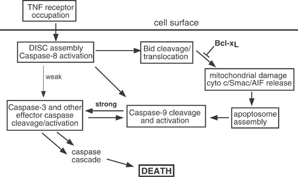
We propose the following model to explain our observations. In addition to cleaving the Bcl-2 protein Bid and small amounts of procaspase-3, active caspase-8 molecules in FL5.12 cells cleave and activate procaspase-9 in response to TNF-receptor crosslinking (see Figure 9). The cleaved Bid (tBid) promotes the release of cyt c, causing rapid amplification of apoptosis via apoptosome formation and caspase-9 activation.8,9,10 In the absence of cyt c release, however, caspase-9 that has been processed directly by caspase-8 cleaves and activates caspase-3, which, in turn, processes more caspase-9 in a positive feedback amplification loop. It may be argued that a cell overexpressing Bcl-xL is not a ‘normal’ cell and cannot, therefore, serve as a model for understanding mechanisms of apoptosis. We believe we have identified a novel pathway of amplification of the caspase cascade; in order to appreciate the cyt c -independent nature of the process, it is important to use a cell line in which cyt c release can be prevented or delayed. Although we have investigated this pathway in a murine pro-B system, similar mechanisms are undoubtedly utilized in other murine cell types. Apaf-1-deficient fibroblasts offer insight into alternate pathways since cyt _c_-induced activation is also effectively shut off in these cells. It is also relevant to note that a number of cancers resistant to apoptotic induction overexpress antiapoptotic Bcl-2 proteins,40 and activating the apoptotic process in such cells by alternative means can be an important goal in cancer therapy.
Figure 9
Simplified model of the apoptotic pathways activated following TNF receptor occupation in murine cells
A recent study showed that caspase-12 was able to process and activate caspase-9 in response to ER stress,20 while another indicated that Sendai virus infection could trigger a novel, yet unidentified, pathway of caspase-9 cleavage.19 We have presented evidence that implicates caspase-8 in the processing and activation of caspase-9 in death receptor-activated pathways. All three studies clearly indicate that caspase-9 can also be activated by mechanisms involving neither cyt c release nor Apaf-1 activation. In this capacity, the activated protease probably contributes to the amplification of death pathways that have already been initiated. A cell programmed to die utilizes all the means within its control to die a quick death. It stands to reason, therefore, that proteins, such as caspase-9, functioning at critical points of apoptotic pathways have the ability to amplify the apoptotic process by more than just one mechanism to ensure the cell's early and unobtrusive demise.
Materials and Methods
Cell lines, antibodies and plasmid constructs
FL5.12 murine pro-B lymphoma cells were cultured as described earlier.41 For plasmid transfections, 1 × 10⁷ FL5.12 cells were electroporated at 960 _μ_F and 250 V with 10 _μ_g of plasmids, pSFFV-Neo or pSFFV-Bcl-xL, described previously.42 Neomycin-resistant cells were selected in medium containing 1 mg/ml G418. Single cell clones were obtained from transfectant pools by limiting dilution cloning in 96-well microtiter plates. Apaf-1 knockout and control primary embryo fibroblasts (a gift from Scott Lowe, Cold Spring Harbor) were grown in Dulbecco's modified Eagle's medium supplemented with 10% fetal calf serum, 2 mM glutamine, 100 U/ml penicillin, 100 _μ_g/ml streptomycin, and 10 mM _β_-mercaptoethanol. Active recombinant caspase-3 was from Pharmingen and active caspase-8 from BioVision, Inc.
Caspase-9 monoclonal antibody, cleaved caspase-3 polyclonal antibody and the Apaf-1 monoclonal antibody were purchased from Stressgen Biotechnologies, Cell Signaling Technology, and Chemicon International, respectively. The cyt c antibody was a gift from R Jemmerson (University of Minnesota, USA). The COX IV antibody was purchased from Molecular Probes and the actin antibody was obtained from Oncogene. The hC-9 construct was a gift from E Alnemri (Thomas Jefferson University). Full-length mC-9 cDNA was synthesized from mouse brain RNA by PCR using specific oligos, and cloned into the _Bam_HI and _Xho_I sites of the pCDNA 3.0 vector (Invitrogen). The myc-tagged construct, mC-9myc, was generated by subcloning mC-9 into _Bam_HI and _Xho_I sites of a pCmyc vector (gift from G Nunez, University of Michigan, USA). The two point mutants of mC-9myc (LDSA and SEPA) converting D349 or D353, respectively, to alanine, were generated by a two-step PCR method.
Cyt c release studies
Mitochondrial and cytosolic (S100) fractions were prepared by resuspending 1 × 10⁷ FL5.12 cells in 0.8 ml ice-cold buffer A (250 mM sucrose, 20 mM HEPES, 10 mM KCl, 1.5 mM EDTA, 1 mM EGTA, 1 mM DTT, 17 _μ_g/ml aprotinin, 2 _μ_g/ml leupeptin (pH 7.4)). Cells were homogenized using a prechilled cylinder cell homogenizer (H&Y Enterprise Redwood City, CA, USA), following which the unlysed cells and nuclei were pelleted at 750 × g for 25 min. The pellet, representing the mitochondrial fraction was resuspended in buffer A, and the supernatant subjected to further centrifugation at 100 000 × g for 1 h. The supernatant from the final centrifugation represented the cytosolic fraction. Equivalent amounts of mitochondrial and cytosolic (S100) fractions were then Western blotted as previously described, first with an antibody against cyt c (gift from R Jemmerson) followed by an antibody against COX IV (Molecular Probes).
Cell viability and caspase activity assays
Viability of FL5.12 cells was measured at 2 or 3 h intervals over a period 24 h following the addition of TNF-α (5 ng/ml) and CHX (20 _μ_g/ml) to the medium, or at 24 h intervals for 4 or 5 days following withdrawal of IL-3 from the medium.41 For the caspase inhibitor studies, FL5.12 cells were incubated with z-DQMD-fmk, z-IETD-fmk or z-LEHD-fmk at 100 _μ_M concentration or z-VAD-fmk (50 _μ_M) for 30 min prior to the addition of TNF-α and CHX. For the IL-3 studies, cells were washed three times in growth medium lacking IL-3 and resuspended in this medium at a concentration of 5 × 10⁵ cells/ml. Aliquots were withdrawn at specified time intervals, and viability determined by flow cytometry using PI exclusion alone or a combination of Annexin V–FITC uptake and PI exclusion using protocol suggested by BioVision, Inc. DEVDase, IETDase and LEHDase activities in cell lysates (from 2 × 10⁶ cells per data point) were determined using colorimetric assay kits (R&D Systems) according to the manufacturer's protocol.
Western blotting
FL5.12 cells were pelleted, washed in phosphate-buffered saline, and then lysed in RIPA buffer containing 1% Nonidet P-40, 1% deoxycholate and 0.1% SDS, supplemented with protease inhibitor cocktail (Calbiochem). Adherent MEFs were detached from culture plates with Accutase (Innovative Cell Technologies, Inc.) and pelleted, together with predetached floaters, as described above. The lysed cells were centrifuged at 14 000 × g to remove cellular debris. Protein concentrations of extracts were determined by the colorimetric bicinchoninic acid method (Pierce Chemical Company). Equal amounts of protein were electrophoretically separated in 14% SDS-PAGE and transferred to nitrocellulose. Membrane blocking, washing, primary and secondary antibody incubations and chemiluminescence reactions were carried out according to the Amersham ECL protocol. Antibody dilutions were carried out as suggested on the data sheet provided by the manufacturing company. Blots were stripped for reuse by washing for 30 min to 2 h in TBS-T buffer (pH 3.0) at room temperature.
siRNA construction and transfection
siRNAs were generated according to the protocol accompanying the Silencer siRNA kit (Ambion, Inc.). Briefly, sense and antisense oligos were synthesized to target a 21-nucleotide mC-9 sequence (AAGCAGGATCCAGAGGCTGTT-3′), with an additional 8-nucleotide leader sequence complementary to a T7 promoter primer. Each oligo was hybridized to the T7 promoter primer, filled in with Klenow, and used as a template for transcription by T7 polymerase. Sense and antisense in vitro transcripts were hybridized, following which overhanging leader sequences and residual DNA templates were removed by treatment with RNase and Dnase, respectively. Finally, the double-stranded RNAs were purified by passing through a filter cartridge and eluted in nuclease-free water. FL5.12 cells were either mock-transfected or transfected with the siRNA (100 nM) in the presence of transfection agent siPORT Amine (Ambion, Inc.) for 6 h. A fraction of the transfected population was lysed and immunoblotted with caspase-9 antibodies, and the remaining cells were used for the viability studies described above.
In vitro caspase cleavage assays
Radiolabeled mC-9-myc protein, LDSA, or SEPA proteins were synthesized in vitro using the TNT transcription/translation system (Promega) and 20 _μ_Ci [35S]methionine (Amersham/Pharmacia) per reaction. Active human recombinant caspase-3 (15 mU/_μ_l), or -8 (150 mU/_μ_l), was used to cleave the in vitro translated proteins at 37°C for 60 min using 5 _μ_l in vitro translation mix as substrate in a 25 _μ_l total reaction volume. Reactions were stopped with an equal volume of 2 × Laemmli sample buffer containing reducing agent. The resulting cleavage products were separated by SDS-PAGE, and the gels were fixed, dried and autoradiographed.
Abbreviations
TNF-α:
tumor necrosis factor-alpha
CHX:
cycloheximide
cyt c:
cytochrome c
DISC:
death-inducing signaling complex
References
- Ellis RE, Yuan JY and Horvitz HR (1991) Mechanisms and functions of cell death. Annu. Rev. Cell Biol. 7: 663–698
Article CAS PubMed Google Scholar - Wyllie AH, Kerr JF and Currie AR (1980) Cell death: the significance of apoptosis. Int. Rev. Cytol. 68: 251–306
Article CAS PubMed Google Scholar - Thornberry NA and Lazebnik Y (1998) Caspases: enemies within. Science 281: 1312–1316
Article CAS PubMed Google Scholar - Budihardjo I, Oliver H, Lutter M, Luo X and Wang X (1999) Biochemical pathways of caspase activation during apoptosis. Annu. Rev. Cell Dev. Biol. 15: 269–290
Article CAS PubMed Google Scholar - Scaffidi C, Fulda S, Srinivasan A, Friesen C, Li F, Tomaselli KJ, Debatin KM, Krammer PH and Peter ME (1998) Two CD95 (APO-1/Fas) signaling pathways. EMBO J. 17: 1675–1687
Article CAS PubMed PubMed Central Google Scholar - Medema JP, Scaffidi C, Kischkel FC, Shevchenko A, Mann M, Krammer PH and Peter ME (1997) FLICE is activated by association with the CD95 death-inducing signaling complex (DISC). EMBO J. 16: 2794–2804
Article CAS PubMed PubMed Central Google Scholar - Muzio M, Stockwell BR, Stennicke HR, Salvesen GS and Dixit VM (1998) An induced proximity model for caspase-8 activation. J. Biol. Chem. 273: 2926–2930
Article CAS PubMed Google Scholar - Li H, Zhu H, Xu CJ and Yuan J (1998) Cleavage of BID by caspase 8 mediates the mitochondrial damage in the Fas pathway of apoptosis. Cell 94: 491–501
Article CAS PubMed Google Scholar - Luo X, Budihardjo I, Zou H, Slaughter C and Wang X (1998) Bid, a Bcl2 interacting protein, mediates cytochrome c release from mitochondria in response to activation of cell surface death receptors. Cell 94: 481–490
Article CAS PubMed Google Scholar - Wei MC, Lindsten T, Mootha VK, Weiler S, Gross A, Ashiya M, Thompson CB and Korsmeyer SJ (2000) tBID, a membrane-targeted death ligand, oligomerizes BAK to release cytochrome c (in process citation). Genes Dev. 14: 2060–2071
CAS PubMed PubMed Central Google Scholar - Zou H, Henzel WJ, Liu X, Lutschg A and Wang X (1997) Apaf-1, a human protein homologous to C. elegans CED-4, participates in cytochrome c-dependent activation of caspase-3 (see comments). Cell 90: 405–413
Article CAS PubMed Google Scholar - Zou H, Li Y, Liu X and Wang X (1999) An APAF-1 cytochrome c multimeric complex is a functional apoptosome that activates procaspase-9. J. Biol. Chem. 274: 11549–11556
Article CAS PubMed Google Scholar - Susin SA, Zamzami N, Castedo M, Hirsch T, Marchetti P, Macho A, Daugas E, Geuskens M and Kroemer G (1996) Bcl-2 inhibits the mitochondrial release of an apoptogenic protease. J. Exp. Med. 184: 1331–1341
Article CAS PubMed Google Scholar - Yin XM, Wang K, Gross A, Zhao Y, Zinkel S, Klocke B, Roth KA and Korsmeyer SJ (1999) Bid-deficient mice are resistant to Fas-induced hepatocellular apoptosis. Nature 400: 886–891
Article CAS PubMed Google Scholar - Johnson BW, Cepero E and Boise LH (2000) Bcl-xL inhibits cytochrome c release but not mitochondrial depolarization during the activation of multiple death pathways by tumor necrosis factor-α. J. Biol. Chem. 275: 31546–31553
Article CAS PubMed Google Scholar - Bratton SB, Walker G, Srinivasula SM, Sun XM, Butterworth M, Alnemri ES and Cohen GM (2001) Recruitment, activation and retention of caspases-9 and -3 by Apaf-1 apoptosome and associated XIAP complexes. EMBO J. 20: 998–1009
Article CAS PubMed PubMed Central Google Scholar - Stennicke HR, Deveraux QL, Humke EW, Reed JC, Dixit VM and Salvesen GS (1999) Caspase-9 can be activated without proteolytic processing. J. Biol. Chem. 274: 8359–8362
Article CAS PubMed Google Scholar - Adrain C, Slee EA, Harte MT and Martin SJ (1999) Regulation of apoptotic protease activating factor-1 oligomerization and apoptosis by the WD-40 repeat region. J. Biol. Chem. 274: 20855–20860
Article CAS PubMed Google Scholar - Bitzer M, Armeanu S, Prinz F, Ungerechts G, Wybranietz W, Spiegel M, Bernlohr C, Cecconi F, Gregor M, Neubert WJ, Schulze-Osthoff K and Lauer UM (2002) Caspase-8 and Apaf-1-independent caspase-9 activation in Sendai virus- infected cells. J. Biol. Chem. 277: 29817–29824
Article CAS PubMed Google Scholar - Morishima N, Nakanishi K, Takenouchi H, Shibata T and Yasuhiko Y (2002) An endoplasmic reticulum stress-specific caspase cascade in apoptosis. Cytochrome _c_-independent activation of caspase-9 by caspase-12. J. Biol. Chem. 277: 34287–34294
Article CAS PubMed Google Scholar - Vander Heiden MG, Chandel NS, Williamson EK, Schumacker PT and Thompson CB (1997) Bcl-xL regulates the membrane potential and volume homeostasis of mitochondria (see comments). Cell 91: 627–637
Article CAS PubMed Google Scholar - Rathmell JC, Vander Heiden MG, Harris MH, Frauwirth KA and Thompson CB (2000) In the absence of extrinsic signals, nutrient utilization by lymphocytes is insufficient to maintain either cell size or viability. Mol. Cell 6: 683–692
Article CAS PubMed Google Scholar - Li P, Nijhawan D, Budihardjo I, Srinivasula SM, Ahmad M, Alnemri ES and Wang X (1997) Cytochrome c and dATP-dependent formation of Apaf-1/caspase-9 complex initiates an apoptotic protease cascade. Cell 91: 479–489
Article CAS PubMed Google Scholar - Slee EA, Adrain C and Martin SJ (1999) Serial killers: ordering caspase activation events in apoptosis. Cell Death Differ. 6: 1067–1074
Article CAS PubMed Google Scholar - Slee EA, Harte MT, Kluck RM, Wolf BB, Casiano CA, Newmeyer DD, Wang HG, Reed JC, Nicholson DW, Alnemri ES, Green DR and Martin SJ (1999) Ordering the cytochrome _c_-initiated caspase cascade: hierarchical activation of caspases-2, -3, -6, -7, -8, and -10 in a caspase-9-dependent manner. J. Cell Biol. 144: 281–292
Article CAS PubMed PubMed Central Google Scholar - Nicholson DW (1999) Caspase structure, proteolytic substrates, and function during apoptotic cell death. Cell Death Differ. 6: 1028–1042
Article CAS PubMed Google Scholar - Kuida K (2000) Caspase-9. Int. J. Biochem. Cell Biol. 32: 121–124
Article CAS PubMed Google Scholar - Srinivasula SM, Ahmad M, Fernandes-Alnemri T, Litwack G and Alnemri ES (1996) Molecular ordering of the Fas-apoptotic pathway: the Fas/ APO-1 protease Mch5 is a CrmA-inhibitable protease that activates multiple Ced-3/ICE- like cysteine proteases. Proc. Natl. Acad. Sci. USA 93: 14486–14491
Article CAS PubMed PubMed Central Google Scholar - Hirata H, Takahashi A, Kobayashi S, Yonehara S, Sawai H, Okazaki T, Yamamoto K and Sasada M (1998) Caspases are activated in a branched protease cascade and control distinct downstream processes in Fas-induced apoptosis. J. Exp. Med. 187: 587–600
Article CAS PubMed PubMed Central Google Scholar - Bossy-Wetzel E and Green DR (1999) Caspases induce cytochrome c release from mitochondria by activating cytosolic factors. J. Biol. Chem. 274: 17484–17490
Article CAS PubMed Google Scholar - Cheng EH, Kirsch DG, Clem RJ, Ravi R, Kastan MB, Bedi A, Ueno K and Hardwick JM (1997) Conversion of Bcl-2 to a Bax-like death effector by caspases. Science 278: 1966–1968
Article CAS PubMed Google Scholar - Clem RJ, Cheng EH, Karp CL, Kirsch DG, Ueno K, Takahashi A, Kastan MB, Griffin DE, Earnshaw WC, Veliuona MA and Hardwick JM (1998) Modulation of cell death by Bcl-XL through caspase interaction. Proc. Natl. Acad. Sci. USA 95: 554–559
Article CAS PubMed PubMed Central Google Scholar - Rodriguez J and Lazebnik Y (1999) Caspase-9 and APAF-1 form an active holoenzyme. Genes Dev. 13: 3179–3184
Article CAS PubMed PubMed Central Google Scholar - Srinivasula SM, Ahmad M, Fernandes-Alnemri T and Alnemri ES (1998) Autoactivation of procaspase-9 by Apaf-1-mediated oligomerization. Mol. Cell 1: 949–957
Article CAS PubMed Google Scholar - Bratton SB, Walker G, Roberts DL, Cain K and Cohen GM (2001) Caspase-3 cleaves Apaf-1 into an approximately 30 kDa fragment that associates with an inappropriately oligomerized and biologically inactive approximately 1.4 MDa apoptosome complex. Cell Death Differ. 8: 425–433
Article CAS PubMed Google Scholar - Lauber K, Appel HA, Schlosser SF, Gregor M, Schulze-Osthoff K and Wesselborg S (2001) The adapter protein apoptotic protease-activating factor-1 (Apaf-1) is proteolytically processed during apoptosis. J. Biol. Chem. 276: 29772–29781
Article CAS PubMed Google Scholar - Cardone MH, Roy N, Stennicke HR, Salvesen GS, Franke TF, Stanbridge E, Frisch S and Reed JC (1998) Regulation of cell death protease caspase-9 by phosphorylation (see comments). Science 282: 1318–1321
Article CAS PubMed Google Scholar - Fujita E, Jinbo A, Matuzaki H, Konishi H, Kikkawa U and Momoi T (1999) Akt phosphorylation site found in human caspase-9 is absent in mouse caspase-9. Biochem. Biophys. Res. Commun. 264: 550–555
Article CAS PubMed Google Scholar - Cain K, Bratton SB and Cohen GM (2002) The Apaf-1 apoptosome: a large caspase-activating complex. Biochimie. 84: 203–214
Article CAS PubMed Google Scholar - Strasser A, O'Connor L, Huang DC, O'Reilly LA, Stanley ML, Bath ML, Adams JM, Cory S and Harris AW (1996) Lessons from bcl-2 transgenic mice for immunology, cancer biology and cell death research. Behring. Inst. Mitt. 101–117
- Boise LH, Gonzalez-Garcia M, Postema CE, Ding L, Lindsten T, Turka LA, Mao X, Nunez G and Thompson CB (1993) bcl-x, a bcl-2-related gene that functions as a dominant regulator of apoptotic cell death. Cell 74: 597–608
Article CAS PubMed Google Scholar - Kelekar A, Chang BS, Harlan JE, Fesik SW and Thompson CB (1997) Bad is a BH3 domain-containing protein that forms an inactivating dimer with Bcl-xL . Mol. Cell. Biol. 17: 7040–7046
Article CAS PubMed PubMed Central Google Scholar
Acknowledgements
The authors are grateful to Craig Thompson for stimulating discussions and for a critical reading of the manuscript. We also thank Manuel Melendez for excellent technical assistance. This work was supported by a grant from the Leukemia Research Fund.
Author information
Author notes
- M G Vander Heiden
Present address: Department of Medicine, Brigham and Women's Hospital, Boston, MA, 02115
Authors and Affiliations
- Department of Laboratory Medicine and Pathology, University of Minnesota, Minneapolis, 55455, MN, USA
M A McDonnell, D Wang, S M Khan & A Kelekar - Pritzker School of Medicine, University of Chicago, Chicago, 60637, IL, USA
M G Vander Heiden - Cancer Center, University of Minnesota, Minneapolis, 55455, MN, USA
A Kelekar
Authors
- M A McDonnell
You can also search for this author inPubMed Google Scholar - D Wang
You can also search for this author inPubMed Google Scholar - S M Khan
You can also search for this author inPubMed Google Scholar - M G Vander Heiden
You can also search for this author inPubMed Google Scholar - A Kelekar
You can also search for this author inPubMed Google Scholar
Corresponding author
Correspondence toA Kelekar.
Rights and permissions
About this article
Cite this article
McDonnell, M., Wang, D., Khan, S. et al. Caspase-9 is activated in a cytochrome _c_-independent manner early during TNF_α_-induced apoptosis in murine cells.Cell Death Differ 10, 1005–1015 (2003). https://doi.org/10.1038/sj.cdd.4401271
- Published: 22 August 2003
- Issue Date: 01 September 2003
- DOI: https://doi.org/10.1038/sj.cdd.4401271
