Cholesterol level affects surface charge of lipid membranes in saline solution (original) (raw)
- Article
- Open access
- Published: 21 May 2014
- Vivek Dhawan2,
- Paraskevi Kallinteri1,
- Tapani Viitala3,
- Mohammed Elmowafy3,
- Tomasz Róg4 &
- …
- Alex Bunker1
Scientific Reports volume 4, Article number: 5005 (2014)Cite this article
- 16k Accesses
- 2 Altmetric
- Metrics details
Subjects
Abstract
Cholesterol is an important component of all biological membranes as well as drug delivery liposomes. We show here that increasing the level of cholesterol in a phospholipid membrane decreases surface charge in the physiological environment. Through molecular dynamics simulation we have shown that increasing the level of cholesterol decreases Na+ ion binding. Complementary experimental ζ – potential measurements have shown a decreased ζ – potential with increasing cholesterol content, indicative of reduced surface charge. Both experiments and simulations have been carried out on both saturated 1,2-distearoyl-sn-glycero-3-phosphocholine (DSPC) and monounsaturated 1-palmitoyl-2-oleoyl-sn-glycero-3-phosphocholine (POPC) membranes. This result is particularly important because membrane surface charge plays an important role in the interactions of biomembranes with peripheral membrane proteins and drug delivery liposomes with the immune system.
Introduction
Cholesterol is a key component of the animal cell membrane. It is present in different proportions based on the type of membrane1 and known to modify membrane properties in a number of important ways. Cholesterol affects the mechanical properties of membranes, increasing their mechanical strength, affecting membrane elasticity and increasing the packing density of lipids via the so called “ordering and condensing” effects2,3,4,5. As a result of the above modifications, the membrane becomes less permeable to water, small molecules and ions6,7,8. Due to this, cholesterol is often included in the formulation of liposomes used in drug delivery. Of the ~15 liposome based drugs in phase III trials, it is present in all of them9. Cholesterol is also a key molecule involved in the formation of lipid nanodomains, known as rafts, which in turn are involved in numerous cellular processes like apoptosis, signaling and cell differentiation10,11,12. Despite its paramount importance and the extensive studies that have been carried out, there are still some gaps in our knowledge concerning the effect of the presence of cholesterol in phospholipid membranes, both in the context of biomembranes and drug delivery liposomes. One of the less studied issues is the effect of cholesterol in the membrane on the properties of the water-membrane interface. It is known that cholesterol affects, for example, the binding of peripheral proteins like, e.g. cholera toxin13, to both artificial and biological membranes, as well as the binding of small molecules, like neurotransmitters, at the water membrane interface14. Little is known, however, about the effect of cholesterol on the interaction between the lipid bilayer and ions found in the intercellular and intracellular media.
It is known that Na+ cations bind to the carbonyl and phosphate groups of lipids15,16. Considering other biologically important cations, the binding of K+ ions to lipids is much weaker, while the divalent cation Ca2+ binds very strongly17. Since the Cl− anions of the salts commonly found in physiological conditions remain only loosely associated with the lipid bilayer17, the result is an effective positive charge on the membrane surface. This will influence the interaction of the membrane with all molecules found in the fluid phase.
While the interaction of ions with lipid membranes has been studied both experimentally18 and computationally19 and rigorous computational study of the ordering effect of cholesterol on lipid membranes has been carried out20, systematic study of the effects of cholesterol in the membrane on the interaction between the membrane and salt ions has, however, up until now not been carried out. In this study we have determined that increasing the level of cholesterol in the lipid membrane reduces Na+ binding to lipid headgroups, reducing the surface charge of the membrane. To achieve this we have combined molecular dynamics simulations of membranes with experimental measurements of the surface (ζ) potentials of liposomes.
Results
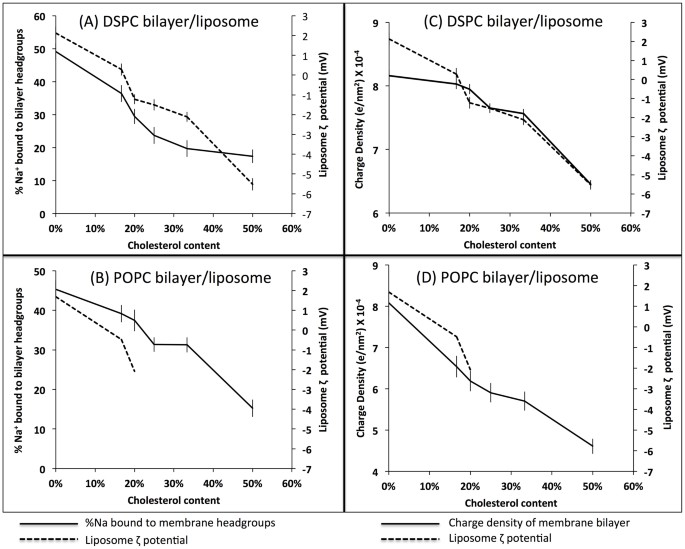
In Figure 1A and 1B, the percentage of all Na+ ions in the systems that are bonded to the lipid headgroups is shown. Ions were defined as bonded if they were within a distance of 0.325 nm from any oxygen atom of the lipid molecule17. Figure 1A and 1B shows a decrease of ions bonded with the lipid headgroups in both bilayer types. In the range of 0 to 50% of cholesterol the percentage of bound ions drops from 45–50% to 20%. In this manuscript all percentages referred in the analysis of the simulation results are percentages of the total number of ions of that particular element in the simulation system, not the molar percentage. This is an interesting observation as the DSPC bilayer at 310 K remains in the gel state while the POPC bilayer remains in the liquid crystalline state. While looking at the area per lipid, DSPC occupies about 0.43 nm2 and is relatively constant, independent of cholesterol concentration while for the case of POPC the area drops from 0.673 to 0.623 nm2 (See supplementary figure S2). This means that the area per lipid or membrane phase is not a primary factor affecting Na+ binding.
Figure 1
(A) and (B) Percentage of cations bound to the phospholipid bilayer in the molecular dynamics simulations and ζ – potential result from the experiment on liposomes vs. cholesterol content in the bilayer for a) DSPC and b) POPC. The plot shows that as the cholesterol content in the phospholipid bilayer increases, the percentage of Na+ ions bound to the membrane and the ζ – potential of the liposomes both decrease. (C) and (D) Charge density (the integral of the charge through the membrane cross section (the region of the membrane where the membrane headgroup is located, according to the mass density profile) divided by the area of the membrane, in units of electron charge per nm2) in the lipid layer as a function cholesterol concentration of cholesterol showing decline parallel to the decline in number of bound ions.
If we look at the charge per unit area of the membrane, shown in figure 1C and 1D, we see a trend in agreement with the result of the number of bound ions. Additionally, if we plot the number of bound ions relative to the number of available PC groups in the membrane, we also see a decline with increasing cholesterol content, as shown in supplementary figure S9. The relationship is, however, not linear, for the POPC the number of bound ions per PC headgroup remains approximately constant between zero and 30% cholesterol.
We have also measured the number of hydrogen bonds between the cholesterol oxygens and the phospholipid headgroups21, shown in supplementary figure S12 A–B. This result shows that the association between cholesterol oxygens and phospholipid headgroups increases as the cholesterol content in the membrane grows.
We then investigated the specific groups to which the bound Na+ ions were associated. We found that binding to phosphate oxygens to be dominant, as shown in supplementary figure S6. Of particular note is the very small level of Na+ binding to the cholesterol oxygens. We thus see that as cholesterol level increases, strongly binding PC headgroups are replaced with more weakly binding cholesterol OH groups. This partly explains the decrease of the number of bound ions and is probably the only factor for the case of POPC in low cholesterol concentration, where the number of Na+ ions bound per PC headgroup is constant. In addition, PC headgroups become increasingly associated with the cholesterol headgroups, further decreasing the number of binding sites for cations. The increase in the level of cholesterol in the membrane increases the hydrophobicity of the membrane, further decreasing the membrane affinity for cations. This mechanism of expulsion of Na+ ions from the water membrane interface is similar to that proposed in the “Umbrella model”22. In this model the changes induced by the cholesterol in the bilayer properties are proposed to result from the tendency of PC headgroups to prevent unfavorable cholesterol interaction with water at the membrane water interface. We have also shown the binding stoichiometry in figure S7, where we see significant bridging between DSPC phosphate groups, some bridging between the POPC phosphate groups and the cholesterol oxygens and some ions binding to more than two groups.
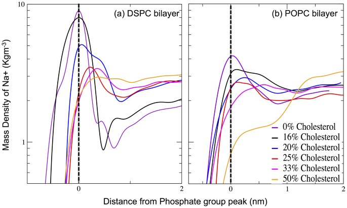
To better show the effect of cholesterol on the interaction between ions and lipids, mass density plots from all simulations for Na+ with the phosphate group peaks aligned are shown in Figure 2. As can be seen, cholesterol affects the amplitude of the Na+ peak in keeping with the observed decrease in the density of ions at the membrane interface. In addition, the position of the peak is gradually shifted towards the water phase. A Mass density plot including the Cl− ion distribution is included in the supplementary materials as supplementary figure S4. The distribution of Cl− ions is in qualitative agreement with previous results16,23, and, as shown in supplementary figure S10 and S11, there is no evidence of chlorine ions binding to the membrane headgroups. We also measured the water ordering along the membrane normal and showed this in figure S13.
Figure 2
Partial mass density profiles comparison: (A) DSPC (B) POPC: The black dashed line represents the position of phosphate head groups. As the cholesterol content in the bilayer increases, the association of Na+ ions with the membrane head group decreases.
To validate the findings from the computational studies, we performed experimental measurement of the ζ – potential of liposomes composed of DSPC or POPC and cholesterol. We have considered this to be crucial due to reported discrepancies in quantitative picture of ions binding to the membrane surface obtained from various force fields. We could formulate the DSPC liposomes with 0%, 16.66%, 20%, 25%, 33.33% and 50% cholesterol. However for POPC only liposomes containing 0%, 16.66% and 20% cholesterol where formulated; attempts at cholesterol density above this resulted in liposome aggregation. The particle size of the liposomes was ~100 nm for the liposomes containing cholesterol and ~200 nm for pure DSPC and POPC liposomes. The surface potential of the Pure DSPC liposome in saline buffer is positive (~2 mV) but as the cholesterol content is increased in the liposomes, the surface potential drops to negative values. The negative ζ – potential result in a system composed of neutral lipids is not a surprising result, in light of previous published results24. This showed that there is a definite trend, which can be attributed to the change in the cholesterol content.
Discussion
Results shown above indicate that the presence of cholesterol decreases Na+ ion binding with the lipid headgroup, releasing ions from the water membrane interface to the water phase. This leads to a more neutral surface charge of the membrane as seen in the observed changes in the ζ – potential. Since we see this effect in both DSPC and POPC lipid systems we see that this is a robust effect that will be present in both biological systems and drug delivery liposomes. The selected lipid compositions cover the whole range of lipid phases present in biological membranes and liposomes, including: 1) the liquid disordered phase for the case of pure POPC 2) the liquid ordered phase for the case of the bilayer with the higher cholesterol concentration and 3) the gel phase for the case of the pure DSPC bilayer. The presence of Na+ ions at the membrane interface plays an important role in both the interaction between 1) biomembranes and peripheral membrane proteins and 2) drug delivery liposomes and serum opsonins20 in the bloodstream. Our results indicate that the addition of cholesterol to the liposome membrane reduces the binding of Na+ ions to the membrane. Thus, this could 1) for the case of biomembranes play a role in governing the interaction between the membrane and peripheral membrane proteins and 2) for the case of drug delivery liposomes, decrease interactions with serum opsonins, hence increasing the circulation time of the liposome25.
We should add that regarding ion binding properties of membranes, it is known that there is some discrepancy between the results obtained using the different potential sets26,27. While these discrepancies will clearly have a quantitative effect, as shown in the referenced papers, we do not expect a qualitative effect, i. e. while the extent of the phenomenon may differ in the experimental system, the phenomenon itself has been correctly identified.
Clearly the blood plasma is more complex than our simple picture of it comprising of water, ions and opsonins and both our experimental and computational models are very simplified models that only capture one aspect of this complex system. In spite of this we propose that our results, however, provide new insight into the effect of cholesterol on a lipid membrane with dissolved salts in its solvent and this in turn provides insight into the role cholesterol plays in lipid membranes both biologically and pharmaceutically.
Methods
In our molecular dynamics simulations, we prepared ten phospholipid bilayers for a total of 288 lipid molecules with varying proportions of saturated 1,2-distearoyl-sn-glycero-3-phosphocholine (DSPC) or monounsaturated 1-palmitoyl-2-oleoyl-sn-glycero-3-phosphocholine (POPC) and cholesterol in proportions: 6:0, 5:1, 4:1, 3:1, 2:1 and 1:1. The exact composition of the simulated systems is given in Table S1. All simulations were performed using GROMACS 4.5.5 software28. The simulation length was 200 ns, with the initial 100 ns considered as the equilibration period based on an analysis of the surface area per lipid and the number of ions binding to the membrane interface, as shown in supplementary figure S3 and S5 respectively. In addition we measured the dynamics of ion exchange between the bonded and unbonded states, shown in supplementary figure S8. These show an increase in the exchange dynamics with increasing level of cholesterol and faster dynamics for POPC than DSPC. The longest binding time for the pure DSPC system is in agreement with results in our previous work16. In all cases the data was fit to a multiexponential29 and our interpretation of this multiexponential fit is found in the supplementary material.
The final 100 ns of the trajectory were used for all analysis. The molecular dynamics simulations were conducted under constant pressure (1 Bar) controlled through a Parrinello−Rahman barostat30 using a semi-isotropic pressure scheme. The temperature (310 K) was controlled by Nosé-Hoover thermostat31,32. The temperatures of the solute and solvent were controlled independently. For parameterization of all molecules and ions we used the OPLS33 all atom force field (OPLS-AA) as described in our previous publications13,14,15. For water we used the TIP3 water model34. Periodic boundary conditions with the usual minimum image convention were used in all three directions. In order to preserve the covalent bond lengths, the linear constraint solver (LINCS) algorithm35 was employed and a 2 fs time step was used in all simulations. The Lennard-Jones Interactions were cut off at 1.0 nm and for the electrostatic interactions we employed the particle mesh Ewald method36. All error bars were calculated using the block method37.
For ζ – potential measurements of liposomes DSPC and POPC liposomes were prepared. Both DSPC and POPC were purchased from Lipoid® GmbH Germany, cholesterol (99% pure) and Sodium Chloride (99% pure) were obtained from Sigma-Aldrich. Other reagents and solvents were of analytical grade. Water obtained from a MilliPore system with a conductivity of 18 MΩ cm−1 was used for the preparation of the liposomes, using the thin film hydration followed by extrusion38 method to achieve a uniform size of liposomes. The hydration media contained 140 mM NaCl. The liposomal suspensions were stored at 4°C and analyzed for particle size and ζ – potential.
References
- van Meer, G., Voelker, D. & Feigenson, G. Membrane lipids: where they are and how they behave. Molecular Cell. Biol. 9, 112–124 (2008).
CAS Google Scholar - Róg, T., Pasenkiewicz-Gierula, M., Vattulainen, I. & Karttunen, M. Biochim. Biophys. Acta. 1788, 97–121 (2009).
Article Google Scholar - Marsh, D. & Smith, I. Headgroup conformation and lipid-cholesterol association in phosphatidylcholine vesicles: a 31P(1H) nuclear overhauser effect study. Biochim. Biophys. Acta 298, 133–144 (1973).
Article CAS Google Scholar - Jacobs, R. & Oldfield, E. Interactions of cholesterol with the membrane lipid matrix. A solid state NMR approach. Biochemistry. 18, 3280–3285 (1979).
Article CAS Google Scholar - Yeagle, P. L. Methyl-β-Cyclodextrins and Liposomes as Water-Soluble Carriers for Cholesterol Incorporation into Membranes and Its Evaluation by a microenzymatic fluorescence assay and Membrane fluidity-sensitive dyes. Biochim. Biophys. Acta. 822, 267–287 (1985).
CAS Google Scholar - Coderch, L. et al. Influence of cholesterol on liposome fluidity by EPR- relationship with percutaneous absorption. J. Control Release. 68, 85–95 (2000).
Article CAS Google Scholar - Vasir, J. K. & Labhasetwar, V. Biodegradable nanoparticles for cytosolic delivery of therapeutics. Adv. Drug. Delivery Rev. 59, 718–728 (2007).
Article CAS Google Scholar - Semple, S. C., Chonn, A. & Cullis, P. R. Influence of cholesterol on association of plasma proteins with liposome. Biochemistry. 35, 2521–2525 (1996).
Article CAS Google Scholar - Barenholz, Y. J. Targeted drug delivery systems mediated by a novel peptide in breast cancer therapy and imaging controlled release. PLOS one. 160, 117–134 (2012).
CAS Google Scholar - Maxfield, F. R. & Tabas, I. Role of cholesterol and lipid organization in disease. Nature. 438, 612–621 (2005).
Article ADS CAS Google Scholar - Zidovska, A., Evans, H. M., Ahmad, A., Ewert, K. K. & Safinya, C. R. The role of cholesterol and structurally related molecules in enhancing transfection by cationic liposome-DNA complexes. J. Phys. Chem. B., 113, 5208–5216 (2009).
Article CAS Google Scholar - Gatfield, J. & Pieters, J. Essential role for cholesterol in entry of mycobacteria into macrophages. Science. 288, 1647–1650 (2000).
Article ADS CAS Google Scholar - Lingwood, D. et al. Cholesterol modulates glycolipid conformation and receptor activity. Nature Chem. Biol. 7, 260–262 (2011).
Article CAS Google Scholar - Orłowski, A. et al. Strong preferences of dopamine and L-dopa towards lipid head group: importance of lipid composition and implication for neurotransmitter metabolism. Neurochem. 122, 681–690 (2012).
Article Google Scholar - Berkowitz, M. L., Bostick, D. L. & Pandit, S. Aqueous solutions next to phospholipid membrane surfaces: insights from simulations. Chem. Rev. 106, 1527–1539 (2006).
Article CAS Google Scholar - Stepniewski, M., Bunker, A., Pasenkiewicz-Gierula, M., Karttunen, M. & Rog, T. Effects of the lipid bilayer phase state on the water membrane interface. J. Phys. Chem. B. 114, 11784–11792 (2010).
Article CAS Google Scholar - Magarkar, A., Karakas, E., Stepniewski, M., Róg, T. & Bunker, A. Molecular dynamics simulation of PEGylated bilayer interacting with salt ions: a model of the liposome surface in the bloodstream. J. Phys. Chem. B. 116, 4212–4219 (2012).
Article CAS Google Scholar - Ferber, U. M., Kaggwa, G. & Jarvis, S. P. Direct imaging of salt effects on lipid bilayer ordering ay sub-molecular resolution Eur. Biophys. J., 40, 329–338 (2011).
CAS Google Scholar - Böckmann, R. A., Hac, A., Heimburg & Grubmüller, T. Effect of sodium chloride on a lipid bilayer. Biophys. J., 85, 1647–1655 (2003).
Article Google Scholar - Martinez-Seara, H., Róg, T., Karttunen, M., Vattulainen, I. & Riegada, R. Cholesterol induces specific spatial and orientational order in cholesterol/phospholipid membranes. PLoS ONE, 5, e11162 (2010).
Article ADS Google Scholar - Pandit, S., Bostick, D. & Berkowitz, L. M. Complexation of phosphotidylcholine lipids with cholesterol. Biophysical Journal. 86, 1345–1356 (2004).
Article ADS CAS Google Scholar - Huang, J. Y. & Feigenson, G. W. A microscopic interaction model of maximum solubility of cholesterol in lipid bilayers. Biophysical Journal. 76, 2142–2157 (1999).
Article ADS CAS Google Scholar - Miettinen, M. S., Gurtovenko, A. A., Vattulainen, I. & Karttunen, M. Ion dynamics in cationic lipid bilayer systems in saline solutions. J. Phys. Chem. B. 113, 9226–9234 (2009).
Article CAS Google Scholar - Makino, K. et al. Temperature and ionic strength induced conformatiomational changes in the lipid head group region of liposomes as suggested by zeta potential data. Biophysical Chemistry. 41, 175–183 (1991).
Article CAS Google Scholar - Scherphof, G. L. & Kamps, J. A. A. M. J. Liposome opsonization. Liposome Res. 15, 109–139 (2005).
Article Google Scholar - Klasczyk, K. & Knecht, V. Validating affinities for ion-lipid association from simulation against experiment. J. Phys. Chem. A. 115, 10587–10595 (2011).
Article CAS Google Scholar - Jurkiewicz, P., Cwiklik, L., Vojtíšková, A., Jungwirth, P. & Hof, M. Structure, dynamics and hydration of POPC/POPS bilayers suspended in NaCl, KCl and CsCl solutions. Biochim. Biophys. Acta. 1818, 609–616 (2012).
Article CAS Google Scholar - Pronk, S. et al. GROMACS 4.5: a high-throughput and highly parallel open source molecular simulation toolkit. Bioinformatics. 29, 845–854 (2013).
Article CAS Google Scholar - Titantah, J. T. & Karttunen, M. Long-time correlations and hydrophobe-modified hydrogen-bonding dynamics in hydrophobic hydration. J. Am. Chem. Soc. 134, 9362–9368 (2012).
Article CAS Google Scholar - Parrinello, M. & Rahman, A. Polymorphic transitions in single crystals: A new molecular dynamics method. J. Appl. Phys. 52, 7182–7190 (1981).
Article ADS CAS Google Scholar - Nosé, S. J. A unified formulation of the constant temperature molecular dynamics methods. Chem. Phys. 81, 511–519 (1984).
ADS Google Scholar - Hoover, W. G. Canonical dynamics: Equilibrium phase-space distributions. Phys. Rev. A. 31, 1695–1697 (1985).
Article ADS CAS Google Scholar - Jorgensen, W. L. & Tirado-Rives, J. The OPLS potential functions for proteins. Energy minimizations for crystals of cyclic peptides and crambin. J. Am. Chem. Soc. 110, 1657–1666 (1988).
Article CAS Google Scholar - Jorgensen, W. L., Chandrasekhar, J., Madura, J. D., Impey, R. & Klein, M. L. Comparison of simple potential functions for simulating liquid water. J. Chem. Phys. 79, 926–935 (1983).
Article ADS CAS Google Scholar - Hess, B., Bekker, H., Berendsen, H. J. C. & Fraaije, J. G. E. M. A linear constraint solver for molecular simulations. J. Comput. Chem. 18, 1463–1472 (1997).
Article CAS Google Scholar - Essmann, U. et al. A smooth particle mesh Ewald method. J. Chem. Phys. 103, 8577–8593 (1995).
Article ADS CAS Google Scholar - Hess, B. Determining the shear viscosity of model liquids from molecular dynamics simulations. J. Chem. Phys. 116, 209–217 (2002).
Article ADS CAS Google Scholar - Olson, F., Hunt, C. A., Szoka, F. C., Vail, W. J. & Papahadjopoulos, D. Preparation of liposomes of defined size distribution by extrusion through polycarbonate membranes. Biochim. Biophys. Acta. 557, 9–23 (1997).
Article Google Scholar
Acknowledgements
Authors would like to thank Centre for scientific computing (CSC), Finland for providing computing resources. The authors thank Prof. Arto Urtti for his advice. The Academy of Finland and the Finnish Cultural Foundation funded this work.
Author information
Authors and Affiliations
- Centre for Drug Research, Faculty of Pharmacy, University of Helsinki, Helsinki, Finland
Aniket Magarkar, Paraskevi Kallinteri & Alex Bunker - Bombay College of Pharmacy, University of Mumbai, Mumbai, India
Vivek Dhawan - Division of Biopharmaceutics and Pharmacokinetics, Faculty of Pharmacy, University of Helsinki, Helsinki, Finland
Tapani Viitala & Mohammed Elmowafy - Department of Physics, Tampere University of Technology, Tampere, Finland
Tomasz Róg
Authors
- Aniket Magarkar
You can also search for this author inPubMed Google Scholar - Vivek Dhawan
You can also search for this author inPubMed Google Scholar - Paraskevi Kallinteri
You can also search for this author inPubMed Google Scholar - Tapani Viitala
You can also search for this author inPubMed Google Scholar - Mohammed Elmowafy
You can also search for this author inPubMed Google Scholar - Tomasz Róg
You can also search for this author inPubMed Google Scholar - Alex Bunker
You can also search for this author inPubMed Google Scholar
Contributions
A.M. and T.R. built the computational models of the systems and performed the simulation V.D. and M.E. performed the experiments and analyzed the data P.K. and T.V. designed the experiments, A.M., T.R. and A.B. wrote the manuscript.
Ethics declarations
Competing interests
The authors declare no competing financial interests.
Electronic supplementary material
Rights and permissions
This work is licensed under a Creative Commons Attribution-NonCommercial-NoDerivs 3.0 Unported License. The images in this article are included in the article's Creative Commons license, unless indicated otherwise in the image credit; if the image is not included under the Creative Commons license, users will need to obtain permission from the license holder in order to reproduce the image. To view a copy of this license, visit http://creativecommons.org/licenses/by-nc-nd/3.0/
About this article
Cite this article
Magarkar, A., Dhawan, V., Kallinteri, P. et al. Cholesterol level affects surface charge of lipid membranes in saline solution.Sci Rep 4, 5005 (2014). https://doi.org/10.1038/srep05005
- Received: 28 November 2013
- Accepted: 01 May 2014
- Published: 21 May 2014
- DOI: https://doi.org/10.1038/srep05005

