Glucocorticoids synergize with IL-1β to induce TLR2 expression via MAP Kinase Phosphatase-1-dependent dual Inhibition of MAPK JNK and p38 in epithelial cells (original) (raw)
- Research article
- Open access
- Published: 04 May 2004
BMC Molecular Biology volume 5, Article number: 2 (2004)Cite this article
Abstract
Background
Despite the importance of glucocorticoids in suppressing immune and inflammatory responses, their role in enhancing host immune and defense response against invading bacteria is poorly understood. Toll-like receptor 2 (TLR2) has recently gained importance as one of the major host defense receptors. The increased expression of TLR2 in response to bacteria-induced cytokines has been thought to be crucial for the accelerated immune response and resensitization of epithelial cells to invading pathogens.
Results
We show that IL-1β, a key proinflammatory cytokine, greatly up-regulates TLR2 expression in human epithelial cells via a positive IKKβ-IκBα-dependent NF-κB pathway and negative MEKK1-MKK4/7-JNK1/2 and MKK3/6-p38 α/β pathways. Glucocorticoids synergistically enhance IL-1β-induced TLR2 expression via specific up-regulation of the MAP kinase phosphatase-1 that, in turn, leads to dephosphorylation and inactivation of both MAPK JNK and p38, the negative regulators for TLR2 induction.
Conclusion
These results indicate that glucocorticoids not only suppress immune and inflammatory response, but also enhance the expression of the host defense receptor, TLR2. Thus, our studies may bring new insights into the novel role of glucocorticoids in orchestrating and optimizing host immune and defense responses during bacterial infections and enhance our understanding of the signaling mechanisms underlying the glucocorticoid-mediated attenuation of MAPK.
Background
Epithelial cells are often situated at host-environment boundaries, and thus act as the first line of defense against bacterial pathogens [1]. The epithelial cells are not merely a passive barrier but can detect foreign pathogens and generate a range of mediators that play important roles in the activation of innate and adaptive immunity. For effective host defense, the epithelial cells recognize highly conserved structural motifs only expressed by microbial pathogens, called pathogen-associated molecular patterns (PAMPs). Toll-like receptors (TLRs) play a critical role in early innate immunity to invading microorganisms by sensing PAMPs [2]. Stimulation of TLRs by PAMPs initiates a signaling cascade that induces the production and secretion of proinflammatory cytokines [3]. Moreover, stimulation of TLRs also induces the production of effector cytokines that leads to activation of adaptive immunity.
Mammalian TLRs were originally found as homologues of the Drosophila Toll [4]. TLRs are type I transmembrane receptors, that possess extracellular leucine-rich repeats domains flanked by cytoplasmic domains [4, 5]. Although at least 10 TLRs have been identified, only two TLRs, TLR2 and TLR4, have been well-studied. While TLR4 is mainly involved in Gram-negative bacteria lipopolysaccharide (LPS) signaling, TLR2 can respond to a variety of Gram-positive bacterial products, including peptidoglycan, lipoprotein, lipoteichoic acid and lipoarabinomannan. In addition to Gram-positive bacterial PAMPs, TLR2 also recognizes factors released by Gram-negative bacteria including nontypeable Haemophilus influenzae (NTHi) [6, 7] and coccobacilli, Neisseria meningitidis [8] as well as the Mycoplasma lipopeptides [9, 10]. The importance of TLR2 in host defense was further demonstrated by studies of knockout mice showing decreased survival of TLR2-deficient mice after infection with Gram-positive Staphylococcus aureus [11]. Thus, it is clear that TLR2 plays a crucial role in host defense against a variety of microbial pathogens.
In contrast to its relatively high level of expression in lymphoid tissues, TLR2 is expressed at low levels in epithelial cells. A key issue that has thus been raised is whether the low amount of TLR2 expressed in epithelial cells is sufficient for mediating the host defense response against invading microbial pathogens. Our recent studies revealed that TLR2, although expressed at very low level in unstimulated human epithelial cells, is greatly up-regulated by NTHi via a positive IKKβ-IκBα-dependent NF-κB pathway and a negative MKK3/6-p38α/β pathway [12]. Moreover, glucocorticoids synergistically enhance NTHi-induced expression of TLR2 via a negative cross-talk with p38 MAP kinase pathway, supporting the notion that glucocorticoids plays an important role in orchestrating and optimizing immune functions, including host defense, during bacterial infections [13, 14]. However, still unclear is whether up-regulation of TLR2 expression in epithelial cells can also be generalized to other key mediators such as proinflammatory cytokines, e.g. interleukin 1-β (IL-1β) and if so, whether the cytokine-mediated up-regulation of TLR2 can also be further enhanced by glucocorticoids.
IL-1β, a proinflammatory cytokine, is produced by various cell types including epithelial cells and can be strongly induced during bacterial infections [15]. It has been recognized as one of the key mediators of the host response to microbial invasion, inflammation, immunological reactions and tissue injury [16]. Although it has been shown to induce the expression of a variety of non-structural, function-associated genes that are expressed during inflammation, particularly other cytokines, whether IL-1β also regulates the expression of host defense receptor TLR2 in human epithelial cells is still unknown. In addition, it is still unclear whether the IL-1β-mediated TLR2 expression can also be enhanced by glucocorticoids.
Here we show that IL-1β up-regulates TLR2 via a positive IKKβ-IκBα-dependent NF-κB signaling pathway and negative MKK3/6-p38α/β and MEKK1-MKK4/7-JNK1/2 pathways in human epithelial cells. Surprisingly, glucocorticoids synergistically enhance the IL-1β-induced TLR2 expression via a MKP-1-dependent dual inhibition of MAPK p38 and JNK signaling pathways. These studies may provide novel insights into the role of IL-1β and glucocorticoids in orchestrating and optimizing host immune and defense responses and lead to new therapeutic strategies for modulating innate immune and inflammatory responses for bacterial infections.
Results
IL-1β selectively up-regulates TLR2, but not TLR4, in human epithelial cells
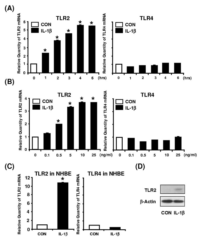
We first examined whether IL-1β up-regulates TLR2 expression in human epithelial cells. Human epithelial HeLa cells were first treated with IL-1β and the relative quantity of TLR2 mRNA was then measured by performing real-time quantitative PCR analysis. As shown in Figure 1a and 1b, IL-1β greatly up-regulated the TLR2 mRNA, but not TLR4 mRNA, in HeLa cells in a time- and dose-dependent manner. Similar results were also observed in primary human bronchial epithelial (NHBE) cells (Figure 1c). To further determine whether up-regulation of TLR2 mRNA is accompanied by elevated TLR2 protein, Western blot analysis was performed using antibodies against human TLR2. As shown in Figure 1d, up-regulation of TLR2 was also observed at protein level. These data demonstrate that TLR2, but not TLR4, is greatly up-regulated by IL-1β in human epithelial cells.
Figure 1
IL-1β up-regulates TLR2, but not TLR4, in human epithelial cells including primary bronchial epithelial cells. A, IL-1β up-regulates TLR2 mRNA, but not TLR4 mRNA, in HeLa cells in a time-dependent manner. Cells were treated with IL-1β (10 ng/ml) for the indicated time periods. The expression of TLR2 and TLR4 at mRNA level was then measured by real-time quantitative PCR in IL-1β-treated and -untreated human cervix epithelial HeLa cells. TLR2 and TLR4 mRNA levels were normalized to the level of cyclophilin that served as an internal control for the amount of RNA used in each reaction. Values are the mean ± SD; n = 3. The symbols (*) indicate results significantly different (p < 0.01) from unstimulated condition. B, IL-1β up-regulates TLR2 mRNA, but not TLR4 mRNA, in HeLa cells in a dose-dependent manner. HeLa cells were treated with the indicated concentration of IL-1β for 3 h. Values are the mean ± SD; n = 3. The symbols (*) indicate results significantly different (p < 0.01) from unstimulated condition. C, IL-1β up-regulates TLR2 mRNA, but not TLR4 mRNA, in primary human bronchial epithelial NHBE cells. NHBE cells were treated with IL-1β (10 ng/ml) for 3 h. Values are the mean ± SD; n = 3. The symbols (*) indicate results significantly different (p < 0.01) from unstimulated condition. D, IL-1β also induces expression of TLR2 at protein level in HeLa cells. HeLa cells were treated for 5 h with IL-1β (10 ng/ml). Protein level of TLR2 was then assessed by Western blot analysis using an anti-hTLR2 antibody. β-Actin was used as a protein loading control.
Activation of NF-κB via IKKβ-IκBα signaling pathway is required for IL-1β-induced TLR2 up-regulation
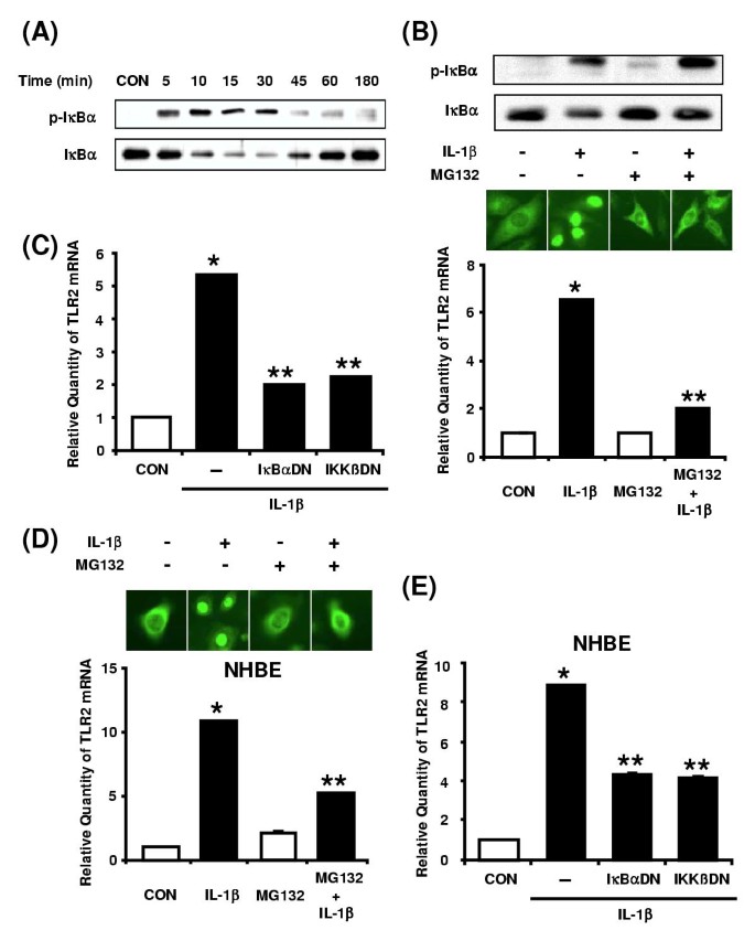
We next sought to determine which intracellular signaling pathways are involved in IL-1β-induced TLR2 up-regulation. Based on the essential involvement of NF-κB in regulating the expression of large numbers of genes involved in immunity and inflammation and the fact that NF-κB can be activated by IL-1β, we investigated the involvement of NF-κB signaling in TLR2 induction. We first investigated whether IL-1β induces phosphorylation and degradation of IκBα by performing Western blot analysis using antibodies against phosphorylated and total IκBα, respectively. IκBα phosphorylation became evident at 5 min, peaked at 10 min, and declined thereafter (Figure 2a). Consistent with this result, the IL-1β-induced degradation of IκBα was observed at 5 min and returned to baseline levels by 3 h (Figure 2a). We then determined the requirement of IκBα degradation in IL-β-induced TLR2 expression. As shown in Figure 2b, MG132, a proteasome inhibitor that prevents the degradation of IκBα, completely blocked the NF-κB nuclear translocation induced by IL-1β (middle panel). As expected, the IL-1β-induced IκBα degradation was also blocked by MG132 (Figure 2b, upper panel). Accordingly, phosphorylated IκBα was no longer degraded in the presence of MG-132, thus persisting in the cytoplasm. Concomitantly, IL-1β-induced TLR2 up-regulation was also inhibited by MG132 (Figure 2b, lower panel). To further confirm the involvement of IκBα degradation in IL-1β-induced TLR2 expression, we next transfected HeLa cells with a transdominant mutant of IκBα (S32A, S36A). As shown in Figure 2c, overexpression of the IκBα transdominant mutant inhibited IL-1β-induced TLR2 up-regulation. On the basis of a recent report that IκB kinase β (IKKβ) acts as an immediate kinase of IκBα, we investigated the role of IKKβ in IL-1β-induced TLR2 induction. Overexpression of a dominant-negative mutant form of IKKβ inhibited IL-1β-induced TLR2 up-regulation (Figure 2c). Likewise, the involvement of IKKβ-IκBα in IL-1β-induced TLR2 expression was also confirmed in human primary bronchial epithelial NHBE cells (Figure 2d and 2e). Taken together, these data demonstrate that IKKβ-IκBα-dependent activation of NF-κB is required for IL-1β-induced TLR2 up-regulation in human epithelial cells.
Figure 2
IKKβ-IκBα-dependent activation of NF-κB is required for IL-1β-induced TLR2 up-regulation. A, IL-1β induces IκBα phosphorylation and degradation in HeLa cells as assessed by Western blot analysis. Cells were treated with IL-1β (10 ng/ml) for the indicated time periods. B, MG 132 inhibits IL-1β-induced NF-κB nuclear translocation, IκBα degradation and TLR2 up-regulation in HeLa cells. For Western blotting and immunofluorescent staining, cells were pretreated with MG132 (1 μM) for 2 h prior to treatment with IL-1β (10 ng/ml) for 20 min. For the measurement of TLR2 mRNA, cells were treated with IL-1β (10 ng/ml) for 3 h after MG132 pretreatment. Values are the mean ± SD; n = 3. The symbols indicate results significantly different (p < 0.01) from unstimulated condition (*) and IL-1β-stimulated condition (without MG132)(**). C, Overexpression of a transdominant mutant of IκBα [IκBα [(S32A, S36A)] and a dominant-negative mutant of IKKβ [IKKβ (K44A)] inhibit IL-1β-induced TLR2 up-regulation in HeLa cells. HeLa cells were treated with IL-1β (10 ng/ml) for 3 h. Values are the mean ± SD; n = 3. The symbols indicate results significantly different (p < 0.01) from unstimulated condition (*) and IL-1β-stimulated condition (without dominant-negative construct)(**). D, MG 132 also inhibits IL-1β-induced NF-κB nuclear translocation and TLR2 up-regulation in NHBE cells. For immunofluorescent staining, cells were pretreated with MG132 (1 μM) for 2 h prior to treatment with IL-1β (10 ng/ml) for 20 min. For the measurement of TLR2 mRNA, cells were treated with IL-1β (10 ng/ml) for 3 h after MG132 pretreatment. Values are the mean ± SD; n = 3. The symbols indicate results significantly different (p < 0.01) from unstimulated condition (*) and IL-1β-stimulated condition (without MG132)(**). E, Overexpression of a transdominant mutant of IκBα (S32A, S36A) and a dominant-negative mutant of IKKβ (K44A) reduce IL-1β-induced TLR2 up-regulation in NHBE cells. Cells were treated with IL-1β (10 ng/ml) for 3 h. Values are the mean ± SD; n = 3. The symbols indicate results significantly different (p < 0.01) from unstimulated condition (*) and IL-1β-stimulated condition (without dominant-negative construct)(**).
Activation of MKK3/6-p38 α/β MAPK pathway is negatively involved in IL-1β-induced TLR2 expression
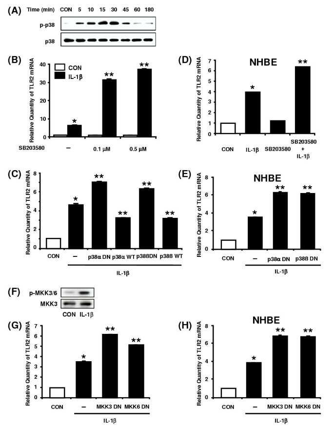
Many cellular stress stimuli can simultaneously activate both NF-κB and p38 MAPK modules. Because of this overlap as well as the evidence for the activation of p38 by IL-1β [17], we explored the possible involvement of p38 MAPK pathway in IL-1β-induced TLR2 up-regulation. We first confirmed whether IL-1β induces phosphorylation of p38 MAPK in HeLa cells. As shown in Figure 3a, p38 MAPK is strongly phosphorylated by IL-1β in a time-dependent manner. To determine whether the activation of p38 MAPK is involved in IL-1β-induced TLR2 up-regulation, we assessed the effect of the pyridinyl imidazole, SB203580, a highly specific inhibitor for p38 MAPK, on TLR2 induction. Interestingly, SB203580 greatly enhanced IL-1β-induced TLR2 up-regulation, suggesting that activation of p38 MAPK is negatively involved in IL-1β-induced TLR2 expression (Figure 3b). Moreover, overexpression of a dominant-negative mutant form of either p38α [fp38α(AF)] or p38β [fp38β(AF)] also enhanced the IL-1β-induced TLR2 expression, whereas overexpression of a wild-type p38α or p38β reduced it, thereby confirming the negative involvement of p38 MAPK in HeLa cells (Figure 3c). Similarly, SB203580 and overexpression of a dominant-negative mutant form of either p38α[fp38α(AF)] or p38β [fp38β(AF)] also enhanced the IL-1b-induced TLR2 expression in primary bronchial epithelial NHBE cells (Figure 3d and 3e). As immediate upstream kinases of p38 α and β, two MAPK kinases (MKK3 and MKK6) have been identified [18–20]. To further investigate whether activation of MKK3/6 is also negatively involved in IL-1β-induced TLR2 expression, we first sought to determine whether IL-1β induces activation of MKK3/6. As shown in Figure 3f, IL-1β strongly induced phosphorylation of MKK3/6 in HeLa cells. We next investigated the effect of overexpressing a dominant-negative mutant form of either MKK3 [MKK3b(A)] or MKK6 [MKK6b(A)] on TLR2 induction in HeLa cells. The IL-1β-induced TLR2 up-regulation was enhanced by blocking MKK3/6 signaling (Figure 3g). Similar results were also obtained in primary NHBE cells (Figure 3h). Collectively, our data suggest that activation of MKK3/6-p38 α/β MAPK signaling pathway is negatively involved in IL-1β-induced TLR2 up-regulation in human epithelial cells.
Figure 3
Activation of MKK3/6-p38 α/β MAPK pathway is negatively involved in IL-1β-induced TLR2 expression. A, IL-1β induces p38 phosphorylation in HeLa cells in a time-dependent manner as assessed by Western blot analysis. Cells were treated with IL-1β (10 ng/ml) for the indicated time periods. B, SB203580 greatly enhances IL-1β-induced TLR2 up-regulation in HeLa cells. Cells were pretreated for 2 h with the indicated concentration of SB203580 prior to treatment with IL-1β (10 ng/ml) for 3 h. Values are the mean ± SD; n = 3. The symbols indicate results significantly different (p < 0.01) from unstimulated condition (*) and IL-1β-stimulated condition (without SB203580)(**). C, Overexpression of a dominant-negative mutants of either p38α [fp38α(AF)] or p38β [fp38β (AF)] enhances, whereas overexpression of a wild-type p38α or p38β reduces, the IL-1β-induced TLR2 expression in HeLa cells. Cells were treated with IL-1β (10 ng/ml) for 3 h. Values are the mean ± SD; n = 3. The symbols indicate results significantly different (p < 0.01) from unstimulated condition (*) and IL-1β-stimulated condition (without wild-type or dominant-negative construct)(**). D, SB203580 greatly enhances IL-1β-induced TLR2 up-regulation in NHBE cells. Cells were pretreated for 2 h with SB203580 (20 μM) prior to treatment with IL-1β (10 ng/ml) for 3 h. Values are the mean ± SD; n = 3. The symbols indicate results significantly different (p < 0.01) from unstimulated condition (*) and IL-1β-stimulated condition (without SB203580)(**). E, Overexpression of a dominant-negative mutant of p38α or p38β enhances the IL-1β-induced TLR2 expression in NHBE cells. Values are the mean ± SD; n = 3. The symbols indicate results significantly different (p < 0.01) from unstimulated condition (*) and IL-1β-stimulated condition (without dominant negative construct)(**). F, IL-1β induces MKK3/6 phosphorylation in HeLa cells. Cells were treated with IL-1β (10 ng/ml) for 20 min. G, Overexpression of a dominant-negative mutant form of either MKK3 [MKK3b (A)] or MKK6 [MKK6b (A)] also enhances IL-1β-induced TLR2 up-regulation in HeLa cells. Cells were treated with IL-1β (10 ng/ml) for 3 h. Values are the mean ± SD; n = 3. The symbols indicate results significantly different (p < 0.01) from unstimulated condition (*) and IL-1β-stimulated condition (without dominant-negative construct)(**). H, Overexpression of a dominant-negative mutant form of either MKK3 or MKK6 also enhances IL-1β-induced TLR2 up-regulation in NHBE cells. Cells were treated with IL-1β (10 ng/ml) for 3 h. Values are the mean ± SD; n = 3. The symbols indicate results significantly different (p < 0.01) from unstimulated condition (*) and IL-1β-stimulated condition (without dominant-negative construct)(**).
Activation of MEKK1-MKK4/7-JNK1/2 pathway is also negatively involved in IL-1β-induced TLR2 expression
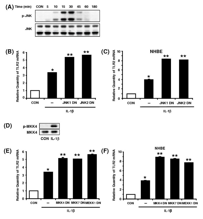
Besides the p38 MAPK pathway, the c-Jun N-terminal kinase (JNK) pathway represents another key MAPK pathway involved in the regulation of immune response genes. JNK is activated by a variety of environmental stresses including pro-inflammatory cytokine IL-1β [18]. Therefore, we next sought to investigate the involvement of JNK pathway in IL-1β-induced TLR2 expression. We first determined whether IL-1β induces phosphorylation of JNK in HeLa cells. As shown in Figure 4a, JNK is strongly phosphorylated by IL-1β in a time-dependent manner. To determine the involvement of JNK pathway in TLR2 up-regulation, a dominant-negative mutant form of either JNK1 [JNK1(AF)] or JNK2 [JNK2(AF)] was transfected into HeLa cells. Overexpression of a dominant-negative mutant of either JNK1 or JNK2 enhanced the IL-1β-induced TLR2 expression (Figure 4b). Similar result was also observed in primary NHBE cells (Figure 4c). These results suggest that JNK1/2 pathway is also negatively involved in IL-1β-induced TLR2 expression in human epithelial cells.
Figure 4
Activation of MEKK1-MKK4/7-JNK1/2 pathway is also negatively involved in IL-1β-induced TLR2 expression. A, IL-1β induces JNK phosphorylation in HeLa cells in a time-dependent manner as assessed by Western blot analysis. Cells were treated with IL-1β (10 ng/ml) for the indicated time periods. B, Overexpression of a dominant-negative mutants of either JNK1 [JNK1 (AF)] or JNK2 [JNK2 (AF)] enhances the IL-1β-induced TLR2 expressionin HeLa cells. Values are the mean ± SD; n = 3. The symbols indicate results significantly different (p < 0.01) from unstimulated condition (*) and IL-1β-stimulated condition (without dominant-negative construct)(**). C, Overexpression of a dominant-negative mutant of either JNK1 or JNK2 enhances the IL-1β-induced TLR2 expression in NHBE cells. Values are the mean ± SD; n = 3. The symbols indicate results significantly different (p < 0.01) from unstimulated condition (*) and IL-1β-stimulated condition (without dominant-negative construct)(**). D, IL-1β induces MKK4 phosphorylation in HeLa cells. Cells were treated with IL-1β (10 ng/ml) for 20 min. E, Overexpression of a dominant-negative mutant form of MKK4 or MKK7 or MEKK1 enhances IL-1β-induced TLR2 up-regulation in HeLa cells. Values are the mean ± SD; n = 3. The symbols indicate results significantly different (p < 0.01) from unstimulated condition (*) and IL-1β-stimulated condition (without dominant negative construct)(**). F, Overexpression of a dominant-negative mutant of MKK4 or MKK7 or MEKK1 also enhances IL-1β-induced TLR2 up-regulation in NHBE cells. Values are the mean ± SD; n = 3. The symbols indicate results significantly different (p < 0.01) from unstimulated condition (*) and IL-1β-stimulated condition (without dominant-negative construct)(**). For all the TLR2 mRNA measurement, cells were treated with IL-1β (10 ng/ml) for 3 h.
On the basis that JNK activation is mediated by two upstream MAPK kinases (MKKs), MKK4 and MKK7, and that both MKKs are activated by the further upstream MAPK/Erk kinase kinase 1 (MEKK1), we next sought to investigate whether the MEKK1-MKK4/7 signaling pathway is also negatively involved in TLR2 induction. We first confirmed whether MKK4 is activated by IL-1β in HeLa cells. Figure 4d showed a strong phosphorylation of MKK4 induced by IL-1β. To further determine the involvement of MEKK1-MKK4/7 pathway, HeLa cells were transfected with a dominant-negative mutant of either MKK4 or MKK7 or MEKK1. As shown in Figure 4e, overexpression of these dominant-negative mutants enhanced IL-1β-induced TLR2 expression (Figure 4e). Similar results were also observed in primary human bronchial epithelial NHBE cells (Figure 4f). Together, these results indicate that activation of MEKK1-MKK4/7-JNK1/2 signaling pathway is also negatively involved in IL-1β-induced TLR2 expression in human epithelial cells.
Glucocorticoids synergistically enhance IL-1β-induced TLR2 expression likely via negative cross-talk with MAPK p38 and JNK pathways
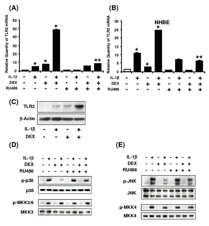
Glucocorticoids are well known for their anti-inflammatory, immune suppressive and anti-allergic activities [21]. They exert their anti-inflammatory effects by inhibiting the activity of transcription factors such as AP-1 and NF-κB that are involved in regulation of a variety of genes involved in the inflammatory responses [22, 23]. Because we have shown that activation of NF-κB is required for IL-1β-induced TLR2 expression, it is logical that glucocorticoids should suppress the IL-1β-induced TLR2 up-regulation. Surprisingly, dexamethasone (DEX), a glucocorticoid analog, synergistically enhanced IL-1β-induced TLR2 expression at the mRNA level in both HeLa cells and primary human bronchial epithelial NHBE cells (Figure 5a and 5b). The enhancing effect of DEX on TLR2 up-regulation was counteracted by RU486, a synthetic antiglucocorticoid that acts as a competitor against binding to the glucocorticoid receptor (GR), suggesting the enhancing effect of DEX is mediated by the GR. Concomitantly, Western blot analysis showed that IL-1β also synergistically enhanced TLR2 expression at protein level, thereby confirming that up-regulation of TLR2 mRNA is indeed accompanied by elevated TLR2 protein (Figure 5c). These results indicate that DEX synergistically enhances IL-1β-induced TLR2 up-regulation at both mRNA and protein levels in human epithelial cells.
Figure 5
Glucocorticoids synergistically enhance IL-1β-induced TLR2 up-regulation via negative cross-talks with MAPK p38 and JNK1/2 pathways. A, Dexamethasone (DEX) synergistically enhances IL-1β-induced TLR2 up-regulation and RU486 counteracts the enhancing effect of DEX on IL-1β-induced TLR2 up-regulation at mRNA level in HeLa cells. HeLa cells were first pretreated with RU486 (10-6 M) for 2 h and were then treated with DEX (10-6 M) for 2 h. The cells were stimulated with IL-1β (10 ng/ml) for 3 h, and the TLR2 mRNA levels were then assessed by real-time quantitative PCR. Values are the mean ± SD; n = 3. The symbols indicate results significantly different (p < 0.01) from unstimulated condition (*) and IL-1β-DEX-stimulated condition (without RU486)(**). B, DEX (10-6 M) synergistically enhances IL-1β-induced TLR2 up-regulation and RU486 (10-6 M) counteracts the enhancing effect of DEX on IL-1β-induced TLR2 up-regulation at mRNA level in NHBE cells. Cells were first pretreated with RU486 (10-6 M) for 2 h and were then treated with DEX (10-6 M) for 2 h. The cells were stimulated with IL-1β (10 ng/ml) for 3 h, and the TLR2 mRNA levels were then assessed. Values are the mean ± SD; n = 3. The symbols indicate results significantly different (p < 0.01) from unstimulated condition (*) and IL-1β-DEX-stimulated condition (without RU486)(**). C, Glucocorticoids also synergistically enhance the induction of TLR2 at protein level in HeLa cells. HeLa cells were first treated for 2 h with DEX (10-6 M). The cells were then stimulated with IL-1β (10 ng/ml) for 5 h. Protein level of TLR2 was assessed by Western blot analysis using an anti-hTLR2 antibody. β-Actin was used as a protein loading control. D, DEX inhibits IL-1β-induced phosphorylation of p38, but not MKK3/6, and the inhibitory effect of DEX is blocked by RU486. HeLa cells were first treated with RU486 (10-6 M) for 2 h and were then treated with DEX (10-6 M) for 2 h. The cells were stimulated with IL-1β (10 ng/ml) for 20 min. IL-1β-induced phosphorylation of p38 MAPK and MKK3/6 was then assessed by Western blot analysis. E, DEX inhibits IL-1β-induced phosphorylation of JNK, but not MKK4, and the inhibitory effect of DEX is blocked by RU486. HeLa cells were first treated with RU486 (10-6 M) for 2 h and were then treated with DEX (10-6 M) for 2 h. The cells were stimulated with IL-1β (10 ng/ml) for 20 min.
Because glucocorticoids have been shown to inhibit a number of MAPK family members, we next sought to determine whether DEX inhibits the IL-1β-induced activation of p38 or JNK by performing Western blot analysis. As shown in Figure 5d and 5e, DEX inhibited IL-1β-induced phosphorylation of both p38 and JNK, but not MKK3/6 or MKK4 and the inhibitory effects of DEX were blocked by GR antagonist RU486. Therefore, it is likely that DEX may synergistically enhance IL-1β-induced TLR2 up-regulation via negative cross-talk with p38 and JNK, but not their upstream kinases MKK3/6 or MKK4.
Glucocorticoids may enhance IL-1β-induced TLR2 up-regulation via up-regulation of MAP kinase phosphatase 1 (MKP-1)
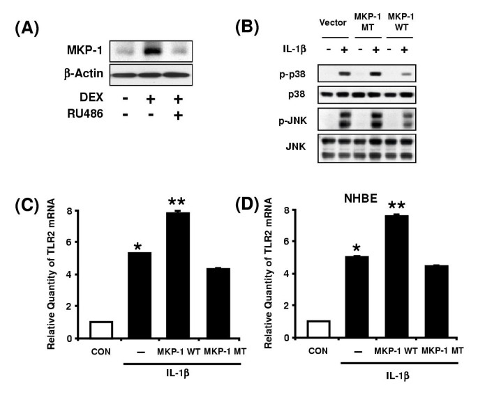
Based on recent reports that glucocorticoids inhibit the activation of p38 and JNK by up-regulating the expression of MKP-1 [24–26], we investigated whether MKP-1 is involved in the enhancing effect of DEX on IL-1β-induced TLR2 expression. We first confirmed that MKP-1 protein expression is indeed induced by DEX, and the DEX-induced MKP-1 expression was counteracted by the GR antogonist RU486 in HeLa cells (Figure 6a). We next investigated whether overexpression of MKP-1 mimics DEX. As expected, overexpression of wild-type MKP-1 inhibited IL-1β-induced p38 and JNK phosphorylation, whereas mutated MKP-1 had almost no effect on their phosphorylation (Figure 6b). Moreover, overexpression of wild-type MKP-1 enhanced IL-1β-induced TLR2 expression, whereas mutated MKP-1 slightly inhibited the TLR2 induction in both HeLa cells (Figure 6c) and primary NHBE cells (Figure 6d). Thus, it is clear that overexpression of wild-type MKP-1 mimics the enhancing effect of DEX on IL-1β-induced TLR2 up-regulation, supporting the hypothesis that glucocorticoids may enhance the IL-1β-induced TLR2 expression via a MKP-1-dependent inhibition of p38 and JNK.
Figure 6
Glucocorticoids enhance IL-1β-induced TLR2 up-regulation likely via upregulation of MKP-1. A, MKP-1 protein expression is induced by DEX and the MKP-1 induction is counteracted by RU486 in HeLa cells. HeLa cells were first treated with RU486 (10-6 M) for 2 h and were then treated with DEX (10-6 M) for 2 h. Protein level of MKP-1 was assessed by Western blot analysis. β-Actin was used as a protein loading control. B, Overexpression of wild-type MKP-1 inhibits IL-1β-induced p38 and JNK phosphorylation, whereas overexpression of MKP-1 mutant has almost no effect on IL-1β-induced p38 and JNK phosphorylation. Transfected HeLa cells were stimulated with IL-1β (10 ng/ml) for 20 min. IL-1β-induced phosphorylation of p38 MAPK and JNK was then assessed by Western blot analysis. C, Overexpression of wild-type MKP-1 enhances the IL-1β-induced TLR2 expression, whereas overexpression of MKP-1 mutant has almost no effect on the IL-1β-induced TLR2 expression in HeLa cells. Values are the mean ± SD; n = 3. The symbols indicate results significantly different (p < 0.01) from unstimulated condition (*) and IL-1β-stimulated condition (without MKP-1 construct)(**). D, Overexpression of wild-type MKP-1 enhances the IL-1β-induced TLR2 expression, whereas overexpression of MKP-1 mutant has almost no effect on the IL-1β-induced TLR2 expression in NHBE cells. Values are the mean ± SD; n = 3. The symbols indicate results significantly different (p < 0.01) from unstimulated condition (*) and IL-1β-stimulated condition (without MKP-1 construct)(**). For all of the TLR2 mRNA measurement, cells were treated with IL-1β (10 ng/ml) for 3 h.
Inhibition of MKP-1 expression attenuates glucocorticoids-mediated inhibition of IL-1β-induced p38 and JNK phosphorylation and enhancement of TLR2 expression
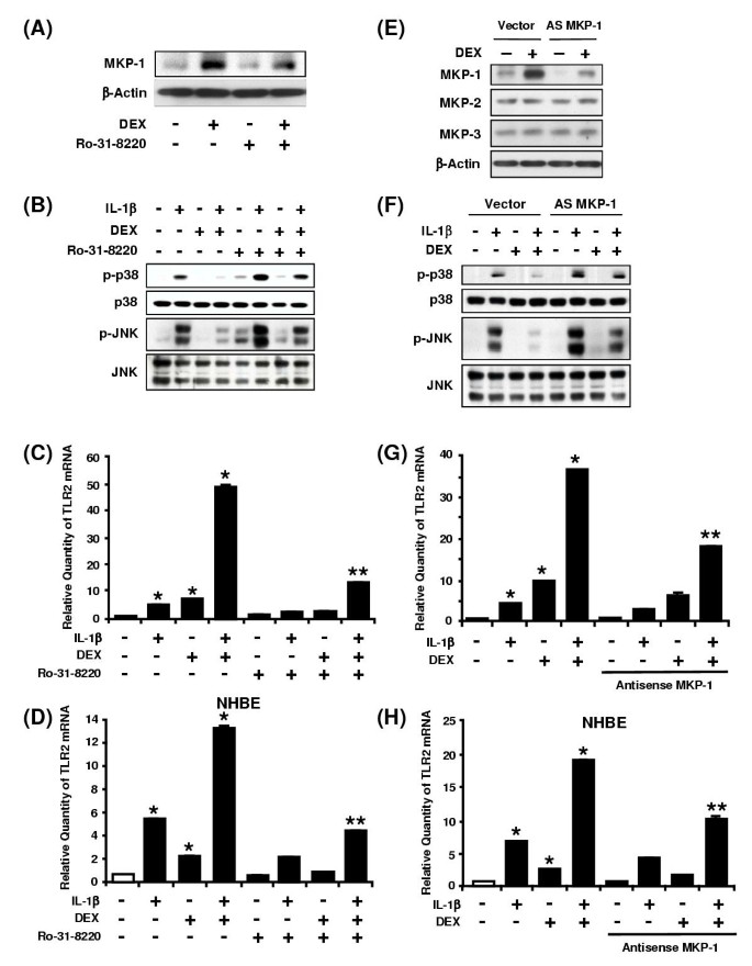
Having demonstrated that overexpression of MKP-1 inhibits IL-1β-induced p38 and JNK phosphorylation and enhances TLR2 induction, we next sought to determine whether inhibition of MKP-1 expression blocks the enhancing effect of DEX on TLR2 induction via negative cross-talks with p38 and JNK. We first confirmed that Ro-31-8220, an inhibitor for MKP-1 expression, inhibited DEX-mediated MKP-1 up-regulation at protein level (Figure 7a). We then determined whether Ro-31-8220 blocks the DEX-mediated inhibition of IL-1β-induced p38 or JNK phosphorylation. As shown in Figure 7b, the inhibitory effect of DEX on IL-1β-induced phosphorylation of p38 or JNK was blocked by Ro-31-8220. Moreover, DEX-mediated synergistic enhancement of IL-1β-induced TLR2 expression was greatly inhibited by Ro-31-8220 in both HeLa cells (Figure 7c) and primary NHBE cells (Figure 7d). These data suggest that DEX-mediated synergistic enhancement of TLR2 induction is mediated by up-regulation of MKP-1.
Figure 7
Inhibition of MKP-1 attenuates dexamethasone-mediated inhibition of IL-1β-induced p38 and JNK phosphorylation and enhancement of TLR2 expression. A, MKP-1 protein expression is induced by DEX and the MKP-1 induction is inhibited by Ro-31-8220, an inhibitor for MKP-1 expression, in HeLa cells. HeLa cells were first treated with Ro-31-8220 (10-6 M) for 2 h, and were then treated with DEX (10-6 M) for 2 h. Protein level of MKP-1 was assessed by Western blot analysis. β-Actin was used as a protein loading control. B, The inhibitory effect of DEX on IL-1β-induced p38 or JNK phosphorylation is antagonized by Ro-31-8220. HeLa cells were first treated with Ro-31-8220 (10-6 M) for 2 h and were then treated with DEX (10-6 M) for 2 h. The cells were stimulated with IL-1β (10 ng/ml) for 20 min. IL-1β-induced phosphorylation of p38 or JNK was then assessed by Western blot analysis. C, DEX-mediated enhancement of IL-1β-induced TLR2 expression is greatly inhibited by Ro-31-8220 in HeLa cells. HeLa cells were first treated with Ro-31-8220 (10-6 M) for 2 h and were then treated with DEX (10-6 M) for 2 h. The cells were stimulated with IL-1β (10 ng/ml) for 3 h, and the TLR2 mRNA levels were then assessed by real-time quantitative PCR. Values are the mean ± SD; n = 3. The symbols indicate results significantly different (p < 0.01) from unstimulated condition (*) and IL-1β-DEX-stimulated condition (without Ro-31-8220)(**). D, DEX-mediated enhancement of IL-1β-induced TLR2 expression is greatly inhibited by Ro-31-8220 in NHBE cells. NHBE cells were first treated with Ro-31-8220 (10-6 M) for 2 h and were then treated with DEX (10-6 M) for 2 h. The cells were stimulated with IL-1β (10 ng/ml) for 3 h. Values are the mean ± SD; n = 3. The symbols indicate results significantly different (p < 0.01) from unstimulated condition (*) and IL-1β-DEX-stimulated condition (without Ro-31-8220)(**). E, Overexpression of the antisense MKP-1 inhibited MKP-1 protein expression, but not MKP-2 or MKP-3 expression. Transfected HeLa cells were treated with DEX (10-6 M) for 2 h. Protein level of MKP-1, -2 and -3 were assessed by Western blot analysis. β-Actin was used as a protein loading control. F, Overexpression of antisense MKP-1 blocks the inhibitory effect of DEX on IL-1β-induced p38 or JNK phosphorylation in HeLa cells. Transfected HeLa cells were first pretreated with DEX (10-6 M) for 2 h. The cells were then stimulated with IL-1β (10 ng/ml) for 20 min. IL-1β-induced phosphorylation of p38 or JNK was next assessed by Western blot analysis. G, DEX-mediated enhancement of IL-1β-induced TLR2 expression is attenuated by overexpression of antisense MKP-1 in HeLa cells. Transfected HeLa cells were pretreated with DEX (10-6 M) for 2 h, and then stimulated with IL-1β (10 ng/ml) for 3 h. Values are the mean ± SD; n = 3. The symbols indicate results significantly different (p < 0.01) from unstimulated condition (*) and IL-1β-DEX-stimulated condition (without antisense MKP-1 construct)(**). H, DEX-mediated enhancement of IL-1β-induced TLR2 expression is also attenuated by overexpression of antisense MKP-1 in NHBE cells. Transfected cells werepretreated with DEX (10-6 M) for 2 h, and then stimulated with IL-1β (10 ng/ml) for 3 h. Values are the mean ± SD; n = 3. The symbols indicate results significantly different (p < 0.01) from unstimulated condition (*) and IL-1β-DEX-stimulated condition (without antisense MKP-1 construct)(**).
To further confirm whether the up-regulation of MKP-1 by glucocorticoids is indeed required for the enhancement of IL-1β-induced TLR2 expression, we investigated the effect of overexpressing an antisense full-length MKP-1 construct on the glucocorticoid-mediated enhancement of TLR2 induction [27]. As shown in Figure 7e, we first confirmed that overexpression of the antisense MKP-1 construct specifically inhibited MKP-1 protein expression, but not MKP-2 or MKP-3 expression. In addition, overexpression of the antisense MKP-1 construct antagonized the inhibitory effect of DEX on IL-1β-induced phosphorylation of p38 or JNK (Figure 7f). Moreover, overexpression of the same antisense MKP-1 also inhibited the DEX-mediated enhancement of IL-1β-induced TLR2 expression in both HeLa cells and primary human bronchial epithelial NHBE cells (Figure 7g and 7h). Taken together, our data demonstrate that glucocorticoids synergistically enhance IL-1β-induced TLR2 expression via up-regulation of MKP-1 that, in turn, leads to dephosphorylation and inactivation of p38 and JNK, the negative regulators for TLR2 expression in human epithelial cells.
Discussion
Despite the importance of glucocorticoids in suppressing immune and inflammatory responses, their role in enhancing host immune and defense response against invading bacterial pathogens is poorly understood [13, 14, 28, 29]. Although there are reports that glucocorticoids may have a beneficial role in certain bacterial infections, the molecular basis for their beneficial effects still remains unclear. In the present study, we provided evidence that glucocorticoids synergistically enhance the TLR2 expression, a key receptor that has been shown to play a crucial role in host defense response, as evidenced by the studies of TLR2 knockout mice showing decreased survival after infection with Gram-positive S. aureus [11]. Thus, it appears that glucocorticoids may not only suppress but also likely enhance host immune and defense response. In addition, our study may provide the molecular basis for the explanation of the beneficial role of glucocorticoids in certain bacterial infections. Given the fact that TLR2 expression is low in unstimulated epithelial cells, the synergistic enhancement of IL-1β-induced TLR2 expression by glucocorticoids will probably contribute to the accelerated immune response by epithelial cells as well as resensitization of epithelial cells to invading pathogens. It should be noted that glucocorticoids enhance the TLR2 expression induced not only by IL-1β, but also by a variety of other pathogenic inducers including the Gram-negative bacterium nontypeable Haemophilus influenzae, the Gram-positive bacterium Streptococcus pneumoniae, and the proinflammatory cytokines TNF-α and IL-1α (data not shown). Therefore, up-regulation of the host defense receptor, TLR2, may be one of the important positive immune-regulatory mechanisms widely involved in the glucocorticoid-mediated host defense against many microbial pathogens. Our studies may bring new insights into the novel role of glucocorticoids in orchestrating and optimizing immune functions, including host defense, during bacterial infections.
Another interesting finding in this study is that glucocorticoids synergistically enhance the IL-1β-induced TLR2 expression via a MKP-1-mediated inactivation of both MAPK p38 and JNK, the negative regulators for TLR2 induction. Although several MAPKs have been identified as potential targets for negative regulation by glucocorticoids [25, 30–36], the signaling mechanisms underlying the inhibition of MAPKs by glucocorticoids still remain largely unknown. Our finding that glucocorticoids inhibit the IL-1β-induced phosphorylation and activation of MAPKs p38 and JNK via up-regulation of MKP-1 may enhance our understanding of the signaling mechanisms underlying the glucocorticoid-mediated attenuation of MAPKs. In agreement with recent reports that glucocorticoids inhibit p38 via up-regulation of MKP-1 [24, 26], our data showed that glucocorticoids enhanced IL-1β-induced TLR2 expression via up-regulating MKP-1 expression, that, in turn, leads to the dephosphorylation and inactivation of p38 in HeLa cells and primary NHBE cells. However, our finding that glucocorticoids inhibit the IL-1β-induced phosphorylation of JNK via an up-regulation of MKP-1 is rather unexpected, because it is in sharp contrast with the previous report that glucocorticoid-mediated inhibition of JNK does not require any de novo protein synthesis and independent of the transactivation function of GR [31]. Similar to the glucocorticoid-mediated inactivation of p38, glucocorticoids require ongoing MKP-1 synthesis to inhibit phosphorylation of ERK1/2 [25, 37]. Hence, multiple signaling mechanisms are likely to underlie the regulation of MAPKs by glucocorticoids.
The molecular mechanisms underlying p38 and JNK-mediated inhibition of TLR2 induction by IL-1β still remain unclear. The human TLR2 promoter contains multiple transcription factor binding elements including Sp1 and Ets-1 [38]. Sp1 and Ets-1 have been shown to be regulated by p38 signaling pathway [39, 40]. Activation of Sp1 and Ets family through JNK has been also reported [41, 42]. Thus, Sp1 and Ets-1 could be involved in p38 and JNK-mediated inhibition of TLR2 expression. TLR2 promoter region also contains some uncharacterized regulatory elements [38]. These elements may also play a role in transcriptional inhibition of TLR2 mediated by p38 and JNK signaling. In addition to these transcription factors, the possible involvement of the regulation of mRNA stability by p38 and JNK can not be ruled out because both have been shown to be involved in regulating the mRNA stability of certain genes [43–46].
Although we have shown the involvement of multiple signaling pathways including the positive IKKβ-IκBα-dependent NF-κB pathway and the negative MKK3/6-p38 α/β and MEKK1-MKK4/7-JNK1/2 pathways, our data do not preclude the involvement of other signaling mechanisms such a direct transcriptional activation of TLR2 by glucocorticoids, or direct cross-talk between GR and other transcription factors that can be activated by the above signaling pathways. Future studies will focus on elucidating the signaling mechanisms underlying the glucocorticoid-mediated synergistic enhancement of IL-1β-induced TLR2 at the transcriptional level by taking advantage of the recent success in cloning the regulatory region of human TLR2. In addition, the role of MKP-1 in mediating the enhancement of TLR2 induction by glucocorticoids will also be confirmed in vivo by using an MKP-1 knockout mouse strain. Moreover, it is also interesting to determine whether glucocorticoids also enhance antibacterial defense responses.
Conclusion
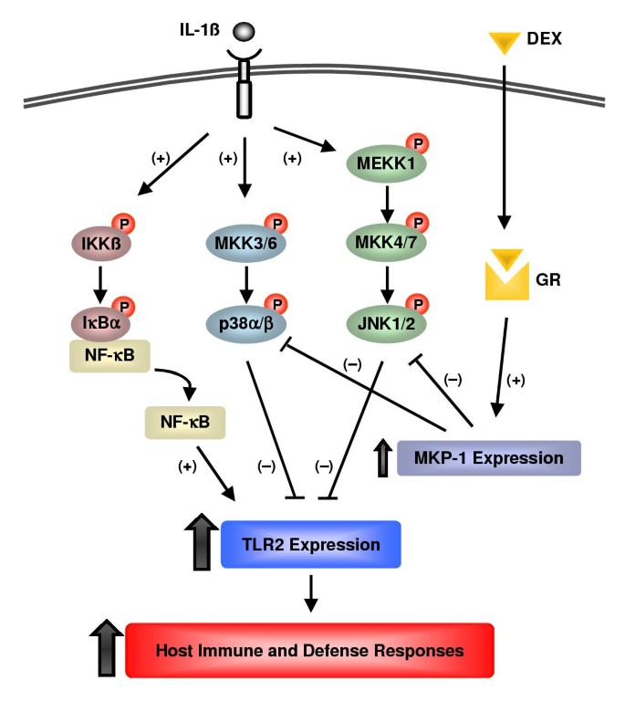
We provided direct evidence that glucocorticoids synergistically enhance the IL-1β-induced TLR2 expression, a key host defense receptor, via specific up-regulation of MKP-1 expression, which, in turn, leads to the inactivation of both p38 and JNK signaling pathways, the negative regulators for TLR2 induction (Figure 8). These studies may bring new insights into the role of glucocorticoids in enhancing host defense response against bacterial invasion and lead to novel therapeutic intervention for modulating host defense and immune responses in bacterial infections.
Figure 8
Schematic representation of the signaling pathways involved in glucocorticoid-mediated enhancement of IL-1β-induced TLR2 expression via MKP-1-dependent inhibition of p38 and JNK in human epithelial cells. IL-1β up-regulates TLR2 expression via a positive IKKβ-IκBα-dependent NF-κB pathway and negative MKK3/6-p38α/β and MEKK1-MKK4/7-JNK1/2 signaling pathways. Glucocorticoids synergistically enhance IL-1β-induced TLR2 expression via up-regulation of MKP-1 that, in turn, leads to dephosphorylation and inactivation of p38 and JNK pathways, the negative regulators for TLR2 expression. Abbreviations: DEX, dexamethasone; GR, glucocorticoids receptor; IκBα, Inhibitor of NF-κBα ; IKKβ, IκB kinase β ; IL-1β, interleukin 1 β ; JNK, c-Jun N-terminal kinase;MAPK, mitogen-activated protein kinase; MKK, MAPK kinase; MEKK1, MAPK/Erk kinase kinase 1; MKP-1, MAP kinase phosphatase-1; NF-κB, nuclear factor κB; TLR2, Toll-like receptor 2.
Methods
Reagents
Human IL-1β was purchased from PIERCE ENDOGEN (Woburn, MA). MG-132, SB203580 and Ro-31-8220 were purchased from Calbiochem (La Jolla, CA). Dexamethasone and RU486 were purchased from Sigma (St. Louis, MO).
Cell culture
The human cervix epithelial cell line, HeLa, was maintained as described [6]. Primary human bronchial epithelial cells (NHBE) (Clonetics, Walkersville, MD) were maintained in Bronchial Epithelial Basal Medium (BEBM) (Clonetics) according to the manufacturer's instructions.
Real-time quantitative PCR analysis of TLR2 and TLR4
Total RNA was isolated from the cells using a TRIzol Reagent (Invitrogen Corporation, Carlsbad, CA) following manufacturer's instruction. For the RT reaction, TaqMan Reverse Transcription Regents (Applied Biosystems, Foster City, CA) were used. The real-time quantitative PCR was performed using an ABI 7700 sequence detector (Applied Biosystems). The primers and probes used for the PCR amplification of TLR2 were; p r i m e r s (5'-GGCCAGCAAATTACCTGTGTG-3' a n d 5'-AGGCGGACATCCTGAACCT-3') and a probe (5'-TCCATCCCATGTGCGTGGCC-3'). The primers and probes used for the PCR amplification of TLR4 were; primers (5'-CCAGTGAGGATGATGCCAGGAT-3' and 5'-GCCATGGCTGGGATCAGAGT-3') and a probe (5'-TGTCTGCCTCGCGCCTGGC-3'). All primers and probes were synthesized by Applied Biosystems Custom Oligo Synthesis Service. Relative quantity of TLR2 and TLR4 mRNA were obtained using Comparative CT Method, and was normalized using Pre-Developed TaqMan Assay Reagent Human Cyclophilin (Applied Biosystems) as an endogenous control as previously described [24].
Plasmids and transfections
The expression plasmids IκBα(S32/36A), IKKβ (K49A), fp38α (AF) and wild-type (WT), fp38β2 (AF) and WT, MKK3b (A), MKK6b (A), JNK1 (AF), JNK2 (AF), MKK4 DN, MKK7 DN and MEKK1 DN were previously described [6, 47]. The expression plasmid of the wild-type and mutant MKP-1 were kindly provided by Dr. N. Tonks (Cold Spring Harbor Laboratory, NY) [48]. The antisense full-length MKP-1 construct was kindly provided by Dr. Desbois-Mouthon (INSERM U-402, Faculté de Médecine Saint-Antoine, 75571 Paris, France) and was described previously [27]. All transient transfections were carried out in duplicate for RT-PCR analysis using TransIT-LT1 reagent (Mirus, Madison, WI) following manufacturer's instruction, unless otherwise indicated. In all transfections with expression plasmids of signaling molecules, an empty vector was used as a control.
Western Blot analysis
Phosphorylation of IκBα, JNK, p38, MKK4 and MKK3/6 was assessed using antibodies against phospho-IκBα (Ser32), IκBα, phospho-JNK (Thr183/Tyr185), JNK, phospho-p38 (Thr180/182), p38, phospho-MKK4 (Thr261), phospho-MKK3/MKK6 (Ser189/207) and MKK3 (New England Biolabs, Beverly, MA). Anti-MKK4 (K-18) was purchased from Santa Cruz Biotechnology, Inc. Santa Cruz, CA). Phosphorylation of of IκBα, JNK, p38, MKK4 and MKK3/6 was detected as described [49]. Anti-MKP-1 (V-15), anti-MKP-2 (H-67) and anti-MKP-3 (C-20) antibodies were purchased from Santa Cruz, and used to assess protein expression. TLR2 expression was assessed with an anti-TLR2 antibody (Imgenex Corporation, San Diego, CA). Monoclonal anti-β-Actin antibody (Sigma) was used as a protein loading control for Western blot analyses of MKP-1, MKP-2, MKP-3 and TLR2.
Immunofluorescent staining
Cells were cultured on 4-chamber microscopeslides. After IL-1β treatment, the cells were fixed in paraformaldehyde solution (4%), incubated with mouse anti-p65 NF-κB monoclonal antibodies for 1 h (Santa Cruz Biotechnology, Inc.). Primary antibody was detected with fluorescein isothiocyanate (FITC)-conjugated goat anti-mouse IgG (Santa Cruz Biotechnology, Inc.). Samples were viewed and photographed using a Zeiss Axiophot microscope.
Statistical analysis
Data were analyzed using Student's t test or ANOVA and Tukey's post hoc test. A value of p < 0.05 was considered significant.
References
- Medzhitov R, Janeway C: The Toll receptor family and microbial recognition. Trends Microbiol. 2000, 8: 452-456. 10.1016/S0966-842X(00)01845-X
Article CAS PubMed Google Scholar - Aderem A, Ulevitch RJ: Toll-like receptors in the induction of the innate immune response. Nature. 2000, 406: 782-787. 10.1038/35021228
Article CAS PubMed Google Scholar - Medzhitov R, Preston-Hurlburt P, Janeway CA: A human homologue of the Drosophila Toll protein signals activation of adaptive immunity. Nature. 1997, 388: 394-397. 10.1038/41131
Article CAS PubMed Google Scholar - Means TK, Golenbock DT, Fenton MJ: Structure and function of Toll-like receptor proteins. Life Sci. 2000, 68: 241-258. 10.1016/S0024-3205(00)00939-5
Article CAS PubMed Google Scholar - O'Neill LA, Dinarello CA: The IL-1 receptor/Toll-like receptor superfamily: crucial receptors for inflammation and host defense. Immunol Today. 2000, 21: 206-209. 10.1016/S0167-5699(00)01611-X
Article PubMed Google Scholar - Shuto T, Xu H, Wang B, Han J, Kai H, Gu XX, Murphy TF, Lim DJ, Li JD: Activation of NF-κB by nontypeable Hemophilus influenzae is mediated by Toll-like receptor 2-TAK1-dependent NIK-IKKα/β-IκBα and MKK3/6-p38 MAP kinase signaling pathways in epithelial cells. Proc Natl Acad Sci U S A. 2001, 98: 8774-8779. 10.1073/pnas.151236098
Article PubMed Central CAS PubMed Google Scholar - Jono H, Xu H, Kai H, Lim DJ, Kim YS, Feng XH, Li JD: Transforming growth factor-β-smad signaling pathway negatively regulates nontypeable _Haemophilus influenzae_-induced MUC5AC mucin transcription via mitogen-activated protein kinase (MAPK) phosphatase-1-dependent inhibition of p38 MAPK. J Biol Chem. 2003, 278: 27811-27819. 10.1074/jbc.M301773200
Article CAS PubMed Google Scholar - Wyllie DH, Kiss-Toth E, Visintin A, Smith SC, Boussouf S, Segal DM, Duff GW, Dower SK: Evidence for an accessory protein function for Toll-like receptor 1 in anti-bacterial responses. J Immunol. 2000, 165: 7125-7132.
Article CAS PubMed Google Scholar - Takeuchi O, Kaufmann A, Grote K, Kawai T, Hoshino K, Morr M, Muhlradt PF, Akira S: Cutting edge: preferentially the R-stereoisomer of the mycoplasmal lipopeptide macrophage-activating lipopeptide-2 activates immune cells through a Toll-like receptor 2- and MyD88-dependent signaling pathway. J Immunol. 2000, 164: 554-557.
Article CAS PubMed Google Scholar - Takeuchi O, Kawai T, Muhlradt PF, Morr M, Radolf JD, Zychlinsky A, Takeda K, Akira S: Discrimination of bacterial lipoproteins by Toll-like receptor 6. Int Immunol. 2001, 13: 933-940. 10.1093/intimm/13.7.933
Article CAS PubMed Google Scholar - Takeuchi O, Hoshino K, Akira S: Cutting edge: TLR2-deficient and MyD88-deficient mice are highly susceptible to Staphylococcus aureus infection. J Immunol. 2000, 165: 5392-5396.
Article CAS PubMed Google Scholar - Shuto T, Imasato A, Jono H, Sakai A, Xu H, Watanabe T, Rixter DD, Kai H, Andalibi A, Linthicum F, Guan YL, Han J, Cato AC, Lim DJ, Akira S, Li JD: Glucocorticoids synergistically enhance nontypeable _Haemophilus influenzae_-induced Toll-like receptor 2 expression via a negative crosstalk with p38 MAP kinase. J Biol Chem. 2002, 277: 17263-17270. 10.1074/jbc.M112190200
Article CAS PubMed Google Scholar - Wilckens T: Glucocorticoids and immune function: physiological relevance and pathogenic potential of hormonal dysfunction. Trends Pharmacol Sci. 1995, 16: 193-197. 10.1016/S0165-6147(00)89021-5
Article CAS PubMed Google Scholar - Wilckens T, De Rijk R: Glucocorticoids and immune function: unknown dimensions and new frontiers. Immunol Today. 1997, 18: 418-424. 10.1016/S0167-5699(97)01111-0
Article CAS PubMed Google Scholar - O'Neill L: The Toll/interleukin-1 receptor domain: a molecular switch for inflammation and host defence. Biochem Soc Trans. 2000, 28: 557-563.
Article PubMed Google Scholar - Bird S, Zou J, Wang T, Munday B, Cunningham C, Secombes CJ: Evolution of interleukin-1β. Cytokine Growth Factor Rev. 2002, 13: 483-502. 10.1016/S1359-6101(02)00028-X
Article CAS PubMed Google Scholar - Bankers-Fulbright JL, Kalli KR, McKean DJ: Interleukin-1 signal transduction. Life Sci. 1996, 59: 61-83. 10.1016/0024-3205(96)00135-X
Article CAS PubMed Google Scholar - Ono K, Han J: The p38 signal transduction pathway: activation and function. Cell Signal. 2000, 12: 1-13. 10.1016/S0898-6568(99)00071-6
Article CAS PubMed Google Scholar - Wang X, McGowan CH, Zhao M, He L, Downey JS, Fearns C, Wang Y, Huang S, Han J: Involvement of the MKK6-p38γ cascade in γ-radiation-induced cell cycle arrest. Mol Cell Biol. 2000, 20: 4543-4552. 10.1128/MCB.20.13.4543-4552.2000
Article PubMed Central CAS PubMed Google Scholar - Ge B, Gram H, Di Padova F, Huang B, New L, Ulevitch RJ, Luo Y, Han J: MAPKK-independent activation of p38α mediated by TAB1-dependent autophosphorylation of p38α. Science. 2002, 295: 1291-1294. 10.1126/science.1067289
Article CAS PubMed Google Scholar - Barnes PJ: Anti-inflammatory actions of glucocorticoids: molecular mechanisms. Clin Sci (Lond). 1998, 94: 557-572.
Article CAS Google Scholar - Konig H, Ponta H, Rahmsdorf HJ, Herrlich P: Interference between pathway-specific transcription factors: glucocorticoids antagonize phorbol ester-induced AP-1 activity without altering AP-1 site occupation in vivo. Embo J. 1992, 11: 2241-2246.
PubMed Central CAS PubMed Google Scholar - Nissen RM, Yamamoto KR: The glucocorticoid receptor inhibits NF-κB by interfering with serine-2 phosphorylation of the RNA polymerase II carboxy-terminal domain. Genes Dev. 2000, 14: 2314-2329. 10.1101/gad.827900
Article PubMed Central CAS PubMed Google Scholar - Imasato A, Desbois-Mouthon C, Han J, Kai H, Cato AC, Akira S, Li JD: Inhibition of p38 MAPK by glucocorticoids via induction of MAPK phosphatase-1 enhances nontypeable _Haemophilus influenzae_-induced expression of Toll-like receptor 2. J Biol Chem. 2002, 277: 47444-47450. 10.1074/jbc.M208140200
Article CAS PubMed Google Scholar - Kassel O, Sancono A, Kratzschmar J, Kreft B, Stassen M, Cato AC: Glucocorticoids inhibit MAP kinase via increased expression and decreased degradation of MKP-1. Embo J. 2001, 20: 7108-7116. 10.1093/emboj/20.24.7108
Article PubMed Central CAS PubMed Google Scholar - Lasa M, Abraham SM, Boucheron C, Saklatvala J, Clark AR: Dexamethasone causes sustained expression of mitogen-activated protein kinase (MAPK) phosphatase 1 and phosphatase-mediated inhibition of MAPK p38. Mol Cell Biol. 2002, 22: 7802-7811. 10.1128/MCB.22.22.7802-7811.2002
Article PubMed Central CAS PubMed Google Scholar - Desbois-Mouthon C, Cadoret A, Blivet-Van Eggelpoel MJ, Bertrand F, Caron M, Atfi A, Cherqui G, Capeau J: Insulin-mediated cell proliferation and survival involve inhibition of c-Jun N-terminal kinases through a phosphatidylinositol 3-kinase- and mitogen-activated protein kinase phosphatase-1-dependent pathway. Endocrinology. 2000, 141: 922-931. 10.1210/en.141.3.922
CAS PubMed Google Scholar - Karin M: New twists in gene regulation by glucocorticoid receptor: is DNA binding dispensable?. Cell. 1998, 93: 487-490. 10.1016/S0092-8674(00)81177-0
Article CAS PubMed Google Scholar - Webster JC, Cidlowski JA: Mechanisms of glucocorticoid-receptor-mediated repression of gene expression. Trends Endocrinol Metab. 1999, 10: 396-402. 10.1016/S1043-2760(99)00186-1
Article CAS PubMed Google Scholar - Rider LG, Hirasawa N, Santini F, Beaven MA: Activation of the mitogen-activated protein kinase cascade is suppressed by low concentrations of dexamethasone in mast cells. J Immunol. 1996, 157: 2374-2380.
CAS PubMed Google Scholar - Caelles C, Gonzalez-Sancho JM, Munoz A: Nuclear hormone receptor antagonism with AP-1 by inhibition of the JNK pathway. Genes Dev. 1997, 11: 3351-3364.
Article PubMed Central CAS PubMed Google Scholar - Swantek JL, Cobb MH, Geppert TD: Jun N-terminal kinase/stress-activated protein kinase (JNK/SAPK) is required for lipopolysaccharide stimulation of tumor necrosis factor alpha (TNF-α) translation: glucocorticoids inhibit TNF-α translation by blocking JNK/SAPK. Mol Cell Biol. 1997, 17: 6274-6282.
Article PubMed Central CAS PubMed Google Scholar - Hirasawa N, Sato Y, Fujita Y, Mue S, Ohuchi K: Inhibition by dexamethasone of antigen-induced c-Jun N-terminal kinase activation in rat basophilic leukemia cells. J Immunol. 1998, 161: 4939-4943.
CAS PubMed Google Scholar - Hulley PA, Gordon F, Hough FS: Inhibition of mitogen-activated protein kinase activity and proliferation of an early osteoblast cell line (MBA 15.4) by dexamethasone: role of protein phosphatases. Endocrinology. 1998, 139: 2423-2431. 10.1210/en.139.5.2423
CAS PubMed Google Scholar - Gonzalez MV, Gonzalez-Sancho JM, Caelles C, Munoz A, Jimenez B: Hormone-activated nuclear receptors inhibit the stimulation of the JNK and ERK signalling pathways in endothelial cells. FEBS Lett. 1999, 459: 272-276. 10.1016/S0014-5793(99)01257-0
Article CAS PubMed Google Scholar - Gewert K, Hiller G, Sundler R: Effects of dexamethasone on mitogen-activated protein kinases in mouse macrophages: implications for the regulation of 85 kDa cytosolic phospholipase A(2). Biochem Pharmacol. 2000, 60: 545-551. 10.1016/S0006-2952(00)00358-0
Article CAS PubMed Google Scholar - Engelbrecht Y, de Wet H, Horsch K, Langeveldt CR, Hough FS, Hulley PA: Glucocorticoids induce rapid up-regulation of mitogen-activated protein kinase phosphatase-1 and dephosphorylation of extracellular signal-regulated kinase and impair proliferation in human and mouse osteoblast cell lines. Endocrinology. 2003, 144: 412-422. 10.1210/en.2002-220769
Article PubMed Central CAS PubMed Google Scholar - Haehnel V, Schwarzfischer L, Fenton MJ, Rehli M: Transcriptional regulation of the human Toll-like receptor 2 gene in monocytes and macrophages. J Immunol. 2002, 168: 5629-5637.
Article CAS PubMed Google Scholar - Tanaka K, Oda N, Iwasaka C, Abe M, Sato Y: Induction of Ets-1 in endothelial cells during reendothelialization after denuding injury. J Cell Physiol. 1998, 176: 235-244. 10.1002/(SICI)1097-4652(199808)176:2<235::AID-JCP2>3.0.CO;2-P
Article CAS PubMed Google Scholar - Ma W, Lim W, Gee K, Aucoin S, Nandan D, Kozlowski M, Diaz-Mitoma F, Kumar A: The p38 mitogen-activated kinase pathway regulates the human interleukin-10 promoter via the activation of Sp1 transcription factor in lipopolysaccharide-stimulated human macrophages. J Biol Chem. 2001, 276: 13664-13674.
CAS PubMed Google Scholar - Tanaka T, Kanai H, Sekiguchi K, Aihara Y, Yokoyama T, Arai M, Kanda T, Nagai R, Kurabayashi M: Induction of VEGF gene transcription by IL-1β is mediated through stress-activated MAP kinases and Sp1 sites in cardiac myocytes. J Mol Cell Cardiol. 2000, 32: 1955-1967. 10.1006/jmcc.2000.1228
Article CAS PubMed Google Scholar - Yordy JS, Muise-Helmericks RC: Signal transduction and the Ets family of transcription factors. Oncogene. 2000, 19: 6503-6513. 10.1038/sj.onc.1204036
Article CAS PubMed Google Scholar - Guan Z, Baier LD, Morrison AR: p38 mitogen-activated protein kinase down-regulates nitric oxide and up-regulates prostaglandin E2 biosynthesis stimulated by interleukin-1β. J Biol Chem. 1997, 272: 8083-8089. 10.1074/jbc.272.12.8083
Article CAS PubMed Google Scholar - Chen CY, Gherzi R, Andersen JS, Gaietta G, Jurchott K, Royer HD, Mann M, Karin M: Nucleolin and YB-1 are required for JNK-mediated interleukin-2 mRNA stabilization during T-cell activation. Genes Dev. 2000, 14: 1236-1248.
PubMed Central CAS PubMed Google Scholar - Frevel MA, Bakheet T, Silva AM, Hissong JG, Khabar KS, Williams BR: p38 Mitogen-activated protein kinase-dependent and-independent signaling of mRNA stability of AU-rich element-containing transcripts. Mol Cell Biol. 2003, 23: 425-436. 10.1128/MCB.23.2.425-436.2003
Article PubMed Central CAS PubMed Google Scholar - Subbaramaiah K, Marmao TP, Dixon DA, Dannenberg AJ: Regulation of cyclooxygenase-2 mRNA stability by taxanes. Evidence for involvement of p38, MAPKAPK-2 and HuR. J Biol Chem. 2003, 278: 37637-37647. 10.1074/jbc.M301481200
Article CAS PubMed Google Scholar - Jono H, Xu H, Kai H, Lim DJ, Gum JR, Kim YS, Yamaoka S, Feng XH, Li JD: Transforming growth factor-β-Smad signaling pathway cooperates with NF-κB to mediate nontypeable _Haemophilus influenzae_-induced MUC2 mucin transcription. J Biol Chem. 2002, 277: 45547-45557. 10.1074/jbc.M206883200
Article CAS PubMed Google Scholar - Sun H, Charles CH, Lau LF, Tonks NK: MKP-1 (3CH134), an immediate early gene product, is a dual specificity phosphatase that dephosphorylates MAP kinase in vivo. Cell. 1993, 75: 487-493. 10.1016/0092-8674(93)90383-2
Article CAS PubMed Google Scholar - Wang B, Lim DJ, Han J, Kim YS, Basbaum CB, Li JD: Novel cytoplasmic proteins of nontypeable Haemophilus influenzae up-regulate human MUC5AC mucin transcription via a positive p38 mitogen-activated protein kinase pathway and a negative phosphoinositide 3-kinase-Akt pathway. J Biol Chem. 2002, 277: 949-957. 10.1074/jbc.M107484200
Article CAS PubMed Google Scholar
Acknowledgements
We are grateful to Dr. N. Tonks and Dr. C. Desbois-Mouthon for various reagents. We thank Dr. A. Andalibi for critically reviewing this manuscript. This work is supported by grants from NIH HL070293, DC004562, and DC005843 (to J.D. Li).
Author information
Authors and Affiliations
- Gonda Department of Cell and Molecular Biology, House Ear Institute, and the Department of Otolaryngology, University of Southern California, Los Angeles, CA, 90057, USA
Akihiro Sakai & Jian-Dong Li - Department of Immunology, the Scripps Research Institute, La Jolla, CA, 92037, USA
Jiahuai Han - Forschungszentrum Karlsruhe, Institute of Toxicology & Genetics, P.O. Box 3640, D-76021, Karlsruhe, Germany
Andrew CB Cato - Department of Host Defense, Research Institute for Microbial Diseases, Osaka University, CREST of Japan Science and Technology Corporation, 3-1 Yamada-oka, Suita, Osaka, 565-0871, Japan
Shizuo Akira
Authors
- Akihiro Sakai
You can also search for this author inPubMed Google Scholar - Jiahuai Han
You can also search for this author inPubMed Google Scholar - Andrew CB Cato
You can also search for this author inPubMed Google Scholar - Shizuo Akira
You can also search for this author inPubMed Google Scholar - Jian-Dong Li
You can also search for this author inPubMed Google Scholar
Corresponding author
Correspondence toJian-Dong Li.
Additional information
Authors' contributions
AS carried all the experiments described. JH was involved in advising and designing the experiments to investigate the role of p38 MAPK and JNK in TLR2 induction; AC was involved in advising the experiments to explore the role of MKP-1 in mediating GCs-mediated enhancement of TLR2; SA was involved in advising the experiments to explore the molecular details of GCs-mediated TLR2 induction. JDL was involved in hypothesizing and designing the experiments to test that IL-1 induces TLR2 via a complex signaling network involving both NF-κB and MPAK pathways. All authors read and approved the final manuscript.
Authors’ original submitted files for images
Rights and permissions
This article is published under license to BioMed Central Ltd. This is an Open Access article: Verbatim copying and redistribution of this article are permitted in all media for any purpose, provided this notice is preserved along with the article's original URL.
About this article
Cite this article
Sakai, A., Han, J., Cato, A.C. et al. Glucocorticoids synergize with IL-1β to induce TLR2 expression via MAP Kinase Phosphatase-1-dependent dual Inhibition of MAPK JNK and p38 in epithelial cells.BMC Molecular Biol 5, 2 (2004). https://doi.org/10.1186/1471-2199-5-2
- Received: 17 December 2003
- Accepted: 04 May 2004
- Published: 04 May 2004
- DOI: https://doi.org/10.1186/1471-2199-5-2
