Toxic Megacolon (Toxic Colitis) Imaging: Practice Essentials, Radiography, Computed Tomography (original) (raw)
Practice Essentials
Toxic megacolon (or toxic colitis) is defined as a severe episode of colitis with segmental or total dilatation of the colon. It is typically a complication of ulcerative colitis, but it may be a complication of Crohn disease, antibiotic-related pseudomembranous colitis, and other colitides. [1] Pathologically, acute fulminant colitis is associated with neuromuscular degeneration and a rapid and extensive colonic dilatation. [2, 3, 4, 5, 6, 7, 8, 9]
Patients with toxic megacolon often present in the emergency department as having abdominal distention superimposed on chronic or acute diarrhea. The diagnosis should be considered in all such patients. The diagnosis is usually based on thorough clinical history taking and physical examination combined with plain abdominal radiography. [10, 11, 12, 13, 14]
On radiographs, the transverse colon is often identified as dilated because of the anterosuperior position. Diameter greater than 6 cm is considered a highly suspicious finding. Images may show a coarse, irregular mucosal pattern of the large bowel known as thumbprinting, which is caused by mucosal edema due to inflammatory infiltration. [15, 1] CT findings can highlight the extent of involvement (eg, edema), inflammation, and other features such as ascites and abscesses. [15] Ultrasonography and radionuclide studies may have a limited role.
Technetium-99m (99mTc) hexamethyl-propyleneamine oxime (HMPAO)–labeled WBC scanning can be used as an alternative to colonoscopy to assess the extent and severity of the disease in critically ill patients with ulcerative colitis. This technique decreases the number and severity of complications that may occur in these patients. However, the role of this method of scintigraphy is limited in the diagnosis of toxic megacolon and in the determination of its severity. [16]
Chagas disease, Hirschsprung disease, and intestinal pseudo-obstruction may superficially resemble toxic megacolon on plain radiographs. [17] However, because they occur in totally different clinical settings, they are unlikely to be confused with toxic megacolon.
(See the images below.)
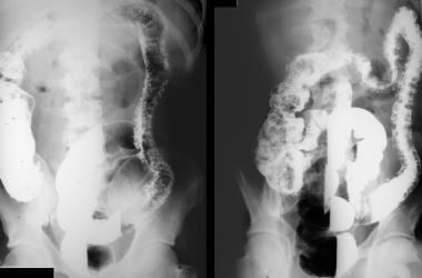
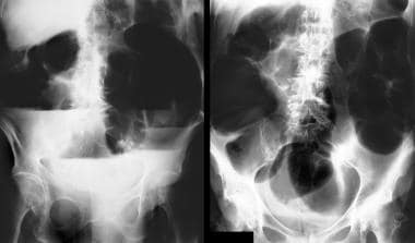
Double-contrast barium enema studies in a 44-year-old man known to have long history of ulcerative colitis. Images show total colitis and extensive pseudopolyposis.
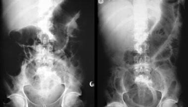
A 22-year-old man presented with abdominal pain, passage of blood and mucus per rectum, abdominal distention, fever, and disorientation. Findings from sigmoidoscopy confirmed ulcerative colitis. Abdominal radiographs obtained 2 days apart show mucosal edema and worsening of the distention in the transverse colon. The patient's clinical condition deteriorated over the next 36 hours despite steroid and antibiotic therapy, and the patient had to undergo a total colectomy and ileostomy.
![]()
Radiography
Toxic megacolon is a clinical diagnosis, one based on thorough history taking and physical examination and supported by plain abdominal radiographic findings. A diagnosis of toxic megacolon can be made fairly confidently by using plain radiography in the appropriate clinical setting, although a series of radiographs may be required.
If toxic megacolon is clinically suspected, patients are usually followed up with plain abdominal radiography every 12-24 hours, depending on the patient's clinical condition. A single abdominal radiograph may not be sufficient and should be combined with a horizontal-beam radiograph, which may better depict large, dilated bowel loops with fluid levels. Also, abdominal perforation is less likely to be missed. (See the images below).
Double-contrast barium enema studies in a 44-year-old man known to have long history of ulcerative colitis. Images show total colitis and extensive pseudopolyposis.
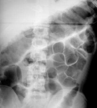
Plain abdominal radiograph. The patient presented with an acute exacerbation of symptoms. Image shows thumbprinting in the region of the splenic flexure of the colon.
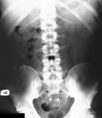
Plain abdominal radiograph that shows distention of the transverse colon associated with mucosal edema. The maximum transverse diameter of the transverse colon is 7.5 cm.
A 22-year-old man presented with abdominal pain, passage of blood and mucus per rectum, abdominal distention, fever, and disorientation. Findings from sigmoidoscopy confirmed ulcerative colitis. Abdominal radiographs obtained 2 days apart show mucosal edema and worsening of the distention in the transverse colon. The patient's clinical condition deteriorated over the next 36 hours despite steroid and antibiotic therapy, and the patient had to undergo a total colectomy and ileostomy.
A 72-year-old woman presented with vomiting and abdominal distention. The supine (right) and erect (left) plain abdominal radiographs show gross dilatation of the colon with multiple air-fluid levels. On further questioning, the patient revealed that she was taking diuretics for hypertension. Blood biochemical tests revealed markedly lowered potassium levels. After potassium replacement therapy, the patient's pseudo-obstruction completely resolved.
Megacolon is considered to be present if the diameter of the colon is 5.5 cm or more, with apparent edema of the bowel wall on plain abdominal radiographs. Rarely, the toxic dilatation may extend to the terminal ileum.
Toxic megacolon is almost always a complication of pancolitis, with occasional sparing of the rectum. Therefore, changes such as strictures and mucosal abnormalities may be seen in association with toxic megacolon.
Toxic megacolon in the setting of Crohn disease is less common, but the plain radiographic findings of toxic megacolon in ulcerative colitis and those of Crohn disease overlap. However, with Crohn disease, the colonic wall tends to be thicker; thus, a thicker colonic wall in the setting of toxic megacolon in a patient with no previous disease should suggest Crohn disease rather than ulcerative colitis.
Marked dilatation is observed in the transverse colon; the upper range of normal for the transverse diameter is 5.5-6.5 cm. This finding has led to the belief that the transverse colon is the area most severely affected. However, if a prone radiograph is obtained, the greatest distention is observed in the ascending colon and descending colon. The apparent prominent involvement simply reflects the movement of the retained gas to the least dependent part of the colon. Serial radiographs may show increasing dilatation of the transverse diameter of the colon.
Images may show a coarse, irregular mucosal pattern of the large bowel. This thumbprinting is caused by mucosal edema due to inflammatory infiltration. The normal haustral pattern is absent in the involved segments, and pseudopolyps often extend into the lumen. [18] These represent mucosal islands in denuded ulcerated colonic wall in ulcerative colitis. Pneumatosis coli is an occasional finding. If perforation occurs, radiographic signs of a pneumoperitoneum may be apparent on the supine and/or lateral decubitus radiographs. [13]
Clinical examination is not accurate in the detection of perforation in the setting of toxic megacolon. The first hint of a colonic perforation may be provided on a plain abdominal radiograph.
The clinical or radiographic features of a toxic megacolon are an absolute contraindication to barium enema examination or the administration of laxatives. Contrast-enhanced studies of the colon should be considered only after the acute symptoms subside and the patient's condition is stabilized.
Dilatation in toxic megacolon may fluctuate or resolve, leaving the patient with toxic colitis. A perforated large bowel in association with a toxic megacolon may be missed on a plain abdominal radiograph.
![]()
Computed Tomography
The large bowel appears distended, with associated fluid levels. The haustral pattern may show edema. In toxic megacolon associated with ulcerative colitis, the bowel wall may be thin. Intramural air in association with small pericolonic fluid collections may be observed. Extraluminal air may be present if a perforation is present as a complication of toxic megacolon. [19]
CT scanning provides better anatomic detail of transmural disease, mesenteric involvement, and intraperitoneal complications of inflammatory bowel disease. [19, 20] Extraluminal air associated with bowel perforation is better seen with CT than with other techniques. [21]
Distinguishing severe acute colitis from toxic megacolon is important in clinical decision making. CT is useful in distinguishing patients with toxic megacolon from patients with severe acute colitis, but not toxic megacolon as a complication. The association of air-filled colonic distension greater than 6 cm, abnormal haustral pattern, and segmental colonic parietal thinning seems pathognomonic of toxic megacolon and should lead to rapid surgery. [20]
None of the CT scan findings is specific; they may also be found in severe forms of colitides.
![]()
- Autenrieth DM, Baumgart DC. Toxic megacolon. Inflamm Bowel Dis. 2012 Mar. 18 (3):584-91. [QxMD MEDLINE Link].
- Tariq S, Farooq A, Ali I, Wijesinghe H. Toxic colonoscopy-how investigating active inflammatory bowel disease can lead to the serious complication of toxic megacolon. BMJ Case Rep. 2015 Jul 22. 2015:[QxMD MEDLINE Link].
- Gan SI, Beck PL. A new look at toxic megacolon: an update and review of incidence, etiology, pathogenesis, and management. Am J Gastroenterol. 2003 Nov. 98(11):2363-71. [QxMD MEDLINE Link].
- Meuwissen SG, Vandenbroucke-Grauls CM, Geboes K. Spectrum of acute self-limiting colitis: role of the clinician and pathologist. Ital J Gastroenterol Hepatol. 1999 Nov. 31(8):807-16. [QxMD MEDLINE Link].
- Monkemuller KE, Wilcox CM. Diagnosis and treatment of colonic disease in AIDS. Gastrointest Endosc Clin N Am. 1998 Oct. 8(4):889-911. [QxMD MEDLINE Link].
- Hanauer SB, Wald A. Acute and chronic megacolon. Curr Treat Options Gastroenterol. 2007 Jun. 10(3):237-47. [QxMD MEDLINE Link].
- Leppkes M, Ganslmayer M, Strauß R, Neurath MF. [Toxic megacolon]. Med Klin Intensivmed Notfmed. 2015 Oct. 110 (7):500-5. [QxMD MEDLINE Link].
- Magallón-Tapia M, Ceniceros RA, Arenas-Osuna J, Juarez-Leal CL, Peralta-Amaro AL. [Frequency, clinical evolution and prognosis of toxic megacolon]. Rev Med Inst Mex Seguro Soc. 2015. 53 Suppl 1:S88-93. [QxMD MEDLINE Link].
- Criscuoli V, Rizzuto MR, Gallo E, Orlando A, Cottone M. Toxic megacolon and human Cytomegalovirus in a series of severe ulcerative colitis patients. J Clin Virol. 2015 May. 66:103-6. [QxMD MEDLINE Link].
- Gore RM, Ghahremani GG. Radiologic investigation of acute inflammatory and infectious bowel disease. Gastroenterol Clin North Am. 1995 Jun. 24(2):353-84. [QxMD MEDLINE Link].
- Kawamoto S, Horton KM, Fishman EK. Pseudomembranous colitis: spectrum of imaging findings with clinical and pathologic correlation. Radiographics. 1999 Jul-Aug. 19(4):887-97. [QxMD MEDLINE Link].
- Plewa MC. Emergency abdominal radiography. Emerg Med Clin North Am. 1991 Nov. 9(4):827-52. [QxMD MEDLINE Link].
- Rothrock SG, Green SM, Harding M. Plain abdominal radiography in the detection of acute medical and surgical disease in children: a retrospective analysis. Pediatr Emerg Care. 1991 Oct. 7(5):281-5. [QxMD MEDLINE Link].
- Cheung O, Regueiro MD. Inflammatory bowel disease emergencies. Gastroenterol Clin North Am. 2003 Dec. 32(4):1269-88. [QxMD MEDLINE Link].
- Ong SCL, Mohaidin N. Imaging features of toxic megacolon. BMJ Case Rep. 2018 Sep 30. 2018:[QxMD MEDLINE Link].
- Bennink R, Peeters M, D''Haens G. Tc-99m HMPAO white blood cell scintigraphy in the assessment of the extent and severity of an acute exacerbation of ulcerative colitis. Clin Nucl Med. 2001 Feb. 26(2):99-104. [QxMD MEDLINE Link].
- Saunders MD. Acute colonic pseudo-obstruction. Best Pract Res Clin Gastroenterol. 2007. 21(4):671-87. [QxMD MEDLINE Link].
- De Backer AI, Van Overbeke LN, Mortele KJ. Inflammatory pseudopolyposis in a patient with toxic megacolon due to pseudomembranous colitis. JBR-BTR. 2001. 84(5):201. [QxMD MEDLINE Link].
- Imbriaco M, Balthazar EJ. Toxic megacolon: role of CT in evaluation and detection of complications. Clin Imaging. 2001 Sep-Oct. 25(5):349-54. [QxMD MEDLINE Link].
- Moulin V, Dellon P, Laurent O, Aubry S, Lubrano J, Delabrousse E. Toxic megacolon in patients with severe acute colitis: computed tomographic features. Clin Imaging. 2011 Nov-Dec. 35(6):431-6. [QxMD MEDLINE Link].
- Miniello S, Marzaioli R, Balzanelli MG, Dantona C, Lippolis AS, Barnabà D, et al. Toxic megacolon in ulcerative rectocolitis. Current trends in clinical evaluation, diagnosis and treatment. Ann Ital Chir. 2014 Jan-Feb. 85 (1):45-9. [QxMD MEDLINE Link].
- Double-contrast barium enema studies in a 44-year-old man known to have long history of ulcerative colitis. Images show total colitis and extensive pseudopolyposis.
- Plain abdominal radiograph. The patient presented with an acute exacerbation of symptoms. Image shows thumbprinting in the region of the splenic flexure of the colon.
- Plain abdominal radiograph that shows distention of the transverse colon associated with mucosal edema. The maximum transverse diameter of the transverse colon is 7.5 cm.
- A 22-year-old man presented with abdominal pain, passage of blood and mucus per rectum, abdominal distention, fever, and disorientation. Findings from sigmoidoscopy confirmed ulcerative colitis. Abdominal radiographs obtained 2 days apart show mucosal edema and worsening of the distention in the transverse colon. The patient's clinical condition deteriorated over the next 36 hours despite steroid and antibiotic therapy, and the patient had to undergo a total colectomy and ileostomy.
- A 72-year-old woman presented with vomiting and abdominal distention. The supine (right) and erect (left) plain abdominal radiographs show gross dilatation of the colon with multiple air-fluid levels. On further questioning, the patient revealed that she was taking diuretics for hypertension. Blood biochemical tests revealed markedly lowered potassium levels. After potassium replacement therapy, the patient's pseudo-obstruction completely resolved.

Author
Ali Nawaz Khan, MBBS, FRCS, FRCP, FRCR Consultant Radiologist and Honorary Professor, North Manchester General Hospital Pennine Acute NHS Trust, UK
Ali Nawaz Khan, MBBS, FRCS, FRCP, FRCR is a member of the following medical societies: American Association for the Advancement of Science, American Institute of Ultrasound in Medicine, British Medical Association, Royal College of Physicians and Surgeons of the United States, British Society of Interventional Radiology, Royal College of Physicians, Royal College of Radiologists, Royal College of Surgeons of England
Disclosure: Nothing to disclose.
Coauthor(s)
Sumaira Macdonald, MBChB, PhD, FRCP, FRCR, EBIR Chief Medical Officer, Silk Road Medical
Sumaira Macdonald, MBChB, PhD, FRCP, FRCR, EBIR is a member of the following medical societies: British Medical Association, British Society of Interventional Radiology, British Society of Endovascular Therapy, Cardiovascular and Interventional Radiological Society of Europe, International Society for Vascular Surgery, Royal College of Physicians, Royal College of Radiologists, Scottish Radiological Society, Vascular Society of Great Britain and Ireland
Disclosure: Nothing to disclose.
Hemalatha Chandramohan, MBBS, MRCGP General Practitioner, General Practice, West Yorkshire, UK
Hemalatha Chandramohan, MBBS, MRCGP is a member of the following medical societies: Royal College of Obstetricians and Gynaecologists
Disclosure: Nothing to disclose.
Specialty Editor Board
Bernard D Coombs, MB, ChB, PhD Consulting Staff, Department of Specialist Rehabilitation Services, Hutt Valley District Health Board, New Zealand
Disclosure: Nothing to disclose.
David Andrew Nicholson, MBBS, FRCR Honorary Lecturer, Department of Radiology, University of Manchester Medical School; Consultant Gastrointestinal Radiologist, Department of Radiology, Hope Hospital, Salford Royal Hospital NHS Trust, UK
Disclosure: Nothing to disclose.
Chief Editor
Additional Contributors
Neela Lamki, MD, FACR, FRCPC Professor, Department of Radiology, Sultan Qaboos University, Oman; Adjunct Professor, Department of Radiology, Baylor College of Medicine
Neela Lamki, MD, FACR, FRCPC is a member of the following medical societies: American College of Radiology, American Institute of Ultrasound in Medicine, American Roentgen Ray Society, Association of University Radiologists, Radiological Society of North America, Royal College of Physicians and Surgeons of Canada, Texas Medical Association, Texas Radiological Society, Society of Abdominal Radiology, Association of Program Directors in Interventional Radiology
Disclosure: Nothing to disclose.