Microtubules: Structure, Composition, Functions (original) (raw)
Microtubules are found in the cytoplasm of all types of eukaryotic cells with rare absence, such as in human erythrocytes. They are tiny, hollow, bead-like tubular structures that help cells maintain their shape. They are microscopic hollow tubes found inside cells that also provide motor functions for the cell.
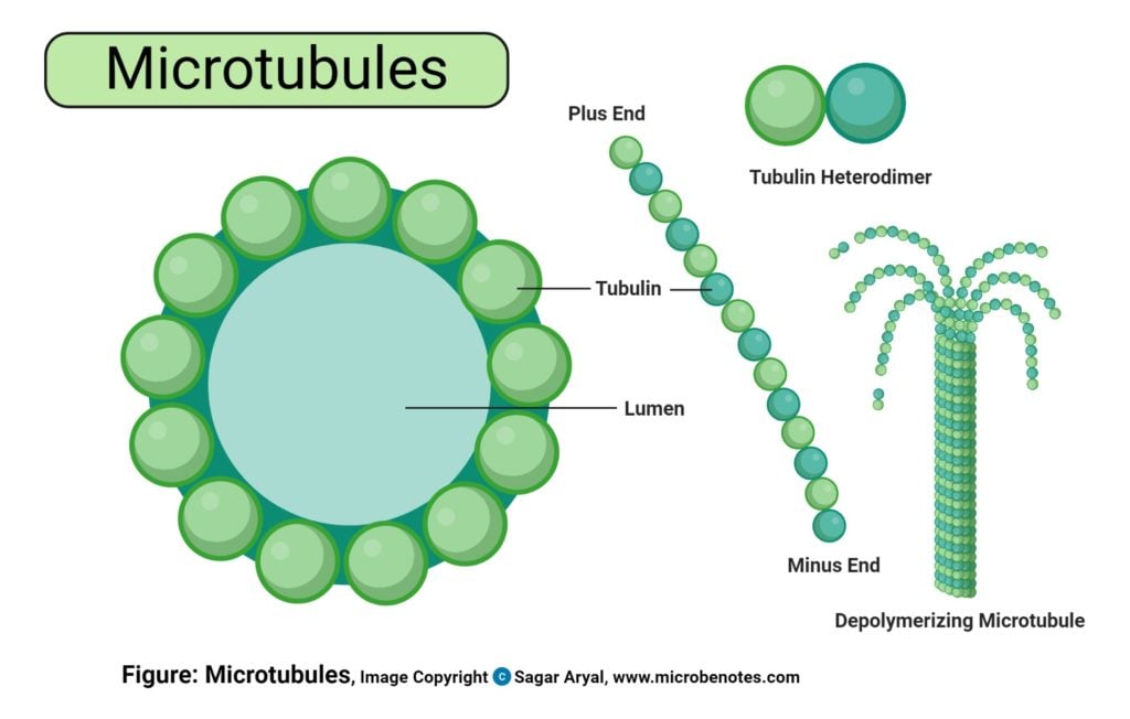
Microtubules Diagram
The cytoskeleton of the eukaryotic cell is made of proteins that give shape to it.
The three types of protein that help in the organization of the cell are microtubules, intermediate filaments, and microfilaments.
This cytoskeleton is absent in the bacteria.
Tubulin is present in the microtubules.
In 1953, Robertis and Franchi observed the microtubules in the axoplasm of the myelinated nerve fibers.
It was called the neurotubules.
In 1963, Ledbetter and Porter described the microtubules of the plant cells.
Microtubule is present in all eukaryotic cells except the human erythrocytes.
It is present in the cytoplasm freely or it can be found forming the parts of centrioles, cilia, and flagella.
In the brain of the vertebrate, the microtubule is present abundantly.
It occurs in the parts of nerve cells like the axons and the dendrites which is helpful in biochemical studies.
In both the animal and plant cells, microtubules are present in the cytoplasm at the following sites:
- cilia
- flagella
- centrioles
- basal bodies
- nerve processes
- the mitotic apparatus
- the cortex of meristematic plant cells
- elongating cells.
- selected structures in Protozoa.
In the protozoa, it is present in:
- the axostyle of parasitic flagellates
- the axoneme of Echinosphaerium
- the fiber systems of Stentor
- the cytopharyngeal basket of Nassula.
There is also a variation in the stability of microtubules of various sites.
Cytoplasmic and the spindle microtubules are labile.
Microtubules at cilia and flagella are resistant to different treatments.
In both the plant and animal cells, microtubules are filamentous rods.
The tubules are long, unbranched, and hollow in structure.
Its diameter is 24-25nm.
It consists of the protofilaments which are of 13 subunits.
13 filamentous structures are present in the wall of the microtubule.
These filaments can be linear or spiral structures having a diameter of 5nm.
They are composed of tubulin.
The center to center spacing of these protofilaments is 4.5nm.
By the negative staining, it reveals the lumen which is 14 nm wide. In the wall, a protofilament or subunit structure can be seen.
Assembly
- Microtubules undergo reversible assembly- disassembly (i.e., polymerization– depolymerization), depending on the need of the cell or organelles.
- Their polymerization is regulated by certain microtubule-associated proteins (MAPs).
- The assembly of microtubules involves preferential addition of subunits (αβ dimers) to one end of the tubule, called A end (or net assembly end); the other end of the tubule is called D end (or net disassembly end). Such an assembly involves the hydrolysis of GTP to GDP. Thus, the assembly of tubulin in the formation of microtubules is a specifically oriented and programmed process.
- Centrioles, basal bodies, and centromeres of chromosomes are the sites of orientation for this assembly. Calcium and calmodulin (an acidic protein having four Ca2+ binding sites) are some other regulating factors in the in vivo polymerization of tubulin.
Microtubules may work alone or join with other proteins to form more complex structures. Cell organelles derived from special assemblies of microtubules include:
- Cilia and flagella
- Basal bodies and Centrioles
Chemical Composition of Microtubules
Tubulin
- Tubulin is the protein that composes the protofilament of the microtubule.
- Nature of protein: Acidic protein
- Molecular weight: 55000
- Sedimentation constant: 6S
- Tubulin is present in two forms: α-tubulin and β-tubulin.
- Each of the tubulins consists of about 450 amino acids.
- The dimer is formed by the tubulin. It then polymerizes into the microtubules.
All 13 protofilaments by maintaining the same polarity are arranged in a parallel way. So, microtubules are polar structures. They have:
- Plus or fast-growing end
- Minus or slow-growing end
There is the presence of the microtubule-organizing centers (MTOCs) to which the minus end is bound tightly. The assembly or the polymerization begins from it. The minus end is also protected from disassembly by the MTOCs. Capping proteins are responsible for the protection of the plus ends from the disassembly. Near the cell margins, there is the termination of the plus ends.
Microtubule-Associated Proteins (MAPs)
- Microtubule-Associated Proteins are the proteins that associate with the surface of tubules.
- Its two classes are: HMW protein and Tau protein
- HMW proteins are high molecular weight proteins.
- The molecular weight of the HMW protein is 200,000 to 300,000 or more.
- The molecular weight of the Tau protein is 40,000 to 60,000.
- Both the HMW and Tau protein consist of the two domains.
- One domain binds to the microtubules. The nucleation process is speeded up.
- Other domain links the microtubule to the other cell components. Recently, several proteins have been identified.
- Both of the HMW and Tau protein binds the cytoplasmic microtubules in the entire length.
Microtubule Organizing Centres (MTOCs)
- To carry out the specific function, microtubules are arranged in a specific pattern.
- MTOCs help in the polymerization of the tubulin by serving as the template for it.
- They are the nucleating centers.
- MTOCs are present in the:
- basal bodies(Example: Chlamydomonas)
- in centrioles (Example: most animal cells)
- on chromosomes (i.e., kinetochore)
- in membranes
- In a study, it was found that the cytoplasmic microtubules are not derived directly from the centrioles.
- The study revealed that it is raised from the densely staining pericentriolar material which surrounds the centrioles.
Microtubule-specific drugs and their action are
- Taxol: It binds and helps to stabilize microtubules.
- Colchicine, colcemid: It binds tubulin dimmers and prevents their polymerization.
- Vinblastine, vincristine: It prevents tubulin dimmers and prevents their polymerization.
Research
Different researches are going on to study the microtubule assembly processes. They are:
- Nanoscale assembly measurements
- Super-resolution microscopy
- Localization of conformation-specific binding proteins
Functions of Microtubules
In the eukaryotic cells, microtubules perform different functions. They are:
a. Mechanical functions
The different orientation of the microtubules and their distribution is related to the:
- the shape of the cell (e.g., red blood cells of non-mammalian vertebrates)
- cell processes or protuberances like axons and dendrites of neurons, microvilli, etc.
b. Morphogenesis
- Microtubules help in determining the shape of the developing cells which occurs during the process of cell differentiation.
- Example: During spermiogenesis, the nucleus of the spermatid gets elongated. Microtubules accompany it.
- Similarly, during the induction of the lens placode in the eye, there is also the elongation of the cells. Numerous microtubules accompany it too.
c. Cellular polarity and motility
- The intrinsic polarity of some cells is also determined by the microtubules.
- Similarly, the directional gliding of cultured cells is also dependent on it.
d. Contraction
- They help in the contraction of the spindle.
- It aids in the movement of chromosomes and centrioles.
- They also help in the ciliary and flagellar motion.
e. Circulation and transport
- For the transportation of the macromolecules, granules, and vesicles within the cell, microtubules get involved in it.
- Examples:
- The cytoplasmic particles migrate in and out in the pseudopodia of Actinosphaerium (Heliozoa) which is a protozoan. In this long and thin pseudopodia, there is the presence of microtubules of about 500.
- The food is driven in the gullet of the protozoan Nassula with the help of the microtubules.
- In the cytoplasmic matrix, channels created by the microtubules help in the movement of the different granules. The melanin granules of the melanocytes can move in these channels.
- In the fish scale, there is the presence of the erythrophores. In between the microtubules, there is the movement of the pigment granules with a speed of 30 µm per second.
- They help in the axoplasmic transportation of proteins, glycoproteins, and enzymes.
References
- Johnson, A., Alberts, B., Bray, D., Hopkin, K., & Raff, M. (2019). Essential Cell Biology (Fifth edition). W. W. Norton & Company.
- Verma, P. S., & Agrawal, V. K. (2006). Cell Biology, Genetics, Molecular Biology, & Ecology (first edition.). S.Chand and Company Ltd
- Goodson, H. V., & Jonasson, E. M. (2018). Microtubules and Microtubule-Associated Proteins. Cold Spring Harbor perspectives in biology, 10(6), a022608.https://doi.org/10.1101/cshperspect.a022608
- Dimitrov A, Quesnoit M, Moutel S, Cantaloube I, Pous C, Perez F.2008. Detection of GTP-tubulin conformation in vivo reveals arole for GTP remnants in microtubule rescues. Science 322:1353–1356.
- Gardner MK, Charlebois BD, Janosi IM, Howard J, Hunt AJ, OddeDJ. 2011. Rapid microtubule self-assembly kinetics. Cell 146: 582–592.
About Author

Sushmita Baniya completed her Master’s degree in Medical Microbiology from the National College of Science and Technology (NIST), Kathmandu, Nepal. She did her Bachelor’s degree in Microbiology from Birendra Multiple Campus, Chitwan, Nepal. She is interested in Genetics and Molecular Biology.