Control of neurotransmitter release by an internal gel matrix in synaptic vesicles (original) (raw)
Abstract
Neurotransmitters are stored in synaptic vesicles, where they have been assumed to be in free solution. Here we report that in Torpedo synaptic vesicles, only 5% of the total acetylcholine (ACh) or ATP content is free, and that the rest is adsorbed to an intravesicular proteoglycan matrix. This matrix, which controls ACh and ATP release by an ion-exchange mechanism, behaves like a smart gel. That is, it releases neurotransmitter and changes its volume when challenged with small ionic concentration change. Immunodetection analysis revealed that the synaptic vesicle proteoglycan SV2 is the core of the intravesicular matrix and is responsible for immobilization and release of ACh and ATP. We suggest that in the early steps of vesicle fusion, this internal matrix regulates the availability of free diffusible ACh and ATP, and thus serves to modulate the quantity of transmitter released.
There is growing evidence that molecular mechanisms regulating exocytosis are practically constant among different cells, tissues, and animal species. A basic mechanism of recognition between proteins on the surface of secretory granules and proteins anchored to the plasma membrane is found in a wide range of cells, from yeast to neurons (1, 2). The fusion between secretory granules and the cell plasma membrane has been recorded in mast cells (3) and chromaffin cells (4) by the simultaneous monitoring of membrane capacitance and amperometry. The membrane capacitance measures the incorporation of the granule membrane as a step jump, and amperometry records the release of amines from a single granule. Granule fusion begins when a 450-pS pore is established between the interior and the extracellular space. This pore grows progressively and allows the complete release of the granule content (5).
In mast cells, before the complete fusion of the granule, the pore flickers and serotonin is released. It has been calculated that, during this period, serotonin does not move freely from the inside of the granule, which suggests that it is retained by an intragranular matrix (3). The physical properties of the intragranular matrices are directly related to osmotic balanced storage of secretory products, and to the molecular mechanism of their release (6). The slow diffusion of secretory products into the matrix limits the velocity of their release, whereas the diameter of the pore is not a limiting factor (7, 8).
In chromaffin cells, during some of the transient fusion events, the granule releases its content completely (9). The duration of transient fusion pore is inversely proportional to its diameter. The transient fusion pore may also occur during synaptic vesicle exocytosis and it has been suggested that a brief fusion may be sufficient to release all or part of its content [the kiss-and-run hypothesis (10–12)]. By analogy to the above, the release of neurotransmitters from synaptic vesicles may also be controlled by an intravesicular matrix. However, as most of the synaptic vesicles in the brain and in the neuromuscular junction have an electrolucent interior when viewed under the electron microscope, the existence of such matrix has not been considered. Of interest here is the fact that ATP is stored in all synaptic vesicles independently of neurotransmitter type, making this molecule a marker for synaptic vesicular content. We chose the electric organ of Torpedo for the present study because the synaptic vesicles fraction is easily isolated. Cholinergic synaptic vesicles of Torpedo electric organ store acetylcholine (ACh) and ATP (13), at a concentration near the molar range (14). These molecules are cosecreted during synaptic transmission (15), and the release of both can be monitored by means of specific luminescence assays. Here we study the release of ACh or ATP from permeabilized synaptic vesicles in conditions that mimic the interior of the synaptic vesicle and in the absence of small ions. In addition, we attempt to identify the vesicular constituent that builds up the vesicular matrix. Because SV2 is highly glycosylated (16), we examined its role in the intravesicular storage and release of neurotransmitters. The results indicate that ACh and ATP are not in free solution inside the synaptic vesicles but are adsorbed to a glycidic matrix, which would be mainly composed of the glycan chains of the SV2 protein.
Materials and Methods
Details of the research methods can be found in Supporting Materials and Methods, which is published as supporting information on the PNAS web site, www.pnas.org. In brief, cholinergic synaptic vesicles were obtained from the electric organ of Torpedo by sucrose gradient centrifugation (17). Ionic concentration of synaptic vesicle fractions was lowered by controlled dialysis in an isosmotic medium. Synaptic vesicles were permeabilized with detergent or by osmotic shock in ultrapure water. The release of vesicle content was recorded by measuring the discharge of ACh or ATP. ACh release was monitored by the luminescence reaction based on an enzymatic chain reaction (18) in which ACh generates oxygen that reacts with luminol (3-aminophthalhydrazide). ATP release was monitored using the luciferin–luciferase luminescence reaction. In some experiments, intravesicular matrix obtained after permeabilization of synaptic vesicles was refilled with ACh or ATP by dialysis. Intravesicular matrix was immunoprecipitated with monoclonal antibodies against SV2. Size change of intravesicular matrix was recorded in water or saline solutions by atomic force microscopy (AFM) in tapping mode. Computational calculations for the complex between vesicular matrix and ATP were done on a Silicon Graphics (Mountain View, CA) work station.
Results
ACh Release from Permeabilized Synaptic Vesicles.
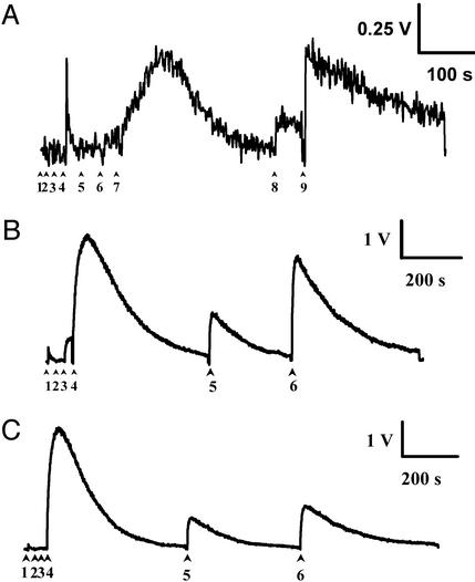
When dialyzed synaptic vesicles were permeabilized with Triton X-100 (Fig. 1A), a small release of ACh was detected (6.9 ± 1.5 nmol of ACh per mg of protein, n = 3). By comparison, when these permeabilized vesicles were further tested with an additional pulse of NaCl (8 mM), a 10-fold increase of ACh was detected (62 ± 12 nmol of ACh per mg of protein, n = 3).
Figure 1.
ACh and ATP release from permeabilized cholinergic synaptic vesicles. Synaptic vesicle fraction was dialyzed against low ionic strength medium, and ACh and ATP release were continuously monitored by light emission of luminescent enzymatic reactions. (A) In a hemolysis tube containing the recording solution (600 mM sucrose/1 mM Hepes/Tris, pH 5.5) the following reagents were added (see arrowheads): 1, choline oxidase; 2, horseradish peroxidase; 3, luminol; 4, synaptic vesicle fraction; 5, acetylcholinesterase; 6, Triton X-100; 7, 8 mM NaCl; 8, 2 nmol of ACh; and 9, 5 nmol of ACh. (B) In a hemolysis tube containing the recording solution (600 mM sucrose/50 mM Hepes/Tris, pH 5.5), the following reagents were added in the following order: 1, luciferin–luciferase mixture; 2, synaptic vesicle suspension; 3, Triton X-100 detergent; 4, 8 mM NaCl; 5, 100 pmol of ATP; and 6, 500 pmol of ATP. The exogenous doses of ATP (5 and 6) were used to calculate the amount of ATP released. (C) The order of addition of the reagents was changed and the NaCl solution was added before the detergent Triton X-100: 1, luciferin–luciferase mixture; 2, synaptic vesicle suspension; 3, 8 mM NaCl; 4, Triton X-100 detergent; 5, 100 pmol of ATP; and 6, 500 pmol of ATP.
ATP Released from Permeabilized Synaptic Vesicles.
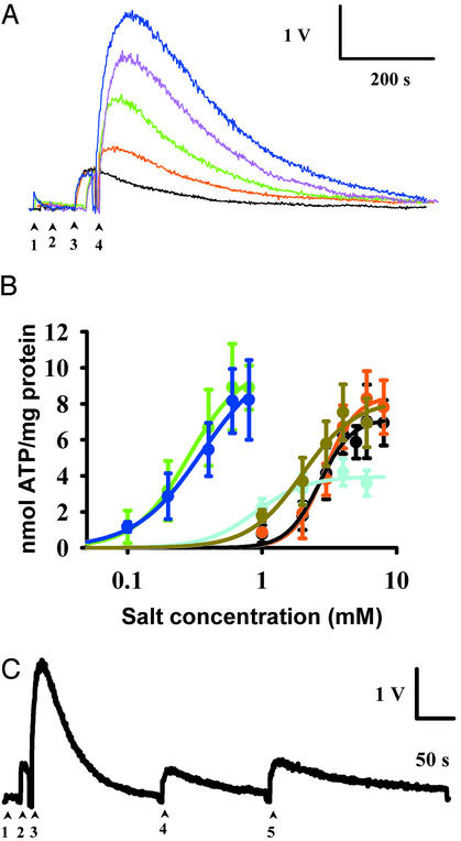
When dialyzed synaptic vesicles were permeabilized with the detergent Triton X-100 (Fig. 1B), 0.45 ± 0.06 nmol of ATP per mg of protein (n = 4) was released. When further challenged with an additional pulse of NaCl (8 mM), additional ATP was released (6.95 ± 0.56 nmol of ATP per mg of protein, n = 10). Similar results were obtained by using other nonionic detergents such as Triton X-114, Nonidet P-40, digitonin, Polidocanol, octyl glucoside, and Tween-20 (data not shown). These results were obtained only when reagents were sequentially added in the order indicated. By contrast, when the NaCl (8 mM) was added first, ATP release was not detected until the subsequent addition of the detergent (Fig. 1C). These results indicate that spontaneous leakage of ATP did not occur and that only a small fraction (4.9 ± 0.5%, n = 8) of the ATP in the synaptic vesicles was free (readily releasable). At higher concentrations of NaCl, up to 8 mM or higher, the entire ATP content of synaptic vesicles was released (Fig. 2A). Solutions of NaCl or KCl (Fig. 2B) were equally effective, with an EC50 of 2.64 and 2.85 mM, respectively. Divalent salts were more powerful than monovalent salts in extracting the ATP (EC50 = 1.45 mM for Na2SO4 and EC50 = 1.85 mM for Na2HPO4). Calcium salts were also much more powerful than sodium and potassium salts (EC50 = 0.26 mM for calcium acetate and EC50 = 0.28 mM for CaCl2). Indeed, cholinergic synaptic vesicles take up calcium (17), in addition, the EC50 for calcium uptake was of the same order as the calcium concentration reached in the synaptic active zones when neuroexocytosis takes place (19, 20). The Hill index for monovalent salts was ≈4, whereas that for divalent salts was ≈2 (Table 1), indicating that the displacing process is highly cooperative. The number of ions needed to displace the entire ATP content from a synaptic vesicle is ≈3,000 for NaCl and ≈300 for CaCl2.
Figure 2.
Salt concentration-dependent release of ATP. (A) Superimposed traces of recordings obtained with increasing concentrations of NaCl. The following reagents were added in the following order: 1, luciferin–luciferase mixture; 2, synaptic vesicle suspension; 3, Triton X-100; and 4, NaCl (in increasing concentrations). Traces: black, ultrapure water; red, 2 mM NaCl; green, 4 mM NaCl; magenta, 6 mM NaCl; and blue, 8 mM NaCl. (B) Salt concentration-dependent release of ATP. Traces: red, KCl; black, NaCl; olive green, sodium phosphate; sky blue, sodium sulfate; green, CaCl2; and dark blue, calcium acetate. (C) In a hemolysis tube containing ultrapure water, the reagents were added in the following order: 1, luciferin–luciferase mixture; 2, synaptic vesicle suspension; 3, 8 mM NaCl; 4, 100 pmol of ATP; and 5, 500 pmol of ATP.
Table 1.
Hill index and EC50 of ATP release from permeabilized synaptic vesicles induced by different salts
| Solution | Hill index | EC50, mM |
|---|---|---|
| NaCl | 3.61 | 2.64 |
| KCl | 3.63 | 2.85 |
| Na2SO4 | 2.09 | 1.45 |
| Na2HPO4/NaH2PO4 | 2.18 | 1.85 |
| CaCl2 | 2.15 | 0.28 |
| Ca(OOC–CH3)2 | 1.74 | 0.26 |
We ruled out the possibility that the detergent may have interfered with the ionic environment or with the luminescence reaction by bursting synaptic vesicles in ultrapure water (Fig. 2C). The amount of ATP released was 0.54 ± 0.07 nmol ATP per mg of protein (n = 3), which corresponded to the free ATP inside the synaptic vesicles. An additional bolus of NaCl (8 mM) evoked a second peak of ATP release (9.9 ± 0.93 nmol of ATP per mg of protein, n = 3), which emptied the synaptic vesicles. The ratio of free ATP/total ATP in synaptic vesicles burst by ultrapure water was similar (5.45%, n = 3) to that measured in detergent-permeabilized vesicles.
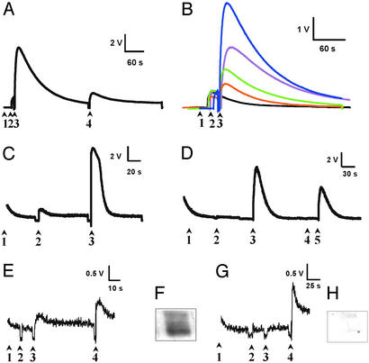
We examined whether the synaptic vesicles lysed by ultrapure water could be refilled after a run of release of ACh and ATP. The water-lysed synaptic vesicle fraction was emptied of ACh and ATP by increasing the NaCl concentration, and was refilled for a second time with ACh or ATP by dialysis combination (see Supporting Materials and Methods). When maintained in ultrapure water, the refilled matrix kept up a high content of ATP at least for few days. However, the addition of low NaCl concentration induced the release of ATP (Fig. 3A). As in the case of intact vesicles, shown before, the release of ATP was also salt concentration-dependent (Fig. 3B). The ATP refilling process was inhibited by 21.5% by incubating the matrix with 10 mM sodium periodate, an agent that breaks C–C bonds in pyranose sugars that have vicinal hydroxyls. Moreover, the pretreatment of lysed vesicles with EGTA (10 mM) completely prevents the process of ATP refilling (see Fig. 6, which is published as supporting information on the PNAS web site). With respect to ACh refilling, lysed and emptied synaptic vesicle fractions were refilled with ACh, and the addition of NaCl induced the release of ACh (Fig. 3 C and D).
Figure 3.
Release of ATP from the matrix of synaptic vesicles. (A) The matrix was refilled according to Materials and Methods. In these conditions, the matrix is able again to release ATP in an ionic force-dependent manner. The reagents were added in the following order: 1, luciferin–luciferase mixture; 2, refilled matrix suspension; 3, 8 mM NaCl; and 4, 100 pmol of ATP. (B) Superimposed traces of recordings obtained with increasing concentrations of NaCl. The reagents were added in the following order: 1, luciferin–luciferase mixture; 2, refilled matrix suspension; and 3, NaCl (in increasing concentrations). Traces: black, ultrapure water; red, 2 mM NaCl; green, 4 mM NaCl; magenta, 6 mM NaCl; and blue, 8 mM NaCl. The matrix was refilled with ACh according to Materials and Methods. In these conditions, the matrix was again able to release ACh when challenged with a NaCl solution. The hemolysis tube contained the refilled matrix and the chemiluminescence reagents (arrowhead 1). (C) To test the presence of free ACh, acetylcholinesterase was added (arrowhead 2). The amount of ACh detected is very small compared with a dose of 5 pmol of ACh (arrowhead 3). (D) NaCl-induced release of ACh. The addition of 20 mM NaCl (arrowhead 2) did not increase the luminescence until acetylcholinesterases (arrowhead 3) were added. The signal is specific to ACh, because a new addition of acetylcholinesterases (arrowhead 4) did not trigger other signal. Finally, the amount of ACh released can be estimated by comparing it to 100 pmol of ACh (arrowhead 5). (E and G) NaCl-dependent release of ATP from immunoprecipitated intravesicular matrix with (E) and without (G) mAb against SV2. The reagents were added in the following order: 1, luciferin–luciferase mixture and, when a baseline was reached, the other reagents were added; 2, immunoprecipitated intravesicular matrix suspension; 3, 8 mM NaCl; and 4, 10 pmol of ATP. (F and H) Western blot detection of SV2 content in immunoprecipitated solution with mAb against SV2 (F) or without the antibody (H).
These results agree with the hypothesis that, as in the case of chromaffin granules or mast cells, synaptic vesicles contain an intravesicular matrix. A study using AFM (21, 22) had shown that intact cholinergic synaptic vesicles have a hard central core, suggesting an intravesicular matrix. If so, ACh and ATP would be released by ionic exchange mechanisms similar to those described for serotonin in mast cell granules (7, 8), or norepinephrine in chromaffin granules (23).
To identify the main, but not only, component of the vesicular matrix, synaptic vesicles lysed in ultrapure water were immunoprecipitated by using a mAb against the synaptic vesicle protein SV2, and Sepharose protein G-conjugated beads (Fig. 3). SV2 was retained in beads, whereas synaptobrevin was below the limit of detection (data not shown). When beads containing the immunoprecipitate were tested with NaCl solution, ATP was released (Fig. 3E). In contrast, those beads obtained without the preincubation with the mAb against SV2 did not release ATP in the presence of NaCl (Fig. 3G).
Intravesicular Matrix Under AFM.
Using the above experimental conditions, synaptic vesicles lysed in ultrapure water were studied under AFM in two different configurations: static measurements of lysed synaptic vesicles were made in air, whereas dynamic measurements were made with samples immersed in water.
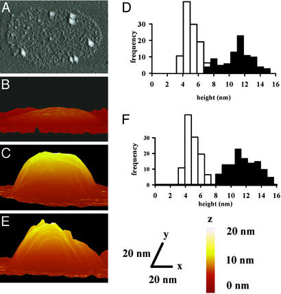
After deposition on mica, lysed vesicles were shaped like a flat disk, 500–700 nm in diameter and 1 nm in height. To detect SV2 protein, we combined AFM with immunogold detection. When synaptic vesicles were pretreated with mAb against synaptic vesicle SV2, and later, with a secondary antibody conjugated with gold particles 5 nm in diameter, disks were decorated with gold particles (Fig. 4A). The number of 5-nm particles per disk varied from 1 to 7. When synaptic vesicles were not treated with the mAb, gold particles were not detected.
Figure 4.
Synaptic vesicle matrix under AFM. (A) Immunogold detection of synaptic vesicle protein SV2 over the intravesicular matrix by AFM. The white spots correspond to 5-nm gold particles. The image was obtained in air. The remaining images were obtained on matrix immersed in liquid. (B) Imaging by AFM of vesicular matrix in ultrapure water. (C) Effect of 8 mM NaCl on the height of the disks. (D) Histogram of the frequency distribution of the height of matrix disks before and after adding NaCl; white, ultrapure water; black, NaCl. (E) Effect of 0.8 mM CaCl2 on the height of the matrix disks. (F) Histogram of the distribution of the height of matrix particles before and after adding CaCl2; white, ultrapure water; black, CaCl2. (Lower) x, y, and z scales for A_–_C and E.
When AFM measurements were made in a water drop, the disks (Fig. 4B) showed a height of 4.97 ± 0.09 nm (n = 105), which was sensitive to changes in ionic strength, and disks swelled when the ionic concentration was increased. When a solution of 8 mM NaCl was placed in the recording chamber, the height of the disk (Fig. 4C) increased to 11.12 ± 0.22 nm (n = 91), and the height distribution was shifted to the right (Fig. 4D). The addition of a solution containing a divalent cation, 0.8 mM CaCl2 (Fig. 4E), induced a similar change. The height increased to 11.71 ± 0.18 nm (n = 120), and again the height distribution was shifted to the right (Fig. 4F).
Discussion
Both ACh and ATP are known to be stored together in the interior of synaptic vesicles (13). Upon stimulation, ATP is released in the same fashion as ACh, either from the neuromuscular junction (15) or from Torpedo electric organ nerve terminals (24, 25). The corelease of ACh and ATP has been measured directly in mouse neuromuscular junction after a single stimulus of the presynaptic nerve trunk (15), reinforcing the view of coincident release of ACh and ATP from single synaptic vesicles. The neuroexocytosis of nerve terminals can be determined by monitoring either ACh or ATP release. Moreover, the release of ATP is a general marker for neuroexocytosis in all kinds of synapses, because ATP is always present inside the vesicles, whereas the main neurotransmitter varies according to the neuron's synthetic machinery.
In the present work, the ratio of ACh and ATP released in permeabilized vesicles is the same as described in intact synaptic vesicles (13, 17), indicating complete and simultaneous release of both molecules after the ionic strength step increase. In our experiments, the records of ATP release had much better resolution than those of ACh. The pH of the reaction was fixed at intravesicular level (5.5). The luminescence reaction for ATP ended in luciferase, whereas the reaction for ACh ended in luminol, which is very inefficient at such low pH. The optimum pH for photon emission of luminol is >10. Because the records of ATP were of better quality, we have used them to quantify the ionic force necessary to displace the soluble substances adhered to the intravesicular matrix. However, under other experimental conditions (pH 7.0, see Supporting Materials and Methods), we were able to measure the release of ACh from lysed and refilled synaptic vesicles. Lysed synaptic vesicles can be refilled with either ACh or ATP after being emptied with increased ionic force. Refilled vesicles retained ACh or ATP while in low ionic strength in solution, but they released ACh or ATP after being challenged with NaCl solutions.
The matrix of mast cells and chromaffin granules is composed of proteoglycans (26). In synaptic vesicles of the electric organ of Torpedo, a proteoglycan was reported (27), containing the SV2 molecule, an integral membrane protein (28), which has a long glycidic intravesicular tail enriched in keratan sulfate residues (16). Thus, the intravesicular matrix of cholinergic synaptic vesicles behaves like an ion-exchanging gel: ACh and ATP3− are displaced by ions and the matrix size increases. This increase in size may be related to the water accompanying the ions (29). The vesicular matrix behaves as a smart gel (30, 31), doubling its original volume when challenged with very low concentrations of ions.
The cholinergic synaptic vesicle proteoglycan is a complex structure that is composed of several proteins (16), with the SV2 molecule being the most conspicuous. Other synaptic vesicle membrane proteins are glycosylated as well, and they include: ACh transporter, synaptophysin, synaptotagmin (32), and svp25 (33). The glycidic part of SV2 and the glycidic part of all or some of these proteins would constitute the vesicular matrix. According to our hypothesis, the vesicular matrix should play a role similar to that of the gels of ion-exchange chromatography. In agreement with this finding, our results indicate that the intravesicular matrix of Torpedo electric organ synaptic vesicles contains the SV2 protein. The number of gold particles found on the matrix is very similar to the calculated number of copies of SV2 per vesicle. The protein SV2 has 12 potential transmembrane regions. The domain between the seventh and the eighth transmembrane region is highly glycosylated, through asparagine residues, and has been related to an osmotic stabilizer function (3, 16) and the control of neurotransmitter release (34). In fact, this protein is known to regulate the number of releasable granules (35). The results obtained in SV2 knock-out mice also suggest that SV2 plays a calcium-buffering role and modulates calcium-stimulated exocytosis (36).
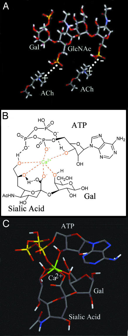
Our calcium salt results demonstrate the high ATP-release sensitivity of the vesicular matrix. The glycidic nature of the matrix offers various sites for the interaction with ACh and ATP. ACh has a quaternary ammonium, conferring a positive charge. The sites for interaction of ACh with the matrix are, most probably, the sulfate groups of the keratan molecules (Fig. 5A). The interaction of positive charges of Na+ or Ca2+ cations would displace ACh in a manner similar to that described in histamine release from mast cell granules (7, 8). ATP can interact through the nitrogen atoms of the purine ring or the phosphate residues, as nitrogen atoms can establish hydrogen bonds with carboxylic radicals (37–39). A second possibility of interaction of ATP is based on the fact that ATP easily complexes with divalent cations (40–42). However, carboxylic groups establish stronger complexes with calcium than with ATP (43). Synaptic vesicles contain sialic acid (from keratan sulfate linked to SV2, and in other synaptic vesicle proteins), ATP, and calcium at high concentrations. A complex between calcium and carboxylic residues from the sialic acid would be formed. However, the coordination of calcium with ATP would still be possible, forming a ternary complex between Ca2+, the sialic-galactose moiety, and ATP trianion (see Fig. 5 B and C). In fact, we have seen that EGTA prevents the refilling of the vesicular matrix. Computational calculations show that the interaction of Ca2+ with ATP3− is expected to be weaker than that with the sialic-galactose moiety (one ionic ligand and three coordinating hydroxy ligands coming from the sialic-galactose disaccharide, and one ionic and one hydroxy ligand arising from the ATP anion). When there are a relatively small number of calcium ions, they are complexed between the sialic-galactose moieties of the vesicle matrix and the ATP trianions (see Fig. 5B). The consequence of this action is that calcium ions and ATP anions are linked to the proteoglycan matrix. When the number of calcium ions increases, the first equilibria is perturbed and new equilibrium are established: the ATP trianion and the additional Ca2+ make a complex, whereas the sialic-galactose moiety retains the initial Ca2+. The mechanism of the equilibria involved is shown in Fig. 7, which is published as supporting information on the PNAS web site. Cations other than Ca2+ can behave similarly, although their efficiency in detaching ATP trianions will likely depend on the relative stability of the corresponding complexes. Thus, cations that form much stronger complexes than Ca2+ with ATP should be more efficient (although, to our knowledge, no studies in this connection have been carried out so far), whereas, as expected, we have shown that a very large excess of monovalent cations is required to cause the detachment of ATP. In addition, synthetic polymers can bind to neurotransmitters such as histamine or serotonin, (44) and also ATP (45). Our results, demonstrating that the matrix can be stressed to an in vitro cycle of depletion and refilled with ATP, are in agreement with the passive nature of the interaction between the matrix and ATP. We conclude from the results of Fig. 2 that the interaction of ACh and ATP with the matrix must involve allosteric properties.
Figure 5.
Model of ACh and ATP interaction with the synaptic vesicle matrix. Keratan sulfate and sialic acid of the vesicular matrix would support the adsorption of ACh and ATP, respectively. (A) Sulfate groups of galactose (Gal) and _N_-acetylglucosamine (GlcNAc) residues from the keratan molecule would be found where electrostatic interaction with the quaternary ammonium of ACh takes place (dashed line). S atoms are yellow, O atoms are red, and N, C, and H atoms are blue, black, and white, respectively. (B) For ATP interaction with the matrix, we propose that a calcium intermediate may be formed of the calcium ions, sialic acid, galactose, and ATP. (C) A simplified tube model of the same complex. The calcium ion, in green for clarity, is coordinated with six O atoms, which are red. P atoms are yellow, and N, C, and H atoms are blue, black, and white, respectively.
In the proposed model of the matrix, two different binding sites are involved: sulfate residues for ACh and sialic acid–Ca2+–galactose complexes for ATP. The existence of both binding sites may explain a differential release of ACh and ATP measured, under special conditions, in cholinergic nerve endings (46, 47) or a differential release of catecholamines and ATP from chromaffin cells (48).
Thus, it is proposed that synaptic vesicles follow the same steps of exocytosis as nonneuronal cells. The interaction of synaptic vesicles and the plasma membrane is led by the association between v- and t-SNARE proteins (2). In the classic pathway of synaptic vesicle exocytosis, neurotransmitter is released after the complete fusion of synaptic vesicles within the nerve-terminal plasma membrane; the content is completely extruded to the extracellular space by diffusion. Alternatively, the kiss-and-run hypothesis (9, 49) suggests that the fusion pore, established between the synaptic vesicle and the plasma membrane, is open for a very short period, in which only a fraction of the content of the synaptic vesicle is quickly released to the synaptic cleft. The pore again closes by the interaction of dynamin with synaptophysin (50), and the content of the vesicle is rapidly restored. In both cases (the classic and the kiss-and-run hypotheses), the limiting step is the accessibility of the neurotransmitters to be released. Our results indicate that only a small fraction of the ACh/ATP vesicular package is free to be released when the fusion pore is established (51). In the classic hypothesis, the entire ATP content of the vesicle would be released only if the totality of the matrix is exteriorized and reconstituted during vesicle recycling. Recent works (11, 12) strongly support the kiss- and-run hypothesis in the readily releasable pool of vesicles, and we suggest that an ionic current through the vesicle membrane would be necessary to detach the neurotransmitters from the matrix (34). Ions can flow through two theoretical pathways: the fusion channel, or the electrogenic molecules of the synaptic vesicle membrane. Using low ionic concentration of the extracellular solution (2 mM KCl) in the frog neuromuscular junction, Van der Kloot (52) and Khanin et al. (53) found that miniature end-plate currents changed polarity but were not abolished, suggesting that ions do not flow through the fusion pore. On the other hand, molecules that could generate ionic currents, such as synaptophysin (54), the proton pump, or other vesicular ionic channels (55, 56), have been described in synaptic vesicles. The conjunction of an intravesicular matrix and ionic channels in synaptic vesicles would result in an adjustable molecular mechanism of neurotransmitter release after vesicle fusion, in which the activation of a single channel would lead to the release of part or all of the content. A theoretical time course of ACh release using an ion-exchange mechanism has been calculated (57). As stated above, the number of ions that would have to enter the vesicle to release its content of ATP is 300–3,000. According to the results reported by Yakir and Rahamimoff (55), the ionic channels in synaptic vesicles would have to be open for 10–100 μs to allow the entry of this number of ions. The intravesicular matrix makes possible the postfusion control of neurotransmitter release (58, 59) and opens the possibility that a quantum does not correspond exactly to the content of one synaptic vesicle, but rather would match a fixed number of molecules displaced by the activity of ionic channels or ionic transporters placed in the vesicle membrane. A single vesicle of Torpedo electric organ contains ≈200,000 ACh molecules (60), whereas the number of ACh molecules that generate a miniature end-plate current is 10,000 (61). The fact that neurotransmitters are adsorbed to an internal matrix indicates that unknown molecular mechanisms regulate, with high precision, the number of molecules released immediately after the fusion of a synaptic vesicle. In Torpedo electric organ, and in vertebrate neuromuscular junction, different kinds of quantal events have been recorded: miniature end-plate currents and giant miniature end-plate currents (which correspond to several times the amplitude of a miniature end-plate current). The first type of current would be the result of a partial depletion of a synaptic vesicle, whereas giant-miniature would correspond to a complete fusion of the membrane and the ejection of the vesicular matrix.
The understanding of how neurons release neurotransmitters is progressing thanks to a deep knowledge of interactions between SNAREs. Recently, Almers (62) and Peters (ref. 63, see also ref. 64) suggested the formation of a proteolipidic pore controlling the fusion of synaptic vesicles, but the role of the internal matrix should also be considered. The matrix should be regarded as an intelligent interface between the nerve terminal and the extracellular medium regulating the fine tuning of neuroexocytosis.
Supplementary Material
Supporting Information
Acknowledgments
We thank Prof. Kathleen Buckley (Harvard University, Cambridge, MA) for the mAb against SV; Dr. E. M. Silinsky (Northwestern University, Chicago) for helpful comments; Dr. R. Pons and Dra. N. Azemar from the Centre d'Investigación i Desenvolupament–Consejo Superior de Investigaciones Científicas, Barcelona, for light scattering measurements; and the Servei d'Assessorament Linguistic of the Universitat de Barcelona for linguistic help. The program WIN WHOLE CELL ANALYSIS 3.0.2 was kindly provided by Dr. J. Dempster (University of Strathclyde, Glasgow, U.K.). This study was funded by the Dirección General de Enseñanza Superior e Investigación Científica of the Spanish Government, the Comisio Interdepartamental de Recerca i Innovacio Tecnologica, and the Generalitat de Catalunya and Fundació La Marató de TV3. D.R. is a Fellow of the Fundació August Pi i Sunyer. P.G. acknowledges a research grant from the Fundación Francisco Cobos.
Abbreviations
ACh
acetylcholine
AFM
atomic force microscopy
References
- 1.Fernandez-Chacón R, Sudhof T C. Annu Rev Physiol. 1999;61:753–776. doi: 10.1146/annurev.physiol.61.1.753. [DOI] [PubMed] [Google Scholar]
- 2.Jahn R, Sudhof T C. Annu Rev Biochem. 1999;68:863–911. doi: 10.1146/annurev.biochem.68.1.863. [DOI] [PubMed] [Google Scholar]
- 3.Álvarez de Toledo G, Fernández-Chacón R, Fernández J M. Nature. 1993;363:554–558. doi: 10.1038/363554a0. [DOI] [PubMed] [Google Scholar]
- 4.Albillos A, Dernick G, Horstmann H, Almers W, Álvarez de Toledo G, Lindau M. Nature. 1997;389:509–512. doi: 10.1038/39081. [DOI] [PubMed] [Google Scholar]
- 5.Monck J R, Fernandez J M. Curr Opin Cell Biol. 1996;8:524–533. doi: 10.1016/s0955-0674(96)80031-7. [DOI] [PubMed] [Google Scholar]
- 6.Nanavati C, Fernández J M. Science. 1993;259:963–965. doi: 10.1126/science.8438154. [DOI] [PubMed] [Google Scholar]
- 7.Marszalek P E, Farrell B, Verdugo P, Fernandez J M. Biophys J. 1997;73:1160–1168. doi: 10.1016/S0006-3495(97)78148-7. [DOI] [PMC free article] [PubMed] [Google Scholar]
- 8.Marszalek P E, Farrell B, Verdugo P, Fernandez J M. Biophys J. 1997;73:1169–1183. doi: 10.1016/S0006-3495(97)78149-9. [DOI] [PMC free article] [PubMed] [Google Scholar]
- 9.Alés E, Tabares L, Poyato J M, Valero V, Lindau M, Álvarez de Toledo G. Nat Cell Biol. 1999;1:40–44. doi: 10.1038/9012. [DOI] [PubMed] [Google Scholar]
- 10.Fesce R, Meldolesi J. Nat Cell Biol. 1999;1:E3–E4. doi: 10.1038/8950. [DOI] [PubMed] [Google Scholar]
- 11.Pyle J L, Kavalali E T, Piedras-Renteria E S, Tsien R W. Neuron. 2000;28:221–231. doi: 10.1016/s0896-6273(00)00098-2. [DOI] [PubMed] [Google Scholar]
- 12.Stevens C F, Williams J H. Proc Natl Acad Sci USA. 2000;97:12828–12833. doi: 10.1073/pnas.230438697. [DOI] [PMC free article] [PubMed] [Google Scholar]
- 13.Dowdall M J, Boyne A F, Whittaker V P. Biochem J. 1974;140:1–12. doi: 10.1042/bj1400001. [DOI] [PMC free article] [PubMed] [Google Scholar]
- 14.Kelly R B, Hooper J E. In: The Secretory Granule, The Secretory Process. Poisner A M, Trifaró J M, editors. Vol. 1. Amsterdam: Elsevier Biomedical; 1982. pp. 81–118. [Google Scholar]
- 15.Silinsky E M. J Physiol (London) 1975;247:145–162. doi: 10.1113/jphysiol.1975.sp010925. [DOI] [PMC free article] [PubMed] [Google Scholar]
- 16.Scranton T W, Iwata M, Carlson S S. J Neurochem. 1993;61:29–44. doi: 10.1111/j.1471-4159.1993.tb03535.x. [DOI] [PubMed] [Google Scholar]
- 17.Israel M, Manaranche R, Marsal J, Meunier F M, Morel N, Frachon P, Lesbats B. J Membr Biol. 1980;54:115–126. doi: 10.1007/BF01940565. [DOI] [PubMed] [Google Scholar]
- 18.Israel M, Lesbats B. J Neurochem. 1981;37:1475–1483. doi: 10.1111/j.1471-4159.1981.tb06317.x. [DOI] [PubMed] [Google Scholar]
- 19.Llinas R, Sugimori M, Silver R B. Science. 1992;256:677–679. doi: 10.1126/science.1350109. [DOI] [PubMed] [Google Scholar]
- 20.Neher E. Neuron. 1998;20:389–399. doi: 10.1016/s0896-6273(00)80983-6. [DOI] [PubMed] [Google Scholar]
- 21.Laney D E, Garcia R A, Parsons S M, Hansma H G. Biophys J. 1997;72:806–813. doi: 10.1016/s0006-3495(97)78714-9. [DOI] [PMC free article] [PubMed] [Google Scholar]
- 22.Garcia R A, Laney D E, Parsons S M, Hansma H G. J Neurosci Res. 1998;52:350–355. doi: 10.1002/(SICI)1097-4547(19980501)52:3<350::AID-JNR11>3.0.CO;2-A. [DOI] [PubMed] [Google Scholar]
- 23.Uvnas B, Aborg C H. Acta Physiol Scand. 1985;124:629–630. doi: 10.1111/j.1748-1716.1985.tb00057.x. [DOI] [PubMed] [Google Scholar]
- 24.Israel M, Dunant Y, Lesbats B, Manaranche R, Marsal J, Meunier F. J Exp Biol. 1979;81:63–73. doi: 10.1242/jeb.81.1.63. [DOI] [PubMed] [Google Scholar]
- 25.Schweitzer E. J Neurosci. 1987;7:2948–2956. doi: 10.1523/JNEUROSCI.07-09-02948.1987. [DOI] [PMC free article] [PubMed] [Google Scholar]
- 26.Verdugo P. Ciba Found Symp. 1984;109:212–225. doi: 10.1002/9780470720905.ch15. [DOI] [PubMed] [Google Scholar]
- 27.Carlson S S, Kelly R B. J Biol Chem. 1983;258:11082–11091. [PubMed] [Google Scholar]
- 28.Feany M B, Lee S, Edwards R H, Buckley K M. Cell. 1992;70:861–867. doi: 10.1016/0092-8674(92)90319-8. [DOI] [PubMed] [Google Scholar]
- 29.Parpura V, Fernandez J M. Biophys J. 1996;71:2356–2366. doi: 10.1016/S0006-3495(96)79483-3. [DOI] [PMC free article] [PubMed] [Google Scholar]
- 30.Sato-Matsuo S, Tanaka T. Nature. 1992;358:482–485. [Google Scholar]
- 31.Tanaka T. Sci Am. 1981;244(1):124–136. doi: 10.1038/scientificamerican0181-124. [DOI] [PubMed] [Google Scholar]
- 32.Volknandt W. Neuroscience. 1995;64:277–300. doi: 10.1016/0306-4522(94)00408-w. [DOI] [PubMed] [Google Scholar]
- 33.Volknandt W, Schlafer M, Bonzelius F, Zimmermann H. EMBO J. 1990;9:2465–2470. doi: 10.1002/j.1460-2075.1990.tb07424.x. [DOI] [PMC free article] [PubMed] [Google Scholar]
- 34.Rahamimoff R, Fernández J M. Neuron. 1997;18:17–27. doi: 10.1016/s0896-6273(01)80043-x. [DOI] [PubMed] [Google Scholar]
- 35.Xu T, Bajjalieh S M. Nat Cell Biol. 2001;3:691–698. doi: 10.1038/35087000. [DOI] [PubMed] [Google Scholar]
- 36.Janz R, Goda Y, Geppert M, Missler M, Sudhof T C. Neuron. 1999;24:1003–1016. doi: 10.1016/s0896-6273(00)81046-6. [DOI] [PubMed] [Google Scholar]
- 37.Zimmerman S C, Wu W, Zeng Z. J Am Chem Soc. 1991;113:196–201. [Google Scholar]
- 38.Shea K J, Spivak D A, Sellergren B. J Am Chem Soc. 1992;115:3368–3369. [Google Scholar]
- 39.Lancelot G. J Am Chem Soc. 1977;99:7037–7042. doi: 10.1021/ja00463a044. [DOI] [PubMed] [Google Scholar]
- 40.Sigel H. Eur J Biochem. 1987;165:65–72. doi: 10.1111/j.1432-1033.1987.tb11194.x. [DOI] [PubMed] [Google Scholar]
- 41.Ramírez F, Marecek J. Biochim Biophys Acta. 1980;589:21–29. doi: 10.1016/0005-2728(80)90129-2. [DOI] [PubMed] [Google Scholar]
- 42.Cini R, Sabat M, Sundaralingam M, Burla M C, Nunzi A, Polidori G, Zanazi P F. J Biomol Struct Dyn. 1983;1:633–637. doi: 10.1080/07391102.1983.10507470. [DOI] [PubMed] [Google Scholar]
- 43.Fenton D E. In: Comprehensive Coordination Chemistry: The Synthesis, Reactions, Properties and Applications of Coordination Compounds. Wilkinson G, Gillard R D, McCleverty J A, editors. Vol. 3. Oxford: Pergamon; 1987. pp. 1–80. [Google Scholar]
- 44.Kiser P F, Wilson G, Needham D. Nature. 1998;394:459–462. doi: 10.1038/28822. [DOI] [PubMed] [Google Scholar]
- 45.Matheu J, Buchardt O. Bioconjugate Chem. 1995;6:524–528. doi: 10.1021/bc00035a004. [DOI] [PubMed] [Google Scholar]
- 46.Marsal J, Egea G, Solsona C, Rabasseda X, Blasi J. Proc Natl Acad Sci USA. 1989;86:372–376. doi: 10.1073/pnas.86.1.372. [DOI] [PMC free article] [PubMed] [Google Scholar]
- 47.Marsal J, Solsona C, Rabasseda X, Blasi J, Casanova A. Neurochem Int. 1987;10:295–302. doi: 10.1016/0197-0186(87)90103-3. [DOI] [PubMed] [Google Scholar]
- 48.Hollins B, Ikeda S R. J Neurophysiol. 1997;78:3069–3076. doi: 10.1152/jn.1997.78.6.3069. [DOI] [PubMed] [Google Scholar]
- 49.Ceccarelli B, Hurlbut W P. Physiol Rev. 1980;60:396–441. doi: 10.1152/physrev.1980.60.2.396. [DOI] [PubMed] [Google Scholar]
- 50.Daly C, Sugimori M, Moreira J E, Ziff E B, Llinas R. Proc Natl Acad Sci USA. 2000;97:6120–6125. doi: 10.1073/pnas.97.11.6120. [DOI] [PMC free article] [PubMed] [Google Scholar]
- 51.Monck J R, Fernandez J M. Neuron. 1994;12:707–716. doi: 10.1016/0896-6273(94)90325-5. [DOI] [PubMed] [Google Scholar]
- 52.Van der Kloot W. Biophys J. 1995;69:148–154. doi: 10.1016/S0006-3495(95)79884-8. [DOI] [PMC free article] [PubMed] [Google Scholar]
- 53.Khanin R, Segel L, Parnas H, Ratner E. Biophys J. 1996;70:2030–2032. doi: 10.1016/S0006-3495(96)79769-2. [DOI] [PMC free article] [PubMed] [Google Scholar]
- 54.Thomas L, Hartung K, Langosch D, Rehm H, Bamberg E, Franke W W, Betz H. Science. 1988;242:1050–1053. doi: 10.1126/science.2461586. [DOI] [PubMed] [Google Scholar]
- 55.Yakir N, Rahamimoff R. J Physiol (London) 1995;485:683–697. doi: 10.1113/jphysiol.1995.sp020762. [DOI] [PMC free article] [PubMed] [Google Scholar]
- 56.Kelly M L, Woodbury D J. Biophys J. 1996;70:2593–2599. doi: 10.1016/S0006-3495(96)79830-2. [DOI] [PMC free article] [PubMed] [Google Scholar]
- 57.Khanin R, Parnas H, Segel L. Biophys J. 1994;67:966–972. doi: 10.1016/S0006-3495(94)80562-4. [DOI] [PMC free article] [PubMed] [Google Scholar]
- 58.Meir A, Ginsburg S, Butkevich A, Kachalsky S G, Kaiserman I, Ahdut R, Demirgoren S, Rahamimoff R. Physiol Rev. 1999;79:1019–1088. doi: 10.1152/physrev.1999.79.3.1019. [DOI] [PubMed] [Google Scholar]
- 59.Murthy V N, Stevens C F. Nat Neurosci. 1999;2:503–507. doi: 10.1038/9149. [DOI] [PubMed] [Google Scholar]
- 60.Whittaker V P. FASEB J. 1989;41:2759–2764. [PubMed] [Google Scholar]
- 61.Girod R, Correges P, Jacquet J, Dunant Y. J Physiol (London) 1993;471:129–157. doi: 10.1113/jphysiol.1993.sp019894. [DOI] [PMC free article] [PubMed] [Google Scholar]
- 62.Almers W. Nature. 2001;409:567–568. doi: 10.1038/35054637. [DOI] [PubMed] [Google Scholar]
- 63.Peters C. Nature. 2001;409:581–586. doi: 10.1038/35054500. [DOI] [PubMed] [Google Scholar]
- 64.Flak-Vairant J, Correges P, Eder-Colli L, Salem N, Roulet E, Bloc A, Meunier F, Lesbats B, Loctin F, Synguelakis M, et al. Proc Natl Acad Sci USA. 1996;93:5203–5207. doi: 10.1073/pnas.93.11.5203. [DOI] [PMC free article] [PubMed] [Google Scholar]
Associated Data
This section collects any data citations, data availability statements, or supplementary materials included in this article.
Supplementary Materials
Supporting Information
