A role for the light-dependent phosphorylation of visual arrestin (original) (raw)
Abstract
Arrestins are regulatory proteins that participate in the termination of G protein-mediated signal transduction. The major arrestin in the Drosophila visual system, Arrestin 2 (Arr2), is phosphorylated in a light-dependent manner by a Ca2+/calmodulin-dependent protein kinase and has been shown to be essential for the termination of the visual signaling cascade in vivo. Here, we report the isolation of nine alleles of the Drosophila photoreceptor cell-specific arr2 gene. Flies carrying each of these alleles underwent light-dependent retinal degeneration and displayed electrophysiological defects typical of previously identified arrestin mutants, including an allele encoding a protein that lacks the major Ca2+/calmodulin-dependent protein kinase site. The phosphorylation mutant had very low levels of phosphorylation and lacked the light-dependent phosphorylation observed with wild-type Arr2. Interestingly, we found that the Arr2 phosphorylation mutant was still capable of binding to rhodopsin; however, it was unable to release from membranes once rhodopsin had converted back to its inactive form. This finding suggests that phosphorylation of arrestin is necessary for the release of arrestin from rhodopsin. We propose that the sequestering of arrestin to membranes is a possible mechanism for retinal disease associated with previously identified rhodopsin alleles in humans.
G protein-coupled receptors are a large family of proteins that have seven transmembrane domains and are activated in response to a large variety of extracellular signals including hormones, light, and odorants (1, 2). Receptor stimulation is followed by the activation of a heterotrimeric G protein and the subsequent activation of specific intracellular effector molecules. Although a great deal of information has been obtained concerning the activation of various G protein-coupled signaling cascades, the mechanisms involved in inactivating components of various cascades in vivo have not been well established. To terminate signaling through the cascade efficiently, all activated intermediates, including the receptor, need to be inactivated.
The inactivation of G protein-coupled receptors, often termed desensitization, has been studied most extensively for rhodopsin and the β-adrenergic receptor (3–6). In both cases, the molecular mechanism of desensitization occurs in a two-step process. The first step involves a rapid phosphorylation of the activated receptor by a specific G protein-coupled receptor kinase (7, 8). Phosphorylation of receptors by G protein-coupled receptor kinases results in only minimal desensitization; however, phosphorylation does increase the affinity of the receptors for another group of proteins, known as arrestins. The binding of arrestin to the phosphorylated, activated receptors quenches signal transduction via its apparent ability to decrease receptor/G protein coupling directly (9–12).
Arrestin was identified originally as an abundant soluble protein in the bovine retina and was called S antigen (13). Subsequently, visual arrestin homologues have been identified in a large number of species, and, in addition, numerous arrestins have been found in nonretinal tissue. It has been established that retinal and nonretinal arrestins mediate the inactivation of G protein-coupled receptors, and this finding has suggested a common mechanism for desensitizing this large class of receptor molecules. In addition to its role in inactivating receptor molecules, a subclass of arrestins also have been shown to be involved in receptor-mediated endocytosis (14, 15).
The Drosophila melanogaster visual system provides an excellent model system for the study of G protein-coupled receptor inactivation in vivo (16). The existence of numerous mutations that affect phototransduction in Drosophila, along with the ability to screen for additional mutations, allows for a genetic dissection of this signaling pathway. Phototransduction in Drosophila is initiated by the activation of the G protein-coupled receptor rhodopsin. Invertebrate rhodopsin consists of the apoprotein, opsin, which is covalently linked to an 11-_cis_-retinal chromophore. Photon absorption converts the chromophore from the 11-cis conformation to all-trans with a subsequent conformational change in the opsin moiety. Activated rhodopsin, or metarhodopsin, activates the alpha subunit of a heterotrimeric G protein, which, in turn, activates an eye-specific phospholipase C molecule. Phospholipase C hydrolyzes the membrane phospholipid phosphatidylinositol-4,5-bisphosphate into two second messengers, inositol trisphosphate and diacylglycerol. Phospholipase C activity is required to open two light-activated, cation-selective ion channels; however, the molecular mechanism involved in the opening of these channels has yet to be determined. Recent work has implicated polyunsaturated fatty acids in gating both light-activated channels (17).
In Drosophila, two visual system arrestins have been identified: Arr1 and Arr2. Whereas mutations in Arr1 have no noticeable phenotype, severe loss-of-function mutations in Arr2 undergo rapid light-dependent retinal degeneration and are defective in rhodopsin inactivation (18, 19). Both Arr1 and Arr2 are phosphorylated in a light-dependent manner, and it has been established that Arr2 is phosphorylated by a Ca2+/calmodulin-dependent protein kinase (20). Although it has been known for quite some time that Arr2 is phosphorylated in a light-dependent manner, the role of this phosphorylation is unknown. Here, we provide evidence that the phosphorylation of Arr2 is necessary for its release from membranes once rhodopsin has been photoconverted back to its inactive form.
MATERIALS AND METHODS
Genetic Screen, Plasmid Construction, and Sequence Characterization.
arr2 mutants were generated by using a noncomplementation screen. Wild-type flies were treated with the mutagen ethyl methanesulfonate and crossed to _arr2_3 homozygotes (18). _arr2_3 heterozygotes undergo retinal degeneration in room light in 8–10 days, so F1 flies were screened for enhanced retinal degeneration (at 3–5 days), indicated by the loss of a deep pseudopupil (21). The S366A allele was generated by using site-directed mutagenesis and introduced into the _arr2_5 null background via P element-induced transposition (22, 23). This construct contained ≈2 kilobases of endogenous promoter sequence. For sequence analysis, the mutant arr2 genes were isolated by PCR amplification of genomic DNA from the mutant stocks. Two independent PCR fragments for each of the mutant alleles were cloned into pBluescript II SK(+) (Stratagene) and sequenced. Nucleotide sequences were obtained on an Applied Biosystems model 373A Automated Sequencer (Dartmouth College Molecular Biology Core Facility) by using the Ready-reaction Dye-Terminator kit with AmpliTaq FS (Applied Biosystems).
Electroretinogram Analysis.
Electroretinograms were performed as described (18). Electrical signals were amplified through a DAM50 amplifier (World Precision Instruments, Sarasota, FL), digitized with a MacAdios ADPO (GW Instruments, Somerville, MA), and viewed with superscope software (GW Instruments). Stimulating light was generated with a 300-W xenon/mercury lamp (Oriel, Stamford, CT) and attenuated through neutral density filters. Blue and orange light was generated by filtering through 480 nm and 580 nm band-pass filters (Oriel), respectively. All light pulses were 1 s in duration and at maximum intensity except where noted. All recordings were performed on newly eclosed adult flies (less than 2 days) that were reared in the dark.
Biochemical Assays.
For Arr2 binding assays, 6–10 Drosophila heads were dissected from newly eclosed (less than 2 days) dark-reared adults and added to a buffer containing 150 mM KCl, 20 mM Tris (pH 7.5), 5 mM DTT, 10 μg/ml PMSF, 0.5 μg/ml leupeptin, 0.5 μg/ml pepstatin, and 0.5 μg/ml aprotinin. The heads were then exposed to 5 min of either orange light (580 ± 10 nm) or blue light (480 ± 10 nm), homogenized in the dark, and centrifuged at 13,000 × g for 5 min. Pellet and supernatant fractions were separated under very dim red light and subjected to SDS/PAGE and Western analysis with antibodies against Arr2 (18) and rhodopsin (Rh1; Developmental Studies Hybridoma Bank, University of Iowa, Iowa City). Arr2-release assays were performed in the same manner, except that the isolated fly heads were exposed to 10 s of blue light followed by 60 s of orange light.
In vitro phosphorylation assays were performed as described (24). Briefly, 15 Drosophila heads from dark-reared newly eclosed (less than 2 days) adults were dissected and homogenized in 20 μl of the above-mentioned binding assay buffer. Phosphorylation reactions were allowed to proceed for 10 min and were assayed in 5 μl of 16 mM Tris (pH 7.5), 5 mM MgCl2, 5 mM β-mercaptoethanol, 10 μM ATP, 3 μCi [γ-32P]ATP (7,000 Ci/mmol), and 1 μl of homogenate. The chelating EGTA concentration was 2 mM, and calcium reactions contained 0.5 mM CaCl2. For the in vitro phosphorylation reactions (shown in Fig. 4), membranes were collected by centrifugation at 13,000 × g for 5 min, and the pellet and supernatant fractions were separated and subjected to SDS/PAGE, autoradiography, and Western analysis with antibodies directed against Arr1, Arr2, and Rh1.
Figure 4.
Phosphorylated Arr2 protein is found as a non-membrane-associated soluble factor. (Left) Autoradiograph of γ-32P-labeled proteins isolated from fractionated wild-type Drosophila head lysates in the presence of 0.5 mM Ca2+ (lanes 1 and 2) or 2 mM EGTA (lanes 3 and 4). S, supernatant fraction; P, pellet fraction. (Right) The same gel transferred to nitrocellulose and probed with antibodies specific for Arr1, Arr2, and Rh1.
For in vivo phosphorylation assays, newly eclosed (less than two days) dark-reared flies that had been starved for 12 h were fed [32P]orthophosphate for 24 h and then dark-adapted for 10 h (25, 26). Flies were then either brought into the light for 30 min or left in the dark, before being frozen in liquid nitrogen and dehydrated in acetone. Retinas (n = 15–20) were dissected and homogenized in sample buffer containing the phosphatase inhibitor okadaic acid (1 μg/ml) and subjected to SDS/PAGE, autoradiography, and Western analysis with antibodies directed against Arr1, Arr2, and Rh1.
RESULTS
Genetic Screen for arr2 Alleles.
To investigate the role of Arr2 in Drosophila phototransduction further, we generated a large collection of arr2 loss-of-function alleles. Based on the observation that arr2 loss-of-function mutations undergo rapid light-dependent retinal degeneration, a noncomplementation screen was conducted to isolate additional arr2 mutations. Wild-type males were treated with the mutagen ethyl methanesulfonate and crossed to females homozygous for the _arr2_3 loss-of-function mutation (18). The resulting F1 flies were screened for enhanced retinal degeneration based on the light-dependent loss of the deep pseudopupil (27, 28). In screening over 150,000 chromosomes, eight previously unidentified alleles of arr2 were found (Table 1). Sequence characterization and immunoblot analysis indicated that six of these alleles were either nonsense or missense mutations that failed to produce detectable levels of Arr2 protein. To our knowledge, apart from the nonsense mutations reported here, no null mutations have been isolated in arrestin. The remaining two arr2 alleles represent missense alleles that make between 30% and 45% of the wild-type levels of protein. All missense alleles identified in the screen encode proteins with mutations in highly conserved amino acids among known arrestins (Table 1). In addition, we have introduced into an arr2 null background a construct that alters a serine residue at position 366 to an alanine; this serine residue is known to be the major light-dependent phosphorylation site in Arr2 (29).
Table 1.
arr2 alleles
| Allele | Protein level, % | Amino acid change |
|---|---|---|
| _arr2_4 | ND | Frameshift + deletion |
| _arr2_5 | ND | Y20 → stop |
| _arr2_6 | ND | Q178 → stop |
| _arr2_7 | ND | Splice donor site mutation |
| _arr2_8 | ND | V52 → D* |
| _arr2_9 | ND | L56 → P* |
| _arr2_10 | 30 ± 8 | P261 → S* |
| _arr2_11 | 45 ± 8 | D388 → V* |
| _arr2_S366A† | 21 ± 7 | S366 → A |
Characterization of arr2 Alleles.
Like the previously characterized loss-of-function arr2 allele, the alleles described in this study showed complete retinal degeneration based on deep pseudopupil analysis after 3–4 days of light exposure. None of the alleles showed any significant degeneration in the dark, even after several weeks of exposure. Arrestin and rhodopsin interact stoichiometrically; therefore, one might assume that decreased levels of Arr2 would result in retinal degeneration. Although the arr2 missense alleles generate less Arr2 protein than wild type, this finding is not a significant factor in the observed retinal degeneration. Arr2 heterozygotes (50% of wild-type levels) require more than 10 days to degenerate (P.G.A. and P.J.D., unpublished observations), and all the missense alleles generated in the screen degenerate in 3–4 days. Therefore, the degeneration in the missense alleles is primarily due to the expression of a nonfunctional Arr2 protein rather than decreased levels of Arr2.
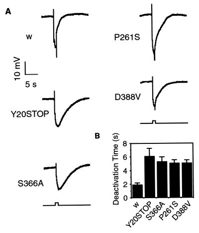
To examine further defects in the phosphorylation mutant and other missense alleles found in the genetic screen, we examined the electrophysiological responses of wild-type and mutant flies by using electroretinogram recordings. An electroretinogram recording represents the sum of the light-induced electrical activity in the photoreceptor cells and their downstream neurons. The photoreceptors of wild-type flies depolarize in response to light and rapidly deactivate the photoresponse once the light stimulus is terminated (Fig. 1A). Consistent with previous work on an arr2 loss-of-function allele (18), the arr2 null mutant (Y20STOP) had marked electrophysiological defects in its inactivation kinetics, with an increase in the time required to reach 85% inactivation (Fig. 1B). Flies carrying the three missense alleles, including the S366A phosphorylation mutation, have deactivation kinetics similar to those of flies carrying the arr2 null mutation. This result indicates further that, although there is Arr2 protein present in flies carrying all three missense alleles, it is essentially nonfunctional.
Figure 1.
Flies carrying arr2 missense mutations have prolonged deactivation kinetics that are indistinguishable from those of an arr2 null allele. (A) Representative electroretinogram recordings of white-eyed control (w) and arr2 mutant flies exposed to a 1-s pulse of blue light (480 nm). Electroretinograms were performed as described (18). (B) Histogram of the time to 85% deactivation for white-eyed control (w) and arr2 mutant flies. For w control flies, _t_85 = 1.87 ± 0.3 s; for Y20STOP flies, _t_85 = 6.07 ± 1.2 s; for S366A flies, _t_85 = 5.27 ± 0.7 s; for P261S flies, _t_85 = 5.05 ± 0.5 s; for D388V flies, _t_85 = 5.03 ± 0.5 s (n = 20–25). Data are means ± SD.
Ser-366 Is the Sole Light-Induced Phosphorylation Site in Arr2.
The Arr2 missense allele that eliminates the serine at position 366 provides a critical test of whether this residue is the sole Ca2+/calmodulin-dependent protein kinase site in Arr2. In lysates prepared from wild-type Drosophila heads, Arr2 shows a high level of phosphorylation that depends on the presence of calcium (Fig. 2A). However, Arr2 lacking the phosphorylation site at position 366 fails to be phosphorylated in vitro. A more stringent test for the presence and absence of phosphorylation is to examine the phosphorylation of Arr2 in vivo. For this in vivo test, flies were labeled with 32P and either exposed to light or kept in the dark; phosphoproteins were then analyzed by PAGE. In wild-type flies, Arr2 is phosphorylated in the light but has only low levels of Arr2 phosphorylation in the dark (Fig. 2B). In contrast to wild type, the Arr2 S366A mutant fails to show light-induced phosphorylation. Therefore, these data suggest that, both in vivo and in vitro, the serine at position 366 represents the sole light-dependent phosphorylation site in Arr2.
Figure 2.
Phosphorylation assays of retinal proteins indicate that the S366A mutant Arr2 protein is not phosphorylated in vitro and fails to undergo light-induced phosphorylation in vivo. (A, Left) Autoradiograph of γ-32P-labeled proteins isolated from Drosophila head lysates in the presence of 0.5 mM Ca2+ (lanes 1, 3, and 4) or 2 mM EGTA, a calcium-specific chelator (lane 2). (A, Right) The same gel transferred to nitrocellulose and probed with antibodies specific for Arr1 and Arr2 (18), as well as Rh1 (Developmental Studies Hybridoma Bank, University of Iowa). Y20STOP represents a null mutation in Arr2. w, white-eyed control flies. (B, Left) Autoradiographs of in vivo γ-32P-labeled retinal proteins from dark-reared white-eyed control (w) and S366A flies either kept in the dark (D) or exposed to light for 30 min (L). (B, Right) The same gels transferred to nitrocellulose and probed with antibodies specific for Arr2 and Arr1. Note the lack of light-induced phosphorylation of Arr2 in the flies expressing the S366A transgene.
Visual system arrestins are known to bind the activated form of rhodopsin (M form or metarhodopsin) and not to the inactive form (R form; refs. 30 and 31). In Drosophila, rhodopsin can be photoconverted between the M and R forms in response to the appropriate light stimulus (32). The major rhodopsin in the Drosophila retina absorbs maximally at 480 nm (blue), converting the rhodopsin to the active M form, and the M form can be photoconverted back to the inactive R form with 580-nm light (orange; refs. 33 and 34). Because Arr2 is known to bind to the M form, we wanted to test whether the unphosphorylated form of Arr2 was defective in binding to the M form. In wild-type flies, when rhodopsin is photoconverted to the M form, Arr2 binds to membranes (Fig. 3A). However, when wild-type flies are treated with orange light, thereby generating the inactive R form of rhodopsin, the arrestin remains largely cytoplasmic. Surprisingly, in spite of the fact that flies carrying the arr2 phosphorylation mutation are electrophysiologically indistinguishable from arr2 null alleles, the Arr2 encoded by this allele shows the same degree of binding to rhodopsin as does wild-type Arr2 (Fig. 3B). Clearly, both wild-type and the unphosphorylated form of Arr2 are interacting with rhodopsin, because the majority of Arr2 fails to bind to membranes that lack the major rhodopsin, Rh1 (Fig. 3B). This result suggests that, although phosphorylation is clearly essential for proper arrestin function, it is not required for rhodopsin binding.
Figure 3.
S366A flies retain the ability to bind to rhodopsin in a light-dependent manner. (A) Isolated wild-type fly heads were exposed to 5 min of either orange (580 nm) or blue (480 nm) light, homogenized in the dark, and centrifuged (13,000 × g for 5 min) Pellet (P) and supernatant (S) fractions were subjected to SDS/PAGE and Western analysis with antibodies directed against Arr2 and Rh1. (B) Western blot of white-eyed control (w), _ninaE_I17, S366A, and S366A;_ninaE_I17 heads that were exposed to 5 min of blue light and then fractionated and centrifuged as described in A. _ninaE_I17 represents a null mutation in the structural gene for the major rhodopsin, Rh1
Arr2 Phosphorylation Is Necessary for Arr2 to Release from Membranes.
Because of the unexpected finding that the unphosphorylated form of Arr2 failed to show binding defects, we reexamined the role of Arr2 phosphorylation and binding to light-treated membranes. If phosphorylation of Arr2 is essential for binding, the phosphorylated form of Arr2 would be found associated with membranes, whereas the unphosphorylated form would be largely cytoplasmic. To test this hypothesis, lysates from wild-type Drosophila heads were incubated with [γ-32P]ATP and then centrifuged to separate the membrane and supernatant fractions. Once again, as expected, Arr2 is phosphorylated in a calcium-dependent manner (Fig. 4). The phosphorylated form of Arr2 is not bound to membranes but, instead, is found as a soluble factor in the supernatant, and the membrane bound form of arrestin is completely unphosphorylated (Fig. 4). These results provide additional evidence that phosphorylation of Arr2 is not necessary for its binding to membranes when rhodopsin is photoconverted to the active form.
Based on the above data, it is clear that phosphorylation of Arr2 is not necessary for rhodopsin binding. Instead, we propose that the phosphorylation mutant is defective in the release of Arr2 from rhodopsin once rhodopsin is photoconverted to its inactive form. To test this idea, we took advantage of the fact that invertebrate arrestin binds to metarhodopsin in response to blue-light stimulus and is released when metarhodopsin is photoconverted back to its inactive R form with orange light (30, 35). As expected, the Arr2 protein in wild-type flies binds to rhodopsin under blue-light conditions, and 70–80% is released after subsequent exposure to orange light (Fig. 5). However, although the mutant S366A Arr2 protein binds to the activated form of rhodopsin, Arr2 fails to release from rhodopsin after orange-light treatment (Fig. 5), suggesting that Arr2 phosphorylation is necessary for arrestin to release from rhodopsin.
Figure 5.
Mutant flies that have reduced phosphorylation of Arr2 have defects in the release of Arr2 from rhodopsin. Western blot of fractionated white-eyed control (w), S366A, _norpA_P41 (norpA), and _cam_352/_cam_n339 (cam) mutant heads that were exposed to 10 s of blue light (B) or 10 s of blue light followed by 60 s of orange light (BO). Pellet (P) and supernatant (S) fractions were subjected to SDS/PAGE and Western analysis with antibodies directed against Arr2 and Rh1. _cam_352 and _cam_n339 represent hypomorphic and null mutations, respectively, in the gene encoding calmodulin (37), whereas _norpA_P41 (43) is a strong loss-of-function mutation in the eye-specific phospholipase C gene.
If the role of Arr2 phosphorylation is to allow its release from rhodopsin, then second site mutations that show decreased levels of arrestin phosphorylation should also show defects in release. It has been shown previously that flies with mutations in the eye-specific phospholipase C (norpA; refs. 29 and 36) and calmodulin (cam; ref. 37) have low levels of Arr2 phosphorylation. As can be seen in Fig. 5, norpA and cam mutants still show Arr2 membrane binding that is identical to wild type. Again, this result indicates that Arr2 phosphorylation does not play a role in rhodopsin binding, and all that is required for Arr2 binding is rhodopsin activation. However, after orange-light stimulus, norpA mutants fail to release Arr2, and cam mutants release <50% of the Arr2 (compared with 70–80% for wild type). The portion of Arr2 that does release in cam mutants can be accounted for by the low levels of Arr2 phosphorylation that has been observed in this genetic background (37).
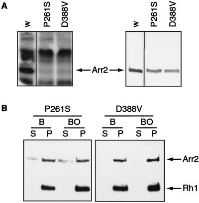
Further evidence supporting this model comes from the two missense alleles, P261S and D388V. Although these two mutations do not alter the Ca2+/Cam dependent protein kinase site directly, both missense alleles fail to show any significant phosphorylation of Arr2 in vitro. (Fig. 6A). It is possible that the mutations generate a structurally altered Arr2 molecule that cannot serve as a substrate for the kinase. In addition, these two missense alleles show no defects in rhodopsin binding; however, biochemical analysis of these two mutants indicates that, like the phosphorylation mutant, they generate Arr2 proteins that are unable to release from rhodopsin (Fig. 6B). Taken together, these data suggest that in the absence of phosphorylation, the arrestin cycle is disrupted; membrane translocation of Arr2 is unaltered, but its subsequent release from rhodopsin is inhibited.
Figure 6.
The P261S and D388V missense Arr2 proteins are not phosphorylated in vitro and have defects in releasing from rhodopsin. (A, Left) Autoradiograph of γ-32P-labeled proteins isolated from Drosophila head lysates in the presence of 0.5 mM Ca2+; (A, Right) The same gel transferred to nitrocellulose and probed with antibodies specific for Arr2. P261S and D388V represent missense mutations in Arr2. w, white-eyed control flies. (B) Western blot of fractionated P261S and D388V mutant heads that were exposed to 10 s of blue light (B) or 10 s of blue light followed by 60 s of orange light (BO). Pellet (P) and supernatant (S) fractions were subjected to SDS/PAGE and Western analysis with antibodies directed against Arr2 and Rh1.
DISCUSSION
Although it has been known for quite some time that Arr2 is phosphorylated in a light-dependent manner (25), it is unclear just what role this phosphorylation serves. The invertebrate phototransduction cascade results in an increase in intracellular calcium. It has been proposed that the calcium- and light-dependent phosphorylation of Arr2 acts as the signal to bind and inactivate metarhodopsin, and, therefore, Arr2 phosphorylation serves to modulate the inactivation of the signaling cascade (20, 30, 38, 39). However, we have found that arrestin is able to bind to activated rhodopsin in the absence of phosphorylation, arguing against this feedback-regulation model. Invertebrate Arr2 is a very basic molecule with a pKa of ≈8.7 (31). This characteristic may allow it to rapidly interact with an exposed acidic surface on activated rhodopsin in the absence of any covalent modification. Instead, posttranslational modification is required to remove arrestin from rhodopsin. Phosphorylation of Arr2 when it is bound to rhodopsin may trigger a conformational change or add negative charge that enables the release of Arr2.
The finding that the phosphorylation of Arr2 is required for proper function brings up an apparent discrepancy: previously, the C terminus of Arr2 was determined to be nonessential (18). In the previous study, a truncated form of Arr2 that lacked the last 45 aa of Arr2, including the serine at position 366, was generated; this mutated form of Arr2 was phenotypically normal. An identical situation occurs in the bovine system, where deletion of the C terminus of arrestin yields a protein that still binds to rhodopsin but has lost its binding specificity, binding to both photoactivated and nonphotoactivated rhodopsin (40). As such, we believe that the truncated form of Arr2 is not defective in release from rhodopsin but instead binds indiscriminately to both the active and inactive forms of rhodopsin. The truncation mutant is phenotypically normal, as it still has a higher binding affinity for the active form of rhodopsin.
In the missense alleles generated from the screen, Arr2 is found associated with membranes under all light conditions. This finding clearly explains the null phenotype of these alleles. Because rhodopsin is approximately five times more abundant than arrestin (18), all of the Arr2 becomes bound to membranes, and no soluble Arr2 is available to bind and inactivate metarhodopsin. However, it is unclear whether the titration of arrestin or the formation of arrestin/rhodopsin complexes is the primary cause of retinal degeneration in these alleles. Interestingly, many recently characterized human dominant rhodopsin alleles that are associated with retinitis pigmentosa and stationary night blindness also have defects in arrestin binding (41, 42). These alleles show continuous activity of rhodopsin in vitro; however, both in vitro and in vivo, these aberrant rhodopsin proteins are constitutively bound to arrestin. One possible mechanism for retinal degeneration in these dominant alleles is the titration of arrestin caused by its increased affinity for the aberrant rhodopsin proteins. In this way, photoreceptor cells undergo degeneration because of insufficient levels of soluble arrestin to quench newly formed metarhodopsin.
Acknowledgments
We thank C. Zuker for providing Drosophila strains, reagents, and advice; M. Dolph for generation and maintenance of Drosophila stocks; and V. Ambros, M. L. Guerinot, N. Grotz, and E. Connolly for comments on the manuscript. This work was supported by National Eye Institute Grant RO1 EY11534-02 and by a grant from the Pew Foundation.
Footnotes
This paper was submitted directly (Track II) to the Proceedings Office.
References
- 1.Baldwin J M. Curr Opin Cell Biol. 1994;6:180–190. doi: 10.1016/0955-0674(94)90134-1. [DOI] [PubMed] [Google Scholar]
- 2.Ji T H, Grossmann M, Ji I. J Biol Chem. 1998;273:17299–17302. doi: 10.1074/jbc.273.28.17299. [DOI] [PubMed] [Google Scholar]
- 3.Ferguson S S, Barak L S, Zhang J, Caron M G. Can J Physiol Pharmacol. 1996;74:1095–1110. doi: 10.1139/cjpp-74-10-1095. [DOI] [PubMed] [Google Scholar]
- 4.Palczewski K, Saari J C. Curr Opin Neurobiol. 1997;7:500–504. doi: 10.1016/s0959-4388(97)80029-3. [DOI] [PubMed] [Google Scholar]
- 5.Carman C, Benovic J. Curr Opin Neurobiol. 1998;8:335–344. doi: 10.1016/s0959-4388(98)80058-5. [DOI] [PubMed] [Google Scholar]
- 6.Zhang J, Ferguson S S, Barek L S, Aber M J, Giros B, Lefkowitz R J. Recept Channels. 1997;5:193–199. [PubMed] [Google Scholar]
- 7.Shichi H, Somers R L. J Biol Chem. 1978;253:7040–7046. [PubMed] [Google Scholar]
- 8.Benovic J L, Strasser R H, Caron M G, Lekkowitz R J. Proc Natl Acad Sci USA. 1986;83:2797–2801. doi: 10.1073/pnas.83.9.2797. [DOI] [PMC free article] [PubMed] [Google Scholar]
- 9.Wilden U, Hall S, Kühn H. Proc Natl Acad Sci USA. 1986;83:1174–1178. doi: 10.1073/pnas.83.5.1174. [DOI] [PMC free article] [PubMed] [Google Scholar]
- 10.Kühn H, Wilden U. J Recept Res. 1987;7:298–383. doi: 10.3109/10799898709054990. [DOI] [PubMed] [Google Scholar]
- 11.Lohse M J, Benovic J L, Condina J, Caron M G, Lefkowitz R J. Science. 1990;248:1547–1550. doi: 10.1126/science.2163110. [DOI] [PubMed] [Google Scholar]
- 12.Lohse M J, Andexinger S, Pitcher J, Trukawinski S, Codina J, Faure J, Caron M G, Lefkowitz R J. J Biol Chem. 1992;267:8558–8564. [PubMed] [Google Scholar]
- 13.Wacker W B, Donoso L A, Kalsow C M, Yankeelov J A, Organisciak D T. J Immunology. 1977;119:1949–1958. [PubMed] [Google Scholar]
- 14.Goodman O B, Jr, Krupnick J G, Santini F, Gurevich V V, Penn R B, Gagnon A W, Keen J H, Benovic J L. Nature (London) 1996;383:447–450. doi: 10.1038/383447a0. [DOI] [PubMed] [Google Scholar]
- 15.Ferguson S S, Downey W E, III, Colapietro A M, Barak L S, Menard L, Caron M G. Science. 1996;271:363–366. doi: 10.1126/science.271.5247.363. [DOI] [PubMed] [Google Scholar]
- 16.Ranganathan R, Maliki D M, Zuker C S. Annu Rev Neurosci. 1995;18:283–317. doi: 10.1146/annurev.ne.18.030195.001435. [DOI] [PubMed] [Google Scholar]
- 17.Chyb S, Raghu P, Hardie R C. Nature (London) 1999;397:255–259. doi: 10.1038/16703. [DOI] [PubMed] [Google Scholar]
- 18.Dolph P J, Ranganathan R, Colley N J, Hardy R W, Socolich M, Zuker C S. Science. 1993;260:1910–1916. doi: 10.1126/science.8316831. [DOI] [PubMed] [Google Scholar]
- 19.Ranganathan R, Stevens C F. Cell. 1995;81:841–848. doi: 10.1016/0092-8674(95)90004-7. [DOI] [PubMed] [Google Scholar]
- 20.Kahn E S, Matsumoto H. J Neurochem. 1997;68:169–175. doi: 10.1046/j.1471-4159.1997.68010169.x. [DOI] [PubMed] [Google Scholar]
- 21.Franceschini N, Kirschfeld K. Kybernetik. 1971;9:159–182. doi: 10.1007/BF02215177. [DOI] [PubMed] [Google Scholar]
- 22.Rubin G R, Spradling A C. Science. 1982;218:348–353. doi: 10.1126/science.6289436. [DOI] [PubMed] [Google Scholar]
- 23.Rubin G R, Spradling A C. Nucleic Acids Res. 1983;11:6341–6351. doi: 10.1093/nar/11.18.6341. [DOI] [PMC free article] [PubMed] [Google Scholar]
- 24.LeVine H, III, Smith D P, Whitney M, Malicki D M, Dolph P J, Smith G F, Burkhart W, Zuker C S. Mech Dev. 1990;33:19–25. doi: 10.1016/0925-4773(90)90131-5. [DOI] [PubMed] [Google Scholar]
- 25.Matsumoto H, Pak W L. Science. 1983;223:184–186. doi: 10.1126/science.6419348. [DOI] [PubMed] [Google Scholar]
- 26.Vinos J, Jalink K, Hardy R W, Britt S G, Zuker C S. Science. 1997;277:687–690. doi: 10.1126/science.277.5326.687. [DOI] [PubMed] [Google Scholar]
- 27.Ondek B, Hardy R, Baker E, Stamnes M, Shieh B, Zuker C. J Biol Chem. 1992;267:16460–16466. [PubMed] [Google Scholar]
- 28.Colley N, Cassill J, Baker E, Zuker C. Proc Natl Acad Sci USA. 1995;92:3070–3074. doi: 10.1073/pnas.92.7.3070. [DOI] [PMC free article] [PubMed] [Google Scholar]
- 29.Matsumoto H, Kurien B T, Takagi Y, Kahn E S, Kinumi T, Komori N, Yamada T, Hayashi F, Isono K, Pak W L, et al. Neuron. 1994;12:997–1010. doi: 10.1016/0896-6273(94)90309-3. [DOI] [PubMed] [Google Scholar]
- 30.Byk T, Bar Y M, Doza Y N, Minke B, Selinger Z. Proc Natl Acad Sci USA. 1993;90:1907–1911. doi: 10.1073/pnas.90.5.1907. [DOI] [PMC free article] [PubMed] [Google Scholar]
- 31.Bentrop J, Plangger A, Paulsen R. Eur J Biochem. 1993;216:67–73. doi: 10.1111/j.1432-1033.1993.tb18117.x. [DOI] [PubMed] [Google Scholar]
- 32.Minke B. In: Photopigment-Dependent Adaptation in Invertebrates: Implications for Vertebrates. Steive H, editor. New York: Springer; 1986. pp. 241–286. [Google Scholar]
- 33.O’Tousa J E, Baehr W, Martin R L, Hirsh J, Pak W L, Applebury M L. Cell. 1985;40:839–850. doi: 10.1016/0092-8674(85)90343-5. [DOI] [PubMed] [Google Scholar]
- 34.Zuker C S, Cowman A F, Rubin G M. Cell. 1985;40:851–858. doi: 10.1016/0092-8674(85)90344-7. [DOI] [PubMed] [Google Scholar]
- 35.Kiselev A, Subramaniam S. Science. 1994;266:1369–1373. doi: 10.1126/science.7973725. [DOI] [PubMed] [Google Scholar]
- 36.Matsumoto H, O’Tousa J, Pak W L. Science. 1982;217:839–841. doi: 10.1126/science.7100927. [DOI] [PubMed] [Google Scholar]
- 37.Scott K, Sun Y, Beckingham K, Zuker C S. Cell. 1997;91:375–383. doi: 10.1016/s0092-8674(00)80421-3. [DOI] [PubMed] [Google Scholar]
- 38.Richard E A, Lisman J. Nature (London) 1992;356:336–338. doi: 10.1038/356336a0. [DOI] [PubMed] [Google Scholar]
- 39.Scott K, Zuker C. Trends Biochem Sci. 1997;22:350–354. doi: 10.1016/s0968-0004(97)01100-6. [DOI] [PubMed] [Google Scholar]
- 40.Palczewski K, Buczylko J, Imami N R, McDowell J H, Hargrave P A. J Biol Chem. 1991;266:15334–15339. [PubMed] [Google Scholar]
- 41.Li T, Franson W K, Gordon J W, Berson E L, Dryja T P. Proc Natl Acad Sci USA. 1995;92:3551–3555. doi: 10.1073/pnas.92.8.3551. [DOI] [PMC free article] [PubMed] [Google Scholar]
- 42.Rim J, Oprian D. Biochemistry. 1995;34:11938–11945. doi: 10.1021/bi00037a035. [DOI] [PubMed] [Google Scholar]
- 43.Lindsley D L, Zimm G G. Genome of Drosophila melanogaster. San Diego: Academic; 1992. [Google Scholar]


