PAK4: a pluripotent kinase that regulates prostate cancer cell adhesion (original) (raw)
Abstract
Hepatocyte growth factor (HGF) is associated with tumour progression and increases the invasiveness of prostate carcinoma cells. Migration and invasion require coordinated reorganisation of the actin cytoskeleton and regulation of cell-adhesion dynamics. Rho-family GTPases orchestrate both of these cellular processes. p21-activated kinase 4 (PAK4), a specific effector of the Rho GTPase Cdc42, is activated by HGF, and we have previously shown that activated PAK4 induces a loss of both actin stress fibres and focal adhesions. We now report that DU145 human prostate cancer cells with reduced levels of PAK4 expression are unable to successfully migrate in response to HGF, have prominent actin stress fibres, and an increase in the size and number of focal adhesions. Moreover, these cells have a concomitant reduction in cell-adhesion turnover rates. We find that PAK4 is localised at focal adhesions, is immunoprecipitated with paxillin and phosphorylates paxillin on serine 272. Furthermore, we demonstrate that PAK4 can regulate RhoA activity via GEF-H1. Our results suggest that PAK4 is a pluripotent kinase that can regulate both actin cytoskeletal rearrangement and focal-adhesion dynamics.
Keywords: PAK, Migration, Prostate cancer
Introduction
A considerable body of evidence exists to suggest that the hepatocyte growth factor (HGF) signalling pathway is involved in the spread of prostate cancer (Gmyrek et al., 2001). HGF induces invasiveness of prostate cancer cells in vitro (Nishimura et al., 1999; Parr et al., 2001) and there is a high level of Met (HGF receptor) expression in malignant prostate epithelium (van Leenders et al., 2002).
Invasion of carcinoma cells into the surrounding stromal tissue requires the coordinated regulation of both actin cytoskeletal rearrangement and turnover of cell-substratum adhesions (Vega and Ridley, 2008). It is well established that the Rho-family GTPases Rho, Rac and Cdc42 orchestrate cell migration and adhesion turnover (Critchley, 2000; Ridley et al., 2003). We have previously shown that HGF-induced cell-cell dissociation and subsequent migration (scattering) of prostate cancer cells leads to disassembly of focal adhesions (Wells et al., 2005), and that HGF activates RhoA, Rac and Cdc42 (Wells et al., 2005).
Focal adhesions are large macromolecular structures that reside at the plasma membrane. They act as both a structural and signalling link between the extracellular matrix and the actin cytoskeleton through interactions with membrane-bound integrins (Zaidel-Bar et al., 2007). Cell adhesions can be highly dynamic and cell migration depends on the continuous formation and disassembly of adhesions at both the front and rear of a migrating cell (Ridley et al., 2003). Paxillin, one of the earliest described intracellular components of focal-adhesion complexes, acts as a scaffolding protein coordinating both the assembly and disassembly of signalling pathways (Deakin and Turner, 2008). Paxillin tyrosine phosphorylation by focal adhesion kinase (FAK) in response to focal adhesion is well documented (Turner, 2000). However, it has recently been reported that paxillin is also serine phosphorylated. It has been reported that phosphorylation of paxillin at serine 273 (chicken) or serine 272 (human) leads to increased turnover of cell adhesions and promotes GIT1 binding, but reduces the affinity of FAK for paxillin (Nayal et al., 2006). However, it is not clear whether this phosphorylation is mediated by PAK1 (Dong et al., 2009); moreover, the potential role of other PAK-family members during paxillin-mediated focal-adhesion turnover has not been investigated.
There are six known mammalian PAK proteins, which have been classified into two groups: group 1 PAKs (PAK1-PAK3) and group 2 PAKs (PAK4-PAK6) (Arias-Romero and Chernoff, 2008). Although there is sequence homology between group 1 and group 2 PAKs, group 1 PAKs bind both Rac and Cdc42, whereas group 2 PAKs bind specifically to Cdc42. Moreover, binding of Cdc42 to group 2 PAKs does not elevate kinase activity (Arias-Romero and Chernoff, 2008). It is likely that PAK-family members are differentially regulated and have specific cellular functions. Indeed, _PAK1_-null mice are viable and fertile, _PAK3_-null mice are viable with a degree of mental retardation and _PAK4_-null mice die during early development (Arias-Romero and Chernoff, 2008), indicating that group 1 PAKs cannot compensate for the loss of group 2 PAK activity, and that PAK4 confers essential and non-redundant functions in tissue development.
In addition to its absolute requirement for normal development, PAK4 is the only PAK-family member that is oncogenic when overexpressed (Callow et al., 2002) and it has recently been reported that PAK4 promotes tumorigenesis in vivo (Liu et al., 2008). PAK4 overexpression has been detected in a number of tumour cell lines, including prostate (Callow et al., 2002). Moreover, PAK4 amplifications have been identified in pancreatic cancer cells (Kim et al., 2008). Cells isolated from _PAK4_−/− mice appear to have increased focal adhesions (Qu et al., 2003) and endogenous PAK4 is localised to adhesive structures (Zhang et al., 2002) where it is thought to interact with β-integrins (Zhang et al., 2002). We have demonstrated that activated PAK4 induces a loss of focal adhesions (Wells et al., 2002) and that PAK4 is activated by HGF (Ahmed et al., 2008; Wells et al., 2002). Moreover, PAK4 was shown to be required for efficient HGF-dependent cell scattering and migration (Ahmed et al., 2008; Wells et al., 2002), and overexpression of PAK4 enhances breast cancer cell migration (Zhang et al., 2002).
A number of PAK4-binding proteins have been identified, including the Rho-family guanine-nucleotide-exchange factor (GEF) GEF-H1. GEF-H1 can act as an exchange factor for both Rac and RhoA but not for Cdc42 (Birkenfeld et al., 2008; Callow et al., 2005). There is evidence to suggest that PAK4 is able to regulate the RhoA exchange activity of GEF-H1. PAK4 is reported to phosphorylate GEF-H1 at serine 885 in vitro (Callow et al., 2005), and phosphorylation of this residue by Aurora inhibits RhoA exchange activity during mitosis (Birkenfeld et al., 2007). However, PAK4 regulation of RhoA activity via an interaction with GEF-H1 has not been demonstrated. Previous work has suggested that GEF-H1 is inactive when bound to microtubules (Krendel et al., 2002) but can activate RhoA when in the cytosol (Birukova et al., 2006; Zenke et al., 2004). The interaction between GEF-H1 and microtubules is predominantly associated with GEF-H1-mediated RhoA activation during late-stage mitosis (Birkenfeld et al., 2007). By contrast, GEF-H1 has also been identified as a tight-junction protein (Guillemot et al., 2008) and overexpression of GEF-H1 promotes focal-adhesion formation (Lim et al., 2008).
We now show here that PAK4 is required for HGF-induced scattering of human prostate cancer cells. We demonstrate that a loss of PAK4 expression leads to increased levels of active RhoA. In parallel, we find that a loss of PAK4 expression leads to a decrease in the level of paxillin phosphorylation at serine 272, a reduction in the turnover rate of focal adhesions and to the development of large focal adhesions. We demonstrate that PAK4 inhibits RhoA activity via GEF-H1 and that PAK4 directly phosphorylates paxillin at serine 272. This mechanism might be relevant to the function of PAK4 in other human cancers, as in prostate cancer presented here. On the basis of these and other supporting data we propose that PAK4 is a pluripotent kinase that can act downstream of HGF to mediate both rearrangement of the actin cytoskeleton and turnover of focal adhesions.
Results
PAK4 is activated by HGF in DU145 cells
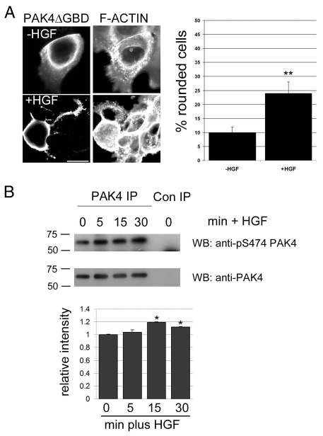
We have previously shown that expression of activated PAK4 can induce cell rounding in MDCK cells through an HGF-dependent pathway (Wells et al., 2002). Moreover, PAK4 is activated by HGF in these cells (Wells et al., 2002). Recently, we developed a model of HGF-induced cell scattering using the DU145 human prostate cancer cell line (Wells et al., 2005). DU145 cells express PAK1, PAK2, PAK4 and PAK6; we have found no evidence for expression of PAK3 or PAK5 (data not shown). Overexpression of activated PAK4 [PAK4ΔGBD; a PAK4 mutant with deleted p21-binding domain (PBD) (Wells et al., 2002)] in DU145 cells induced a significant increase in cell rounding in the presence of HGF (Fig. 1A) and HGF increased the level of endogenous phospho-PAK4 (Fig. 1B). This relatively low fold activation of endogenous PAK4 is probably due to only the more peripheral cells in the colonies responding to HGF stimulation. A similar activation of kinase activity by HGF has also been reported for PAK4 (Wells et al., 2002), SGK1 (serum- and glucocorticoid-inducible kinase 1) and PAK1 (Royal et al., 2000; Shelly and Herrera, 2002). Thus, PAK4-induced cytoskeletal changes are not restricted to MDCK cells. Having established that PAK4 is activated downstream of HGF and can induce cytoskeletal changes in an HGF-dependent manner, we investigated the effect of downregulating PAK4 expression on the ability of DU145 cells to respond to HGF.
Fig. 1.
PAK4 is active in prostate cancer cells. (A) DU145 cells were maintained in low serum for 24 hours and then microinjected with PAK4ΔGBD-HA. Cells were then either maintained in low serum or stimulated with 10 ng/ml HGF for a further 16 hours. All cells were then fixed and stained for F-actin and the HA-tagged protein. HA-positive cells were imaged by confocal microscopy and scored for cell rounding. Over 30 cells were scored for each population in each of three separate experiments. **P<0.005 using Student's _t_-test. (B) DU145 cells were maintained in low serum for 24 hours and then stimulated with HGF (250 ng/ml) for the given times. Endogenous PAK4 was immunoprecipitated from cell lysates using a rabbit anti-PAK4 antibody and the western blot probed for PAK4-phosphoserine474. The blot was re-probed for total PAK4 using the IP antibody. Con IP, rabbit anti-HA IP (control). Autoradiographs were quantified and the relative intensity of phospho-PAK4 signal was normalised to total PAK4. The results shown are the means ± s.e.m. of three independent experiments. Statistical significance compared to time 0 was calculated using Student's _t_-test, *P<0.05. Scale bar: 10 μm.
PAK4 is required for HGF-induced cell scattering
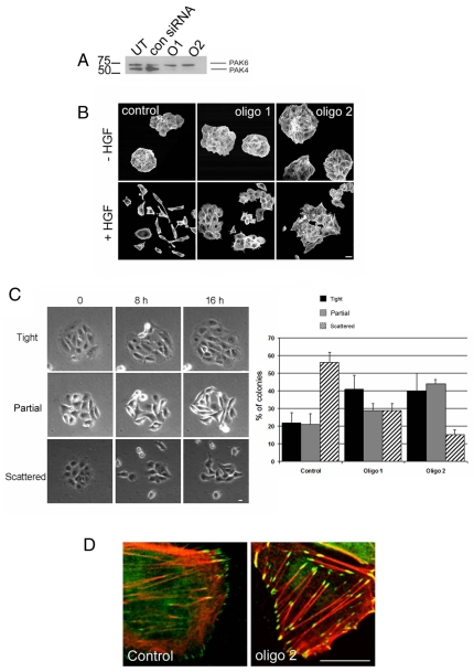
Using two different siRNA oligonucleotides, we reduced the level of PAK4 expression in cells without affecting PAK1, PAK2 or PAK6 expression (Fig. 2A and data not shown). Control-siRNA-transfected cells displayed a normal level of cell scattering, but the ability of PAK4-knockdown populations to scatter in response to HGF was significantly diminished (Fig. 2B and see supplementary material Movies 1-3 for examples of different responses). This is consistent with recent reports that MDCK cells with reduced PAK4 expression have an inhibited scattering response (Paliouras et al., 2009). We observed that, although a few cells were still able to undergo cell-cell dissociation, there was a marked increase in the number of colonies that did not fully dissociate (Fig. 2C). Closer examination of the unscattered colonies in the PAK4-knockdown population revealed an increased prominence of actin stress fibres and paxillin-containing adhesions compared with control cells (Fig. 2D). For the purposes of clarity we will refer to these large paxillin-, zyxin- (see Fig. S1A in the supplementary material) and actin-stress-fibre-associated adhesions at the periphery of DU145 cells as focal adhesions. We and others have previously shown that HGF induces the disassembly of actin stress fibres and focal adhesions (Wells et al., 2005), and that overexpression of activated PAK4 induces stress-fibre disassembly and loss of focal adhesions (Wells et al., 2002). These previous results and the data presented here provide strong evidence that PAK4 acts downstream of HGF to regulate the disassembly of actin stress fibres and/or focal adhesions.
Fig. 2.
PAK4 knockdown in DU145 prostate cancer cells. (A) Untransfected (UT), control-siRNA-transfected (con) and _PAK4_-siRNA-transfected [oligo 1 (O1) and oligo 2 (O2)] DU145 cell lysates were blotted with antibody that recognises both PAK4 and PAK6. (B) Control-siRNA cells and _PAK4_-siRNA knockdown cells (oligo 1 and oligo 2) were starved in low serum for 24 hours. Cells were then either maintained in low serum or stimulated with HGF (10 ng/ml) for 24 hours. All cells were fixed and stained for F-actin. Confocal images were taken from the basal plane. (C) Control-siRNA cells and _PAK4_-siRNA knockdown cells (oligo 1 and oligo 2) were starved in low serum for 24 hours. Cells were then stimulated with HGF (10 ng/ml) and filmed for 16 hours. Still images are shown of a time series from a PAK4-knockdown film depicting an example of a colony that did not scatter (tight), a colony that had partially scattered (partial) and a colony from which clear cell-cell dissociation and independent cell migration could be observed (scattered). Movies of cell colonies from both control-siRNA- and _PAK4_-siRNA-treated cells were scored as either tight, partial or scattered as described above. Data was collated from more than 20 colonies for each condition over three separate experiments. (D) Control and PAK4-knockdown cells were serum starved for 24 hours, fixed and stained for F-actin (red) and paxillin (green). Scale bars: 20 μm (B); 10 μm (C,D).
PAK4 depletion leads to an increased number of cell adhesions
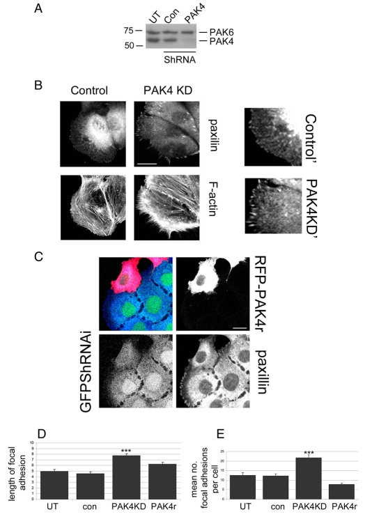
To further investigate the role of PAK4 in DU145-cell adhesion and migration we generated stable control-shRNA- and _PAK4_-shRNA-transfected DU145 cell lines in which normal levels of PAK1, PAK2 and PAK6 expression were maintained (Fig. 3A and data not shown). These cells express a bis-cistronic vector in which we found that the degree of knockdown efficiency is directly linked to the level of GFP expression (see supplementary material Fig. S1B). Consistent with our siRNA knockdown data, we found that PAK4 stable knockdown led to an increased prominence of actin stress fibres and focal adhesions (Fig. 3B), and to a significantly reduced migratory response to HGF (control cell mean speed ± s.e.m.=0.65±0.001 μm/minute; PAK4-knockdown cell mean speed ± s.e.m.=0.41±0.05 μm/minute; P<0.0025). Although control and PAK4-knockdown cells had the same spread area (see supplementary material Fig. S2A), PAK4-knockdown cells had a significantly higher number of focal adhesions per cell (Fig. 3E and supplementary material Table S1) and these focal adhesions were significantly larger (Fig. 3D) in comparison to control cells. Furthermore, the rise in number and size of focal adhesions could be experimentally rescued by the overexpression of PAK4r, a protein resistant to RNAi (Fig. 3C-E and supplementary material Fig. S1B).
Fig. 3.
Reduced PAK4 expression leads to an increase in focal adhesions. (A) Cell lysates from control and PAK4 stable cell lines were blotted for PAK4 and PAK6 expression. (B) Control and _PAK4_-shRNAi-knockdown cells were maintained in low serum for 24 hours, fixed and stained for F-actin and paxillin. On the right (control' and PAK4KD') are higher-magnification images of the periphery of the cell. (C) GFP-positive PAK4 stable knockdown cells expressing an RFP-PAK4 rescue vector (PAK4r; see Materials and Methods) were maintained in low serum for 24 hours, fixed and stained for paxillin. (D) The mean length of focal adhesion per cell was calculated using ImageJ (NIH) software. >40 cells were analysed for each population over three separate experiments. The results shown are the means ± s.e.m. Statistical significance compared to control shRNA cells was calculated using Student's _t_-test; ***P<0.0005. (**E**) The number of focal adhesions per cell (counting only cells at the colony periphery) was calculated for all cell populations. >40 cells were analysed for each population over three separate experiments. The results shown are the means ± s.e.m. Statistical significance compared to wild-type cells was calculated using Student's _t_-test; ***P<0.0005. Scale bars: 10 μm.
PAK4 regulates adhesion dynamics
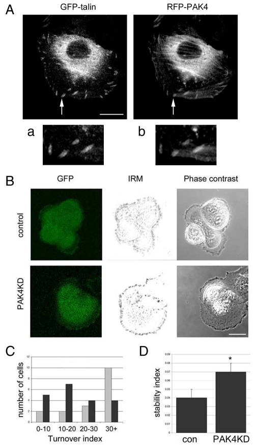
These data led us to consider whether PAK4 is required to disassemble focal adhesions. To test this hypothesis we sought to localise endogenous PAK4 in DU145 cells. Owing to the technical limitations of available antibodies it was not possible to localise endogenous PAK4 protein. However, exogenously expressed PAK4 clearly localised with talin at focal adhesions (Fig. 4A) and with endogenous paxillin (supplementary material Fig. S3A). We then used interference reflection microscopy to determine the adhesion turnover rate in real time of control and PAK4-knockdown cells (Fig. 4B). PAK4-knockdown cells had a significantly slower rate of focal adhesion turnover compared with control cells (Fig. 4C,D). However, suspended control and PAK4-knockdown cells were able to adhere to a substratum equally well (supplementary material Fig. S2B and data not shown).
Fig. 4.
PAK4 affects focal-adhesion turnover. (A) DU145 cells were co-transfected with RFP-PAK4wt and GFP-talin, serum starved for 24 hours then fixed and imaged. Arrow indicates focal adhesions. (a,b) Higher-magnification image of the area indicated by the arrows. (B) Control and _PAK4_-shRNA-knockdown (PAK4KD) cells were serum starved for 24 hours then focal adhesions were imaged in real time using live cell confocal IRM. Still images of GFP expression, the composite IRM image and phase-contrast image from representative control and _PAK4_-shRNA-knockdown cells. (C) Analysis of turnover index and adhesion stability of control-shRNA (grey bars) and _PAK4_-knockdown-shRNA (black bars) cells. (D) Stability index of control-shRNA (con) and _PAK4_-knockdown-shRNA (PAK4KD) cells. The results shown are the means ± s.e.m. Statistical significance compared to control cells was calculated using Student's _t_-test; *P<0.05. In C and D, >15 cells were analysed for each population over three separate experiments (see Materials and Methods for details of analysis).
PAK4 regulates adhesion turnover via paxillin phosphorylation
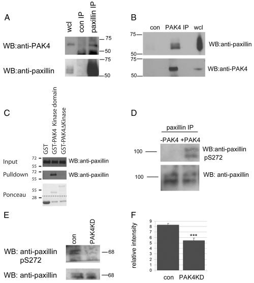
We previously reported that activated PAK4 leads to a loss of focal adhesions (Wells et al., 2002), and report here that a loss of PAK4 expression increases the number and size of PAK4 associated focal adhesions (Fig. 3). Given that PAK4 is localised to focal adhesions (Fig. 4A and supplementary material Fig. S3A), we investigated whether there was a direct association between PAK4 and paxillin, and found that we were able to co-immunoprecipitate endogenous PAK4 and paxillin (Fig. 5A,B). Moreover, using recombinant GST-tagged truncations of PAK4 we were able to show that the interaction between PAK4 and paxillin requires the presence of the PAK4 kinase domain (Fig. 5C). Focal-adhesion turnover is thought to be regulated in part by the phosphorylation of paxillin at serine 272 (Nayal et al., 2006); therefore, using a recombinant-PAK4 in vitro kinase assay (Wells et al., 2002), we tested whether PAK4 is capable of phosphorylating paxillin at serine 272. Paxillin was immunoprecipitated from cells and treated with calf intestinal phosphatase (CIP) to remove any existing phosphorylation. Immunoprecipitated paxillin was then incubated with recombinant PAK4. By using a paxillin-phosphoserine-272-specific antibody (supplementary material Fig. S3B) (Nayal et al., 2006), de-phosphorylated paxillin incubated with recombinant PAK4 was found to be phosphorylated at serine 272 (Fig. 5D and supplementary material Fig. S3B). PAK4-mediated phosphorylation of endogenous paxillin at serine 272 could also be detected (supplementary material Fig. S3D,E). Moreover, we detected no paxillin-phosphoserine-272 signal when using recombinant N-terminal PAK4 (PAK4Δkinase) or when overexpressing GFP-paxillinS272A (Dong et al., 2009) (supplementary material Fig. S4).
Fig. 5.
PAK4 phosphorylates paxillin at serine 272. (A) Endogenous paxillin was immunoprecipitated from DU145 cell lysates and probed for PAK4. Blots were re-probed for paxillin. wcl, whole cell lysate. Molecular mass (kDa) is shown to the right. (B) Endogenous PAK4 was immunoprecipitated from DU145 cell lysates and probed for paxillin. Blots were re-probed for PAK4. (C) GST alone, GST-PAK4 kinase domain and GST-PAK4Δkinase beads were used in an endogenous paxillin pulldown assay from cell lysates. Only GST-PAK4 kinase domain was able to pull down paxillin. (D) RFP-tagged paxillin was immunoprecipitated from HEK293 cell lysates, treated with calf intestinal phosphatase and incubated with or without recombinant active PAK4, then blotted for serine-272 phosphorylation. Serine-272 phosphorylation was only detected in the presence of recombinant PAK4. (E) Lysates from control (con) and PAK4-knockdown (PAK4KD) cell lines were probed for endogenous paxillin serine-272 phosphorylation and re-probed for total paxillin. (F) Autoradiographs were quantified and the relative intensity of phospho-serine-272-paxillin signal was normalised to total paxillin. The results shown are the means ± s.e.m. of three independent experiments. Statistical significance compared to control was calculated using Student's _t_-test; ***P<0.0005.
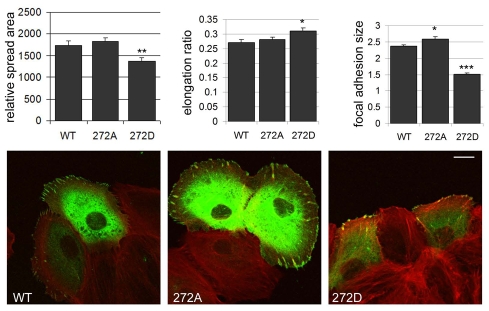
It has been suggested that phosphorylation of paxillin at serine 272 can drive paxillin localisation to the nucleus (Dong et al., 2009) and/or regulate the disassembly of focal adhesions (Nayal et al., 2006). To further characterise the significance of PAK4-mediated phosphorylation of paxillin at serine 272, we analysed the effect of expression of GFP-paxillin (wild type), GFP-paxillinS272A mutants (non-phosphorylated) and GFP-paxillinS272D mutants (phosphomimetic) in DU145 cells (Dong et al., 2009). We found expression of all three proteins in focal adhesions and did not detect a significant nuclear localisation, consistent with previous studies using full-length protein (Dong et al., 2009). Moreover, we also found that expression of paxillinS272D induced a significant reduction in spread area and increased the cell-elongation ratio, suggesting a change in adhesion dynamics in these cells. The difference in spread area and cell shape between populations made the quantification of the number of focal adhesions per cell unviable. However, in agreement with previous reports (Nayal et al., 2006), we were able to establish that expression of GFP-paxillinS272A resulted in larger focal adhesions at the cell periphery, whereas expression of GFP-paxillinS272D resulted in significantly smaller cellular adhesions at the cell periphery compared with expression of GFP-paxillin (wild type) (Fig. 6 and supplementary material Table S2). Thus, phosphorylation of paxillin at serine 272 mediates focal-adhesion disassembly in DU145 cells. Taken together, these results support the hypothesis that PAK4 phosphorylates paxillin at serine 272, which drives the disassembly of cellular adhesions. Consistent with this hypothesis we found significantly reduced levels of paxillin serine 272 phosphorylation in PAK4-knockdown cells (Fig. 5E,F).
Fig. 6.
Adhesions in DU145 cells expressing a serine-272 phospho-mimic are reduced in size. DU145 cells expressing GFP-wt (wild-type), GFP-S272A and GFP-S272D paxillin (green) were fixed and stained for F-actin (red). Cells were imaged by confocal microscopy and the cell-spread area, elongation ratio and mean length of focal adhesion per cell was calculated using ImageJ (NIH) software. >30 cells were analysed for each population over three separate experiments. The results shown are the means ± s.e.m. Statistical significance compared to control shRNA cells was calculated using Student's _t_-test; *P<0.05. Scale bar: 10 μm.
PAK4 regulates the activity of RhoA via GEF-H1
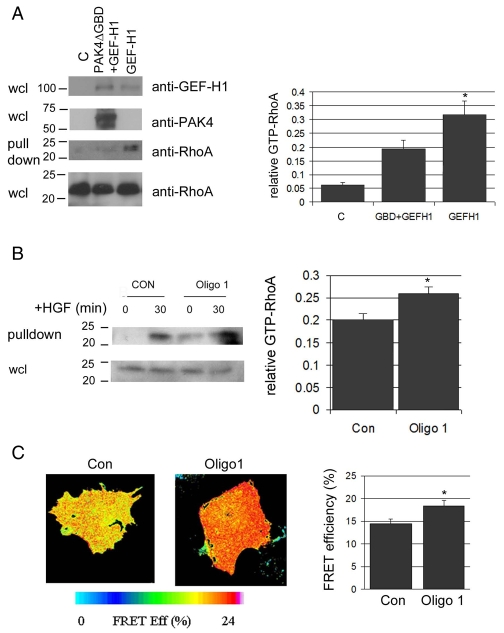
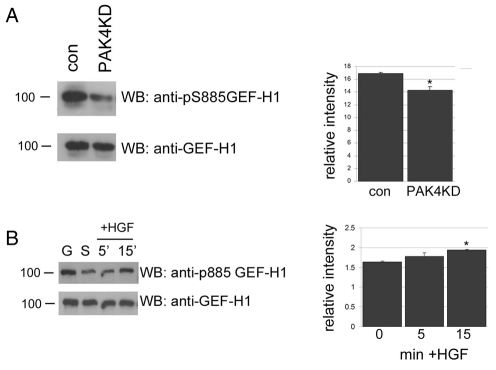
Concomitant with an increased number of paxillin-associated focal adhesions, PAK4-knockdown cells had more prominent actin stress fibres when compared with control cells (Figs 2 and 3), suggesting that RhoA-GTP levels are increased in the absence of PAK4. It had previously been reported that PAK4 can bind to the RhoA GEF GEF-H1 (Callow et al., 2005) and phosphorylate GEF-H1 at an inhibitory residue (Birkenfeld et al., 2007), although PAK4 inhibition of GEF-H1 exchange activity was not directly investigated. We first confirmed an interaction between PAK4 and GEF-H1 in prostate cancer cells (supplementary material Fig. S5). Subsequently, we were able to demonstrate that coexpression of active PAK4 (Wells et al., 2002) and GEF-H1 significantly reduced the GEF-H1-mediated increase in RhoA-GTP levels (Fig. 7A). Moreover, we found that coexpression of GEF-H1 and activated PAK4 significantly reduced the cell-rounding effect of activated PAK4 overexpression downstream of HGF (supplementary material Fig. S6). These results suggest that PAK4 regulates RhoA activity in cells via GEF-H1. Therefore, we sought to establish the level of RhoA activity in our control and PAK4-knockdown cell lines using a Rhotekin pulldown assay (Wells et al., 2005). In cells with reduced levels of PAK4 we detected an increase in the level of active GTP-RhoA in the absence of HGF (Fig. 7B); however, the level of active RhoA following HGF stimulation was not significantly different between control and PAK4-knockdown cells (Fig. 7B and data not shown), consistent with our previous observations (Wells et al., 2005). Furthermore, using a fluorescence resonance energy transfer (FRET) biosensor for RhoA (Carmona-Fontaine et al., 2008; Matthews et al., 2008) we were able to detect a significant increase in RhoA activity in PAK4-knockdown cells (Fig. 7C). Our results suggest that PAK4 regulates RhoA activity in a spatial- and temporal-dependent manner. PAK4 phosphorylates GEF-H1 on serine 885 (Callow et al., 2005), which inactivates RhoA exchange activity (Birkenfeld et al., 2007). We analysed the level of GEF-H1 phosphorylation in stable PAK4-knockdown DU145 cell lines compared with control cells (Fig. 8A) and found that the level of phosphorylation of GEF-H1 serine 885 was reduced. Our results point to a PAK4-mediated regulation of GEF-H1 activity. In support of this hypothesis, the level of phosphorylation of GEF-H1 serine 885 was increased following HGF stimulation of DU145 cells (Fig. 8B).
Fig. 7.
PAK4 regulates RhoA activity. (A) GST-Rhotekin RhoA pulldown assay from HEK293 cell lysates expressing GFP–GEF-H1 or co-expressing RFP-PAK4ΔGBD and GFP–GEF-H1. Whole-cell lysates (wcl) were probed for GEF-H1 expression, PAK4ΔGBD expression and total RhoA. C, untransfected cells. Autoradiographs of the level of active RhoA in all cells were quantified and the signal normalised to total RhoA. The results shown are the means ± s.e.m. of three independent experiments. Statistical significance compared to control was calculated using Student's _t_-test; *P<0.05. (B) GST-Rhotekin RhoA pulldown assay from DU145 control-siRNA and _PAK4_-siRNA knockdown (oligo 1) cell lysates in serum-starved and HGF-stimulated (30 minutes) conditions. Autoradiographs of the level of active RhoA-GTP in serum-starved cells were quantified and the signal normalised to total RhoA. The results shown are the means ± s.e.m. of three independent experiments. Statistical significance compared to control was calculated using Student's _t_-test; *P<0.05. (C) DU145 cells were transfected with a CFP/YFP FRET RhoA biosensor, incubated for 24 hours then transfected with control siRNA or PAK4 siRNA (oligo 1), incubated for a further 24 hours and then fixed and imaged using confocal microscopy (see Materials and Methods for details of FRET acquisition and analysis). Pseudocolour images of FRET efficiency in representative control and PAK4-knockdown cells are shown. The FRET-efficiency histogram represents the mean ± s.e.m. of three independent experiments. Statistical significance compared to control was calculated using Student's _t_-test; *P<0.05.
Fig. 8.
PAK4 regulates GEF-H1 phosphorylation. (A) Lysates from serum-starved control and _PAK4_-shRNA-knockdown DU145 cells were blotted for serine-885 GEF-H1 phosphorylation and re-probed for total GEF-H1. Autoradiographs of the level of phospho-serine-885-GEF-H1 in serum-starved cells were quantified using Andor IQ software (Andor UK) and the signal normalised to total GEF-H1. The results shown are the means ± s.e.m. of three independent experiments. Statistical significance compared to control was calculated using Student's _t_-test; *P<0.05. (B) Lysates from DU145 cells that had been maintained in low serum for 24 hours then stimulated by HGF (10 ng/ml) for the times indicated were probed for serine-885-GEF-H1 phosphorylation and re-probed for total GEF-H1. G, growing cells; S, serum-starved cells. Autoradiographs of the level of phospho-serine-885-GEF-H1 were quantified and the signal normalised to total GEF-H1 levels. The results shown are the means ± s.e.m. of three independent experiments. Statistical significance compared to time 0 was calculated using Student's _t_-test; *P<0.05.
PAK4 phosphorylation is increased in prostate-derived cancer cells
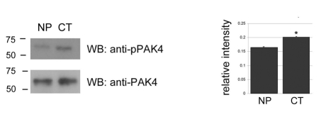
Our previous work (Ahmed et al., 2008) and the results presented here suggest that PAK4 is a key regulator of prostate cancer cell migration and can act in multiple pathways that lead to regulation of actin cytoskeletal rearrangement and cell-adhesion dynamics. We therefore sought to compare the level of PAK4 phosphorylation between a normal and cancer cell line derived from the same radical prostectomy (Bright et al., 1997). Immunoprecipitated PAK4 from normal and cancer cell populations was probed for the level of serine-474 phosphorylation using phosphospecific antibodies (Fig. 9). We found a significant increase in the level of phospho-PAK4 in prostate cancer cells compared with the patient-matched normal cells.
Fig. 9.
PAK4 is activated in prostate cancer. Endogenous PAK4 was immunoprecipitated from a matched patient pair of normal prostate cells (NP) and prostate cancer (CT) cell lines derived from the same radical prostectomy and probed for serine-474 phosphorylation. Immunoprecipitates were re-probed for total PAK4. Autoradiographs of the level of phospho-PAK4 were quantified and the signal normalised to total PAK4 levels. The results shown are the means ± s.e.m. of three independent experiments. Statistical significance compared to normal was calculated using Student's _t_-test; *P<0.05.
Discussion
We show here that PAK4 is required for HGF-induced scattering of human prostate cancer cells. It is becoming apparent that members of the group 2 PAK family play significant roles in cancer progression (Dummler et al., 2009). Indeed, PAK4 activity is correlated with cancer progression (Ahmed et al., 2008; Callow et al., 2002; Chen et al., 2008; Liu et al., 2008), and PAK4 is upregulated in a number of cancer cell lines and tissues (Callow et al., 2002; Liu et al., 2008), is amplified in pancreatic cancer (Chen et al., 2008) and is able to drive tumorigenesis in vivo (Liu et al., 2008). Moreover, PAK4 point mutations have been identified in colorectal cancer (Parsons et al., 2005).
We demonstrate that a decrease in PAK4 expression leads to increased levels of active RhoA. In parallel, we find that a loss of PAK4 expression leads to a decrease in the level of paxillin phosphorylation at serine 272, to a reduction in the turnover rate of cell adhesions and to the appearance of large focal adhesions. We also report that the level of PAK4 autophosphorylation, which is correlated with kinase activity (Wells et al., 2002), is increased in prostate cancer cells compared with normal cells derived from the same radical prostectomy. This is consistent with previous reports that PAK4 kinase activity is increased in pancreatic cancer cells (Chen et al., 2008; Kimmelman et al., 2008). Given these significant observations it would seem pertinent to discover the mode of action of PAK4 in cancer.
We show here that a decrease in PAK4 expression leads to an increase in the size and number of focal adhesions. PAK4 localises to the cell periphery of MDCK cells (Wells et al., 2002) and to peripheral cell adhesions in breast cancer cells (Zhang et al., 2002). We report here that PAK4 is also localised to peripheral focal adhesions in prostate cancer cells. However, we have not detected a direct interaction between PAK4 and β-integrins in prostate cancer cells (data not shown) as has once been described in the MCF-7 breast cancer cell line (Zhang et al., 2002). Interestingly, our previous work (Wells et al., 2002) demonstrated that overexpression of PAK4 induced a loss of focal adhesions. These studies and those of others (Zhang et al., 2002) suggest that PAK4 can regulate the turnover of cell adhesions. Our new data demonstrates that a decrease in PAK4 expression leads to an increased number of cell adhesions, which could be explained by a reduction in the adhesion turnover rate, i.e. increased adhesion stability. We predict that this increase in focal adhesions results in the inability of these DU145 cells to migrate in response to HGF, because HGF-induced cell scattering requires adhesion turnover (Wells et al., 2005). PAK4 binds specifically to Cdc42, and Cdc42 signalling pathways are associated with cell-adhesion disassembly (Chan et al., 2008; Yang et al., 2001). Future studies will seek to elucidate the role of a PAK4-Cdc42 interaction during cell-adhesion turnover.
Both control and PAK4-knockdown cells were equally able to initiate adhesions to the substratum following a period of suspension, and both populations were equally adherent following prolonged shaking of the supporting substratum. These results suggest that integrin activation upon adhesion (Worth and Parsons, 2008) occurs normally in PAK4-knockdown cells and argues against an absolute requirement for an association between β-integrins and PAK4 at this stage. Our results therefore suggest that PAK4 is not required for adhesion formation but rather for adhesion disassembly; however, we cannot rule out the possibility that PAK4 might inhibit the maturation of focal contacts. However, the cell-rounding effect induced by overexpression of active PAK4 would suggest a rapid loss of adhesion rather than an inhibition of maturation (Wells et al., 2002).
Adhesion turnover is in part mediated by the serine phosphorylation of paxillin, a major component of focal adhesions (Deakin and Turner, 2008). We report here for the first time that PAK4 and paxillin co-immunoprecipitate and that the interaction between PAK4 and paxillin is mediated by the kinase domain of PAK4. Given that the kinase domain of PAK4 is peripherally localised and PAK4 binds paxillin via its kinase domain, we would predict that the paxillin-PAK4 interaction recruits PAK4 to focal adhesions. These results suggested that paxillin might be a novel PAK4 substrate and we were able to demonstrate that PAK4 can phosphorylate paxillin on serine 272. Taken together, our data on adhesion dynamics in PAK4-knockdown cells and on the novel interaction between paxillin and PAK4 suggest that PAK4 is required for efficient adhesion turnover and that PAK4 drives adhesion turnover via phosphorylation of paxillin
Although phosphorylation of paxillin at serine 272 is proposed to reduce its affinity for FAK and might lead to the recruitment of GIT1, a protein that can promote adhesion disassembly (Zhao et al., 2000), phosphorylation of paxillin at serine 272 has also been reported, independently, to mediate paxillin nuclear localisation and regulation of gene transcription (Dong et al., 2009). Our work presented here supports a role for serine-272 phosphorylation in the regulation of adhesion turnover but does not exclude the possibility that phosphorylation of paxillin 272 might also induce relocalisation of paxillin from adhesions to the nucleus with a concomitant disassembly of adhesions and subsequent regulation of gene transcription.
PAK1 can phosphorylate paxillin at serine 272 as part of a GIT1-PIX-PAK complex (Nayal et al., 2006). However, recent findings have suggested that phosphorylation of paxillin serine 272 is not mediated by PAK1 (Dong et al., 2009). The ability of PAK4 to phosphorylate paxillin was not tested in either of these studies and PAK4 does not contain a PIX-binding motif (Abo et al., 1998), suggesting that PAK1 and PAK4 might function differently and/or be differentially localised in adhesions. Because a reduction in the expression of PAK1 and/or PAK2 in DU145 cells does not lead to an increase in actin stress fibres or focal adhesions (Bright et al., 2009) (and Michael Bright and Anne Ridley, King's College London, personal communication), we can conclude that PAK4 is the predominant PAK isoform regulating cell-adhesion turnover in DU145 cells. PAK4-knockdown cells contain normal levels of PAK6, and PAK6 does not seem to be able to compensate for loss of PAK4. This is perhaps not surprising given the differences between PAK4 and PAK6 in terms of subcellular localisation and limited sequence similarity outside the kinase domain. Indeed, whereas PAK5-knockout, PAK6-knockout and PAK5/PAK6-knockout mice are viable and fertile, PAK4-knockout mice are embryonic lethal despite retaining good levels of PAK1, PAK2, PAK5 and PAK6 expression, again suggesting that other PAKs cannot compensate for PAK4 function (Dummler et al., 2009).
We have previously reported that overexpression of active PAK4 induces the dissolution of actin stress fibres in MDCK cells; however, the mechanism remained unclear (Wells et al., 2002). We show here that a reduction in PAK4 expression increases the level of active RhoA. PAK4 binds to and phosphorylates GEF-H1 (a GEF for RhoA) and it has recently been shown that phosphorylation of GEF-H1 at serine 885 inhibits guanine-nucleotide-exchange activity (Birkenfeld et al., 2007). We predict that PAK4 phosphorylation of GEF-H1 at serine 885 inhibits RhoA exchange activity leading to a reduction in the level of GTP-RhoA. Indeed, a reduction in GEF-H1 expression leads to a reduction in RhoA activity and an inhibition of cell migration (Nalbant et al., 2009). Thus, in the absence of PAK4, GEF-H1 is permitted to activate RhoA, leading to higher levels of active RhoA. In agreement with our hypothesis, we have detected an increase in active RhoA and a reduction in the level of GEF-H1 serine-885 phosphorylation in PAK4-knockdown cells. However, serine-885 phosphorylation was not completely abolished, a fact we attribute to the continued presence of Aurora and PAK1, kinases also known to phosphorylate GEF-H1 at serine 885 (Birkenfeld et al., 2007; Zenke et al., 2004).
PAK4 also binds to and phosphorylates a second RhoA GEF, PDZRhoGEF; however, this interaction does not inhibit PDZRhoGEF exchange activity (Barac et al., 2004) and therefore the reported PAK4-dependent inhibition of RhoA activation (Barac et al., 2004) might be attributable, at least in part, to inactivation of GEF-H1. PAK4 binds specifically to Cdc42 (Abo et al., 1998) and an antagonistic relationship between the activities of Cdc42 and RhoA has been widely reported (Lim et al., 1996; Matozaki et al., 2000; Roberts et al., 2003). It is possible that Cdc42 inhibition of RhoA activity is mediated by a PAK4–GEF-H1 pathway.
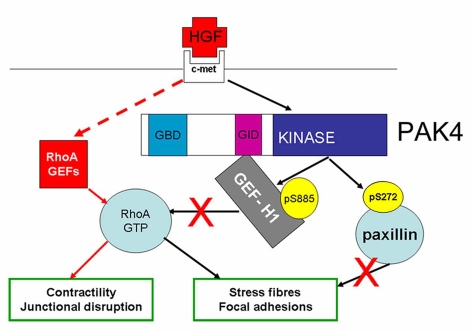
Our previous work demonstrated that RhoA becomes activated downstream of HGF within a similar timeframe to the activation of PAK4 and inactivation of GEF-H1 we report here (Wells et al., 2005). Indeed, in both control and PAK4-knockdown cells HGF stimulated an increase in the level of RhoA-GTP, as we have previously described (Wells et al., 2005). However, these assays only measure global levels of active RhoA. HGF stimulation of DU145 cells not only induces dissolution of focal adhesions but also simultaneously causes the disassembly of cell-cell junctions and an increase in cell contractility as cells begin to migrate (Wells et al., 2005), and RhoA activity has been linked to junctional disruption (Chang et al., 2006). Thus, it might be imagined that within the same temporal phase of response a spatial segregation of RhoA activity can be seen with inactivation of RhoA required in some cellular compartments while in others activation of RhoA will be necessary. Regulation of RhoA activity is likely to involve many signalling pathways that are dependent on the activity and specific localisation of regulatory proteins. Our results suggest that RhoA inactivation at focal adhesions is mediated by a PAK4–GEF-H1 interaction (see Fig. 10).
Fig. 10.
PAK4 regulates adhesion and migration via multiple signalling pathways. RhoA is locally regulated during HGF-induced DU145 cell scattering with both activating (red lines) and inactivating (black lines) pathways. RhoA activation downstream of HGF might be the result of a direct or indirect interaction with the Met receptor (broken line), and is likely to be required for junctional disruption and cell contractility during migration. PAK4 mediates an inactivation pathway via GEF-H1. HGF-activated PAK4 phosphorylates GEF-H1 on serine 885; this phosphorylation inhibits exchange activity towards RhoA at localised areas within the cell. In parallel, active PAK4 phosphorylates paxillin on serine 272. Reduced levels of RhoA at the cell periphery and increased phosphorylation of paxillin at serine 272 drive the dissolution of actin stress fibres and focal adhesions required for HGF-induced cell scattering.
PAK4 knockdown has also been shown to inhibit the migration of PC3 cells (Ahmed et al., 2008). PC3 cells are a highly metastatic prostate cancer cell variant, isolated from a bone metastasis; these cells do not have prominent actin stress fibres or large focal adhesions (Wells et al., 2005) (and our unpublished observations). Reduction of PAK4 expression in PC3 cells leads to a change in cell polarity and to reduced levels of phospho-cofilin (Ahmed et al., 2008). Cofilin is postulated to drive protrusion and migration, and phosphorylation of cofilin is thought to regulate cofilin activity (Bailly and Jones, 2003). However, we detected no changes in cell adhesions or actin-filament organisation in PC3 cells with reduced PAK4 expression (Ahmed et al., 2008). Conversely, we did not detect changes in cell shape nor in the levels of cofilin phosphorylation in PAK4-knockdown DU145 cells. These differences between DU145 and PC3 cells are reflected in the specific responses of these cells to HGF-stimulation. PC3 cells are more mesenchymal in phenotype and respond to HGF stimulation by increasing their speed of migration (Ahmed et al., 2008). DU145 cells are more epithelial in character and respond with an initial cell-cell dissociation step followed by a subsequent cell-migration response (scattering) (Wells et al., 2005). Our data suggest that PAK4 activity is cell-type specific and we have noted that mesenchymal-like MDA-MB-231 breast cancer cells with reduced PAK4 expression display a phenotype similar to PC3 cells, whereas epithelial-like A375 melanoma cells with reduced PAK4 expression have a phenotype similar to DU145 cells (our unpublished data).
In summary, our data point to the importance of PAK4-specific signalling pathways downstream of HGF in DU145 prostate cancer cells (Fig. 10). We have identified a novel interaction between PAK4 and paxillin, and provide evidence that PAK4 regulates cell-adhesion turnover via serine phosphorylation of paxillin. We also demonstrate that PAK4 can negatively regulate RhoA exchange activity via GEF-H1 and that GEF-H1 serine phosphorylation correlates with HGF stimulation in cells. Data presented here highlight the therapeutic potential of targeting the PAK4 signalling pathway. However, as we have demonstrated, PAK4 has multiple substrates. To exploit the therapeutic potential of PAK4 we will first need to understand the temporal and spatial regulation of PAK4 activity and the prevalence of different PAK4 signalling pathways in different cancer cell types.
Materials and Methods
Antibodies and reagents
Rabbit anti-PAK4 [which cross reacts with PAK6 in westerns (Ahmed et al., 2008)], rabbit anti-phospho-PAK4/5/6 (S474), rabbit anti-GEF-H1 and rabbit anti-phospho-S885-GEF-H1 were obtained from Cell Signalling Technology. Mouse anti-paxillin was obtained from Transduction Laboratories and rabbit anti-paxillin-phosphoS273(272) from BioSource International USA. Mouse anti-RhoA was obtained from Santa Cruz Biotechnology. HRP-conjugated secondary antibodies were obtained from DAKO. TRITC-conjugated phalloidin and anti-β-actin were obtained from Sigma, UK. For immunoprecipitation an anti-PAK4 antibody was raised in rabbit using a peptide sequence unique to PAK4 (CRRAGPEKRPKSSREG); this antibody specifically recognises PAK4 and does not cross react with PAK5 or PAK6 as tested by western analysis using overexpressed proteins (data not shown). Recombinant human HGF was purchased from R&D Systems and used at a final concentration of 10 ng/ml. Recombinant active PAK4 was purchased from Upstate Biotechnology. PAK4ΔGBD-HA, mRFP-PAK4ΔGBD and mRFP-PAK4-wt have been previously described (Abo et al., 1998; Ahmed et al., 2008). eGFP–GEF-H1 was a kind gift of Gary Bokoch, Scripps Research Institute, CA and eGFP-talin was a kind gift of Kenneth Yamada, NIH, Bethesda, MD. mRFP-PAK4r was generated by site-directed mutagenesis using a QuikChange Multisuite II kit (Stratagene) according to the manufacturer's instructions. Primers to introduce silent mutations were designed using the QuikChange mutagenic primer design program (Stratagene). Plasmid pENTR-PAK4 (Ahmed et al., 2008) was used as a template for the mutagenic reaction. Clones were screened by sequencing and alignment to wild-type sequences to confirm mutagenesis, prior to Gateway (Invitrogen) recombination to generate an expression vector encoding mRFP-PAK4r. GFP-wt, -S272A and -S272D paxillin (Dong et al., 2009) were a kind gift of Edward Manser, IMCB, Singapore. Expression plasmids encoding GST-tagged PAK4 kinase domain and PAK4Δkinase were generated using Gateway Technology (Invitrogen). Briefly, an 807-bp fragment encoding amino acids 323-591 of PAK4 was amplified by PCR from PAK4 cDNA using specific primers 5′-GGGGACAAGTTTGTACAAAAAAGCAGGCTTGTTCATCAAGATTGGCGAGGGCTCC-3′ and 5′-GGGGACCACTTTGTACAAGAAAGCTGGGTCTCATCTGGTGCGGTTCTGGCGCAT-3′. A 966-bp fragment encoding amino acids 1-322 of PAK4 was amplified in the same manner using specific primers 5′-GGGGACAAGTTTGTACAAAAAAGCAGGCTTGATGTTTGGGAAGAGGAAGCGG-3′ and 5′-GGGGACCACTTTGTACAAGAAAGCTGGGTCTCAGTTGTCCAGGTAGGAGCGGGGGTC-3′. The 868-bp (kinase domain) and 1030-bp (PAK4Δkinase) PCR products, containing terminal attB sites, were used in Gateway recombination to generate entry clones that were sequenced prior to further recombination to generate an expression vector encoding GST-PAK4 kinase domain and -PAK4Δkinase. The fidelity of these plasmids was subsequently confirmed by sequencing.
Cell culture
DU145 cells (European Tissue Culture Collection) were grown in RPMI-1640 (Sigma), supplemented with 10% FBS (Helena Biosciences), L-glutamine and 100 U/ml penicillin-streptomycin. In all cases, pre-plated cells were serum starved for 24 hours in low-serum media consisting of RPMI-1640 (Sigma), supplemented with 0.5% FBS, L-glutamine and 100 U/ml penicillin-streptomycin prior to HGF (10 ng/ml) stimulation. DU145 cells were transiently transfected using Fugene-6 transfection reagent according to the manufacturer's protocol (Roche). HeLa cells and HEK293 cells (European Tissue Culture Collection) were grown in DMEM-GluMAx (Sigma) supplemented with 10% FBS (Helena Biosciences), L-glutamine and 100 U/ml penicillin-streptomycin, and transfected by calcium-phosphate transfection according to the manufacturer's protocol (Invitrogen). HeLa cells (European Tissue Culture Collection) were grown in DMEM (Sigma) supplemented with 10% FBS (Helena Biosciences), L-glutamine and 100 U/ml penicillin-streptomycin. Matched cell lines of normal human prostate (1535-NPTX) and primary cancer (1535-CP3TX) derived from the same radical prostatectomy were grown in keratinocyte serum-free medium (Gibco) supplemented with 10% FBS (Helena Biosciences), L-glutamine, 100 U/ml penicillin-streptomycin, bovine pituitary extract and EGF as previously described (Bright et al., 1997).
Knockdown of PAK4 expression
PAK4 siRNA oligonucleotide 1 (O1) was purchased from Ambion, Austin, TX. The sense sequence was 5′-GGTGAACATGTATGAGTGT-3′. PAK4 siRNA oligonucleotide O2 was purchased from Qiagen, Crawley, West Sussex, UK as a validated PAK4 RNAi oligo (cat. no. SI02660315). Control-RNA oligonucleotides were purchased from Qiagen (cat. no. 1022076). Control and _PAK4_-specific oligos were added to cells using HiPerFect Transfection Reagent (Qiagen) according to the manufacturer's instructions to a final concentration of 25 nM. Routinely we detected a 70-80% reduction in PAK4 protein levels in O1- and O2-transfected cells compared with control cells by western blot. To generate stable control and PAK4-knockdown DU145 cell lines, cells were transfected with control or _PAK4_-specific pGIPz lentiviral vectors that also express TurboGFP (Open Biosystems, Huntsville, AL). Stable mixed clones were puromycin selected (700 ng/ml) and maintained in medium supplemented with 700 ng/ml puromycin.
Immunofluorescence and image analysis
Cells were seeded at a density of 1×10⁴ cells/ml on glass or human-fibronectin (Sigma, UK; 10 μg/ml)-coated coverslips and allowed to form colonies. Cells were maintained in low serum for 24 hours. Following serum starvation, cells were either maintained in low serum or stimulated by HGF for the times indicated in figure legends. All cells were subsequently fixed with 4% paraformaldehyde in PBS for 20 minutes at room temperature and then permeabilised with 0.2% Triton X-100 in PBS for 5 minutes. For F-actin staining, cells were incubated with TRITC-conjugated phalloidin diluted in PBS for 1 hour at room temperature. Following incubation, cells were washed six times in PBS. For detection of paxillin, primary and secondary antibodies were diluted in PBS containing 0.5% bovine serum albumin and all incubations were for 1 hour at room temperature. Following incubation with the primary antibody, cells were washed six times in PBS and then incubated with the secondary antibody and phalloidin (1:1000). For counts of cell rounding, all PAKΔGBD-expressing cells were objectively scored as spread or rounded on the basis of their actin cytoskeletal staining; where rounded cells had a spherical shape and were not polarised. For cell-shape analysis, images of cells were obtained using a Zeiss LSM510 confocal laser-scanning microscope (Zeiss, Welwyn Garden City, UK), using the accompanying LSM 510 software. Images were processed in Adobe Photoshop 7.0. Cell area, elongation ratio (Wells et al., 2005), and the number and size of focal adhesions were quantified using ImageJ (NIH). Data are presented as mean ± s.e.m. The Student paired _t_-test was used to compare differences between groups. Statistical significance was accepted for _P_≤0.05.
Immunoprecipitation
Cells were lysed as previously described (Wells et al., 2002). For immunoprecipitation experiments, cell lysates were pre-cleared with IgG-coupled protein-A or -G–Sepharose beads (GE Healthcare) for 1 hour at 4°C. The pre-cleared lysates were then mixed with primary antibody overnight at 4°C followed by 1-hour incubation with protein-A or -G–Sepharose beads. The immune complexes were washed three times with lysis buffer and resuspended in 2× SDS loading buffer. Proteins were resolved by SDS-PAGE as previously described (Wells et al., 2002). Autoradiographs were quantified using Andor IQ software (Andor UK).
Phosphorylation assay
The immune complexes were washed three times with cold lysis buffer and resuspended in cold kinase buffer (Wells et al., 2002) or treated with calf intestinal phosphatase according to the manufacturer's instructions (New England Biolabs) for 30 minutes at 37°C. Following phosphatase treatment, immune complexes were washed three times with cold lysis buffer and resuspended in cold kinase buffer (Wells et al., 2002). Recombinant PAK4 (80 ng/ml; Upstate Biotechnology) or recombinant PAK4Δkinase (80 ng/ml) was added to the kinase buffer and the reaction mix was incubated at 30°C for 30 minutes. Kinase activity was stopped by the addition of 6× gel sample buffer. Samples were resolved by SDS-PAGE as previously described (Wells et al., 2002). Autoradiographs were quantified using Andor IQ software (Andor UK).
GST pulldown
GST proteins were purified from BL21-A1 bacteria (Invitrogen) as previously described (Ahmed et al., 2008). HeLa cells were lysed in 20 mM Tris-HCl, pH 7.6, 150 mM NaCl, 2 mM EDTA, 0.1% Triton X-100, 50 mM NaF, 1 mM Na3VO4 and protease-inhibitor cocktail (Roche). Lysates were then pre-cleared by incubation with GST-coupled Glutathione Sepharose 4 Fast Flow beads (Amersham) for 1 hour at 4°C. The pre-cleared lysates were incubated with the GST-fusion-protein beads for 3 hours at 4°C, collected by centrifugation, washed three times with wash buffer (20 mM Tris-HCl, pH 7.6, 300 mM NaCl, 2 mM EDTA, 1% Triton X-100, 10% glycerol, 50 mM NaF, 1 mM Na3VO4 and protease-inhibitor cocktail) and resuspended in 2× SDS loading buffer.
Time-lapse microscopy
Six-well plates, containing control or experimental cells as described in the figure legends, were placed on the automated stage of an Axiovert 100 microscope in the presence of 10% CO2. Cell images were collected using a Sensicam (PCO Cook) CCD camera, taking a frame every 10 minutes for 24 hours from each of the six wells using AQM acquisition software (Andor Technology, Belfast, UK). Subsequently, all the acquired time-lapse sequences were displayed as a movie and cells were tracked for the whole of the time-lapse sequence using Motion Analysis software (Andor Technology, Belfast, UK) or colonies were scored according to the level of scattering response; tight = no evidence of cell-cell dissociation, partial = some cell-cell dissociation but little migration, scatter = successful cell-cell dissociation and a migratory response (see supplementary material Movies 1-3). This resulted in the generation of a sequence of position co-ordinates relating to each cell in each frame. 100 cells were tracked over two separate films for each experimental condition. Mathematical analysis was then carried out using Mathematica 6.0 notebooks developed in house by Graham Dunn and G.E.J. Statistical significance was accepted for _P_≤0.05.
Interference reflection microscopy
Cells were seeded on fibronectin-coated coverslips and allowed to form colonies. Cells were maintained in low serum for 24 hours. Following serum starvation, coverslips were mounted onto glass viewing chambers (made in house) in the presence of low-serum medium and placed on the pre-heated stage of a Zeiss LSM 510 confocal microscope. Interference reflection microscopy (IRM) images of GFP-positive cells were captured at 1-minute intervals over 7 minutes and processed as previously described (Chou et al., 2006; Holt et al., 2008). The eight images were then overlapped and re-inverted. A composite image with eight relevant grey levels was obtained. The lightest grey level represented pixels that were present in one of the eight images (adhesion points last for 1 minute), and the darkest grey level represented pixels that were present in eight out of eight images. Therefore, the areas of light grey pixels represent dynamic adhesions whereas areas of dark grey and black pixels represent stable adhesions over the selected interval of time. Turnover index = (% of pixels present in grey level 6, 7, 8) / (% of pixels present in grey level 1, 2, 3). Thus, a ratio of unstable adhesion over stable adhesion in each live cell was obtained. The higher value of the turnover index represents the more dynamic of the cell adhesion. Stability index = (% of pixels present in grey level 1, 2, 3) / (% of pixels present in grey level 6, 7, 8). Thus, a higher value of stability represents less-dynamic cell adhesions. Unpaired Student's _t_-test was used to assess the significance of experimental results.
RhoA pulldown assay
The Rho-activation assay was performed using GST-Rhotekin-PBD-coupled beads (Ren and Schwartz, 2000). Briefly, after treatment, DU145 cells were washed with cold PBS and lysed in 1× lysis buffer (Ren and Schwartz, 2000), with the addition of 10% glycerol, followed by immediate centrifugation at 13,000 g for 10 minutes. A small proportion of the lysate was removed for protein concentration assay (Bio-Rad) and western blot analysis of total protein levels. Cleared lysates were then incubated for 45 minutes with pre-washed GST-Rhotekin-PBD beads at 4°C. The beads were pelleted by centrifugation (6000 g for 1 minute) and washed three times with 1× cold wash buffer (Ren and Schwartz, 2000). The beads were finally resuspended in 30 μl of 2× gel sample buffer. Samples were separated by 12.5% SDS-PAGE and western blotted with an anti-RhoA antibody (Santa Cruz).
FRET analysis
DU145 cells were seeded on coverslips, FuGene6-transfected with the CFP/YFP RhoA biosensor (Carmona-Fontaine et al., 2008; Matthews et al., 2008) incubated for 24 hours then transfected with control and PAK4 siRNA oligonucleotide O1 as described above in low serum conditions. Following a further 24-hour incubation cells were fixed and imaged using a Zeiss lSM 510 META laser scanning confocal microscope and a 63× Plan Apochromat NA 1.4 Ph3 oil objective. The CFP and YFP channels were excited using the 405-nm blue diode laser and the 514-nm argon line, respectively. The two emission channels were split using a 545-nm dichroic mirror, which was followed by a 475- to 525-nm bandpass filter for CFP and a 530-nm longpass filter for YFP. Pinholes were opened to give a depth of focus of 3 mm for each channel. Scanning was performed on a line-by-line basis with zoom level set to two. The gain for each channel was set to approximately 75% of dynamic range (12-bit, 4096 grey levels) and offsets set such that backgrounds were zero. Time-lapse mode was used to collect one pre-bleach image for each channel followed by bleaching with 50 scans of the 514-nm argon laser line at maximum power (to bleach YFP). A second post-bleach image was then collected for each channel. Pre- and post-bleach CFP and YFP images were then imported into Mathematica 7 for processing. Briefly, images were smoothed using a 3×3 box mean filter, background subtracted and post-bleach image fade compensated for. A FRET-efficiency ratio map over the whole cell was calculated using the following formula: CFPpostbleach CFPprebleach / CFPpostbleach. Ratio values were then extracted from pixels falling inside the bleach region as well as an equally sized region outside of the bleach region and the mean ratio determined for each region and plotted on a histogram. The non-bleach ratio was then subtracted from the bleach region ratio to give a final value for the FRET efficiency ratio. Data from images were used only if YFP bleaching efficiency was greater than 70%.
Supplementary Material
[Supplementary Material]
Acknowledgments
This work was supported by grants from Cancer Research UK (G.E.J., J.R.W.M. and C.M.W.), MRC (G.E.J.) and Guys and St Thomas Charity (C.M.W.). C.M.W. was in part supported by a Wellcome Trust VIP fellowship. The authors would like to thank S. Fram for technical assistance and Anne Ridley (Randall Division, King's College London) for critical reading of the manuscript and communication of unpublished results. Deposited in PMC for release after 6 months.
Footnotes
References
- Abo A., Qu J., Cammarano M. S., Dan C., Fritsch A., Baud V., Belisle B., Minden A. (1998). PAK4, a novel effector for Cdc42Hs, is implicated in the reorganization of the actin cytoskeleton and in the formation of filopodia. EMBO J. 17, 6527-6540 [DOI] [PMC free article] [PubMed] [Google Scholar]
- Ahmed T., Shea K., Masters J. R. W., Jones G. E., Wells C. M. (2008). A PAK4-LIMK1 pathway drives prostate cancer cell migration downstream of HGF. Cell. Signal. 20, 1320-1328 [DOI] [PubMed] [Google Scholar]
- Arias-Romero L. E., Chernoff J. (2008). A tale of two Paks. Biol. Cell 100, 97-108 [DOI] [PubMed] [Google Scholar]
- Bailly M., Jones G. E. (2003). Polarised migration: cofilin holds the front. Curr. Biol. 13, R128-R130 [DOI] [PubMed] [Google Scholar]
- Barac A., Basile J., Vazquez-Prado J., Gao Y., Zheng Y., Gutkind J. S. (2004). Direct interaction of p21-activated kinase 4 with PDZ-RhoGEF, a G protein-linked Rho guanine exchange factor. J. Biol. Chem. 279, 6182-6189 [DOI] [PubMed] [Google Scholar]
- Birkenfeld J., Nalbant P., Bohl B. P., Pertz O., Hahn K. M., Bokoch G. M. (2007). GEF-H1 modulates localized RhoA activation during cytokinesis under the control of mitotic kinases. Dev. Cell 12, 699-712 [DOI] [PMC free article] [PubMed] [Google Scholar]
- Birkenfeld J., Nalbant P., Yoon S. H., Bokoch G. M. (2008). Cellular functions of GEF-H1, a microtubule-regulated Rho-GEF: is altered GEF-H1 activity a crucial determinant of disease pathogenesis? Trends Cell Biol. 18, 210-219 [DOI] [PubMed] [Google Scholar]
- Birukova A. A., Adyshev D., Gorshkov B., Bokoch G. M., Birukov K. G., Verin A. D. (2006). GEF-H1 is involved in agonist-induced human pulmonary endothelial barrier dysfunction. Am. J. Physiol. Lung Cell Mol. Physiol. 290, L540-L548 [DOI] [PubMed] [Google Scholar]
- Bright R. K., Vocke C. D., Emmert-Buck M. R., Duray P. H., Solomon D., Fetsch P., Rhim J. S., Linehan W. M., Topalian S. L. (1997). Generation and genetic characterization of immortal human prostate epithelial cell lines derived from primary cancer specimens. Cancer Res. 57, 995-1002 [PubMed] [Google Scholar]
- Bright M. D., Garner A. P., Ridley A. J. (2009). PAK1 and PAK2 have different roles in HGF-induced morphological responses. Cell Signal. 21, 1738-1747 [DOI] [PubMed] [Google Scholar]
- Callow M. G., Clairvoyant F., Zhu S., Schryver B., Whyte D. B., Bischoff J. R., Jallal B., Smeal T. (2002). Requirement for PAK4 in the anchorage-independent growth of human cancer cell lines. J. Biol. Chem. 277, 550-558 [DOI] [PubMed] [Google Scholar]
- Callow M. G., Zozulya S., Gishizky M. L., Jallal B., Smeal T. (2005). PAK4 mediates morphological changes through the regulation of GEF-H1. J. Cell Sci. 118, 1861-1872 [DOI] [PubMed] [Google Scholar]
- Carmona-Fontaine C., Matthews H. K., Kuriyama S., Moreno M., Dunn G. A., Parsons M., Stern C. D., Mayor R. (2008). Contact inhibition of locomotion in vivo controls neural crest directional migration. Nature 456, 957-961 [DOI] [PMC free article] [PubMed] [Google Scholar]
- Chan P. M., Lim L., Manser E. (2008). PAK is regulated by PI3K, PIX, CDC42, and PP2Calpha and mediates focal adhesion turnover in the hyperosmotic stress-induced p38 pathway. J. Biol. Chem. 283, 24949-24961 [DOI] [PMC free article] [PubMed] [Google Scholar]
- Chang Y. W., Marlin J. W., Chance T. W., Jakobi R. (2006). RhoA mediates cyclooxygenase-2 signaling to disrupt the formation of adherens junctions and increase cell motility. Cancer Res. 66, 11700-11708 [DOI] [PubMed] [Google Scholar]
- Chen S., Auletta T., Dovirak O., Hutter C., Kuntz K., El-Ftesi S., Kendall J., Han H., Von Hoff D. D., Ashfaq R., et al. (2008). Copy number alterations in pancreatic cancer identify recurrent PAK4 amplification. Cancer Biol. Ther. 7 Epub [DOI] [PMC free article] [PubMed] [Google Scholar]
- Chou H. C., Anton I. M., Holt M. R., Curcio C., Lanzardo S., Worth A., Burns S., Thrasher A. J., Jones G. E., Calle Y. (2006). WIP regulates the stability and localization of WASP to podosomes in migrating dendritic cells. Curr. Biol. 16, 2337-2344 [DOI] [PMC free article] [PubMed] [Google Scholar]
- Critchley D. R. (2000). Focal adhesions-the cytoskeletal connection. Curr. Opin. Cell Biol. 12, 133-139 [DOI] [PubMed] [Google Scholar]
- Deakin N. O., Turner C. E. (2008). Paxillin comes of age. J. Cell Sci. 121, 2435-2444 [DOI] [PMC free article] [PubMed] [Google Scholar]
- Dong J. M., Lau L. S., Ng Y. W., Lim L., Manser E. (2009). Paxillin nuclear-cytoplasmic localization is regulated by phosphorylation of the LD4 motif: evidence that nuclear paxillin promotes cell proliferation. Biochem. J. 418, 173-184 [DOI] [PubMed] [Google Scholar]
- Dummler B., Ohshiro K., Kumar R., Field J. (2009). Pak protein kinases and their role in cancer. Cancer Metastasis Rev. 28, 51-63 [DOI] [PMC free article] [PubMed] [Google Scholar]
- Gmyrek G. A., Walburg M., Webb C. P., Yu H. M., You X., Vaughan E. D., Vande Woude G. F., Knudsen B. S. (2001). Normal and malignant prostate epithelial cells differ in their response to hepatocyte growth factor/scatter factor. Am. J. Pathol. 159, 579-590 [DOI] [PMC free article] [PubMed] [Google Scholar]
- Guillemot L., Paschoud S., Jond L., Foglia A., Citi S. (2008). Paracingulin regulates the activity of Rac1 and RhoA GTPases by recruiting Tiam1 and GEF-H1 to epithelial junctions. Mol. Biol. Cell 19, 4442-4453 [DOI] [PMC free article] [PubMed] [Google Scholar]
- Holt M. R., Calle Y., Sutton D. H., Critchley D. R., Jones G. E., Dunn G. A. (2008). Quantifying cell-matrix adhesion dynamics in living cells using interference reflection microscopy. J. Microsc. 232, 73-81 [DOI] [PubMed] [Google Scholar]
- Kim J. H., Kim H. N., Lee K. T., Lee J. K., Choi S. H., Paik S. W., Rhee J. C., Lowe A. W. (2008). Gene expression profiles in gallbladder cancer: the close genetic similarity seen for early and advanced gallbladder cancers may explain the poor prognosis. Tumour Biol. 29, 41-49 [DOI] [PubMed] [Google Scholar]
- Kimmelman A. C., Hezel A. F., Aguirre A. J., Zheng H., Paik J. H., Ying H., Chu G. C., Zhang J. X., Sahin E., Yeo G., et al. (2008). Genomic alterations link Rho family of GTPases to the highly invasive phenotype of pancreas cancer. Proc. Natl. Acad. Sci. USA 105, 19372-19377 [DOI] [PMC free article] [PubMed] [Google Scholar]
- Krendel M., Zenke F. T., Bokoch G. M. (2002). Nucleotide exchange factor GEF-H1 mediates cross-talk between microtubules and the actin cytoskeleton. Nat. Cell Biol. 4, 294-301 [DOI] [PubMed] [Google Scholar]
- Lim L., Manser E., Leung T., Hall C. (1996). Regulation of phosphorylation pathways by p21 GTPases. The p21 Ras-related Rho subfamily and its role in phosphorylation signalling pathways. Eur. J. Biochem. 242, 171-185 [DOI] [PubMed] [Google Scholar]
- Lim Y., Lim S. T., Tomar A., Gardel M., Bernard-Trifilo J. A., Chen X. L., Uryu S. A., Canete-Soler R., Zhai J., Lin H., et al. (2008). PyK2 and FAK connections to p190Rho guanine nucleotide exchange factor regulate RhoA activity, focal adhesion formation, and cell motility. J. Cell Biol. 180, 187-203 [DOI] [PMC free article] [PubMed] [Google Scholar]
- Liu Y., Xiao H., Tian Y., Nekrasova T., Hao X., Lee H. J., Suh N., Yang C. S., Minden A. (2008). The pak4 protein kinase plays a key role in cell survival and tumorigenesis in athymic mice. Mol. Cancer Res. 6, 1215-1224 [DOI] [PMC free article] [PubMed] [Google Scholar]
- Matozaki T., Nakanishi H., Takai Y. (2000). Small G-protein networks: their crosstalk and signal cascades. Cell Signal. 12, 515-524 [DOI] [PubMed] [Google Scholar]
- Matthews H. K., Marchant L., Carmona-Fontaine C., Kuriyama S., Larrain J., Holt M. R., Parsons M., Mayor R. (2008). Directional migration of neural crest cells in vivo is regulated by Syndecan-4/Rac1 and non-canonical Wnt signaling/RhoA. Development 135, 1771-1780 [DOI] [PubMed] [Google Scholar]
- Nalbant P., Chang Y. C., Birkenfeld J., Chang Z. F., Bokoch G. M. (2009). Guanine Nucleotide Exchange Factor-H1 Regulates Cell Migration via Localized Activation of RhoA at the Leading Edge. Mol. Biol. Cell 20, 4070-4082 [DOI] [PMC free article] [PubMed] [Google Scholar]
- Nayal A., Webb D. J., Brown C. M., Schaefer E. M., Vicente-Manzanares M., Horwitz A. R. (2006). Paxillin phosphorylation at Ser273 localizes a GIT1-PIX-PAK complex and regulates adhesion and protrusion dynamics. J. Cell Biol. 173, 587-589 [DOI] [PMC free article] [PubMed] [Google Scholar]
- Nishimura K., Kitamura M., Miura H., Nonomura N., Takada S., Takahara S., Matsumoto K., Nakamura T., Matsumiya K. (1999). Prostate stromal cell-derived hepatocyte growth factor induces invasion of prostate cancer cell line DU145 through tumor-stromal interaction. Prostate 41, 145-153 [DOI] [PubMed] [Google Scholar]
- Paliouras G. N., Naujokas M. A., Park M. (2009). Pak4, a novel Gab1 binding partner, modulates cell migration and invasion by the Met receptor. Mol. Cell. Biol. 29, 3018-3032 [DOI] [PMC free article] [PubMed] [Google Scholar]
- Parr C., Davies G., Nakamura T., Matsumoto K., Mason M. D., Jiang W. G. (2001). The HGF/SF-induced phosphorylation of paxillin, matrix adhesion, and invasion of prostate cancer cells were suppressed by NK4, an HGF/SF variant. Biochem. Biophys. Res. Commun. 285, 1330-1337 [DOI] [PubMed] [Google Scholar]
- Parsons D. W., Wang T. L., Samuels Y., Bardelli A., Cummins J. M., DeLong L., Silliman N., Ptak J., Szabo S., Willson J. K., et al. (2005). Colorectal cancer: mutations in a signalling pathway. Nature 436, 792 [DOI] [PubMed] [Google Scholar]
- Qu J., Li X., Novitch B. G., Zheng Y., Kohn M., Xie J. M., Kozinn S., Bronson R., Beg A. A., Minden A. (2003). PAK4 kinase is essential for embryonic viability and for proper neuronal development. Mol. Cell. Biol. 23, 7122-7133 [DOI] [PMC free article] [PubMed] [Google Scholar]
- Ren X. D., Schwartz M. A. (2000). Determination of GTP loading on Rho. Methods Enzymol. 325, 264-272 [DOI] [PubMed] [Google Scholar]
- Ridley A. J., Schwartz M. A., Burridge K., Firtel R. A., Ginsberg M. H., Borisy G., Parsons J. T., Horwitz A. R. (2003). Cell migration: integrating signals from front to back. Science 302, 1704-1709 [DOI] [PubMed] [Google Scholar]
- Roberts L. A., Glenn H., Hahn C. S., Jacobson B. S. (2003). Cdc42 and RhoA are differentially regulated during arachidonate-mediated HeLa cell adhesion. J. Cell Physiol. 196, 196-205 [DOI] [PubMed] [Google Scholar]
- Royal I., Lamarche-Vane N., Lamorte L., Kaibuchi K., Park M. (2000). Activation of cdc42, rac, PAK, and rho-kinase in response to hepatocyte growth factor differentially regulates epithelial cell colony spreading and dissociation. Mol. Biol. Cell 11, 1709-1725 [DOI] [PMC free article] [PubMed] [Google Scholar]
- Shelly C., Herrera R. (2002). Activation of SGK1 by HGF, Rac1 and integrin-mediated cell adhesion in MDCK cells: PI-3K-dependent and -independent pathways. J. Cell Sci. 115, 1985-1993 [DOI] [PubMed] [Google Scholar]
- Turner C. E. (2000). Paxillin and focal adhesion signalling. Nat. Cell Biol. 2, E231-E236 [DOI] [PubMed] [Google Scholar]
- van Leenders G., van Balken B., Aalders T., Hulsbergen-van de Kaa C., Ruiter D., Schalken J. (2002). Intermediate cells in normal and malignant prostate epithelium express c-MET: implications for prostate cancer invasion. Prostate 51, 98-107 [DOI] [PubMed] [Google Scholar]
- Vega F. M., Ridley A. J. (2008). Rho GTPases in cancer cell biology. FEBS Lett. 582, 2093-2101 [DOI] [PubMed] [Google Scholar]
- Wells C. M., Abo A., Ridley A. J. (2002). PAK4 is activated via PI3K in HGF-stimulated epithelial cells. J. Cell Sci. 115, 3947-3956 [DOI] [PubMed] [Google Scholar]
- Wells C. M., Ahmed T., Masters J. R., Jones G. E. (2005). Rho family GTPases are activated during HGF-stimulated prostate cancer-cell scattering. Cell Motil. Cytoskeleton 62, 180-194 [DOI] [PubMed] [Google Scholar]
- Worth D. C., Parsons M. (2008). Adhesion dynamics: mechanisms and measurements. Int. J. Biochem. Cell Biol. 40, 2397-2409 [DOI] [PubMed] [Google Scholar]
- Yang W., Lin Q., Zhao J., Guan J. L., Cerione R. A. (2001). The nonreceptor tyrosine kinase ACK2, a specific target for Cdc42 and a negative regulator of cell growth and focal adhesion complexes. J. Biol. Chem. 276, 43987-43993 [DOI] [PubMed] [Google Scholar]
- Zaidel-Bar R., Itzkovitz S., Ma'ayan A., Iyengar R., Geiger B. (2007). Functional atlas of the integrin adhesome. Nat. Cell Biol. 9, 858-867 [DOI] [PMC free article] [PubMed] [Google Scholar]
- Zenke F. T., Krendel M., DerMardirossian C., King C. C., Bohl B. P., Bokoch G. M. (2004). p21-activated kinase 1 phosphorylates and regulates 14-3-3 binding to GEF-H1, a microtubule-localized Rho exchange factor. J. Biol. Chem. 279, 18392-18400 [DOI] [PubMed] [Google Scholar]
- Zhang H., Li Z., Viklund E. K., Stromblad S. (2002). P21-activated kinase 4 interacts with integrin alpha v beta 5 and regulates alpha v beta 5-mediated cell migration. J. Cell Biol. 158, 1287-1297 [DOI] [PMC free article] [PubMed] [Google Scholar]
- Zhao Z. S., Manser E., Loo T. H., Lim L. (2000). Coupling of PAK-interacting exchange factor PIX to GIT1 promotes focal complex disassembly. Mol. Cell. Biol. 20, 6354-6363 [DOI] [PMC free article] [PubMed] [Google Scholar]
Associated Data
This section collects any data citations, data availability statements, or supplementary materials included in this article.
Supplementary Materials
[Supplementary Material]
