TREM2 Variants in Alzheimer's Disease (original) (raw)
. Author manuscript; available in PMC: 2013 Jul 10.
Published in final edited form as: N Engl J Med. 2012 Nov 14;368(2):117–127. doi: 10.1056/NEJMoa1211851
Abstract
BACKGROUND
Homozygous loss-of-function mutations in TREM2, encoding the triggering receptor expressed on myeloid cells 2 protein, have previously been associated with an autosomal recessive form of early-onset dementia.
METHODS
We used genome, exome, and Sanger sequencing to analyze the genetic variability in TREM2 in a series of 1092 patients with Alzheimer's disease and 1107 controls (the discovery set). We then performed a meta-analysis on imputed data for the TREM2 variant rs75932628 (predicted to cause a R47H substitution) from three genomewide association studies of Alzheimer's disease and tested for the association of the variant with disease. We genotyped the R47H variant in an additional 1887 cases and 4061 controls. We then assayed the expression of TREM2 across different regions of the human brain and identified genes that are differentially expressed in a mouse model of Alzheimer's disease and in control mice.
RESULTS
We found significantly more variants in exon 2 of TREM2 in patients with Alzheimer's disease than in controls in the discovery set (P = 0.02). There were 22 variant alleles in 1092 patients with Alzheimer's disease and 5 variant alleles in 1107 controls (P<0.001). The most commonly associated variant, rs75932628 (encoding R47H), showed highly significant association with Alzheimer's disease (P<0.001). Meta-analysis of rs75932628 genotypes imputed from genomewide association studies confirmed this association (P = 0.002), as did direct genotyping of an additional series of 1887 patients with Alzheimer's disease and 4061 controls (P<0.001). Trem2 expression differed between control mice and a mouse model of Alzheimer's disease.
CONCLUSIONS
Heterozygous rare variants in TREM2 are associated with a significant increase in the risk of Alzheimer's disease. (Funded by Alzheimer's Research UK and others.)
Alzheimer's disease is the most common cause of dementia, typically presenting with a progressive loss of cognitive function and memory. It is a complex disorder with a strong genetic component. In the past, genetic studies have identified mutations in three genes — APP (encoding amyloid precursor protein), PSEN1 (encoding presenilin 1), and PSEN2 (encoding presenilin 2) — as the cause of disease in several families, most of whom have early-onset disease. Expansions in C9orf72 are found in families with mixed types of disease. In late-onset disease, the most common form of Alzheimer's disease, the ε4 allele of the apolipoprotein E gene (APOE) is the major known genetic risk factor.1–5 Several genomic loci have been identified in genomewide association studies as low-risk factors for late-onset disease (implicating CLU, PICALM, CR1, BIN1, MS4A, CD2AP, CD33, EPHA1, and ABCA76–9).
Advances in sequencing techniques have allowed for the assessment of entire exomes and genomes. These techniques have the potential to identify rare mutations in families or patients in whom linkage analysis cannot be performed and to identify rare variants with moderate-to-strong effects in complex diseases.
Homozygous loss-of-function mutations in TREM2, encoding the triggering receptor expressed on myeloid cells 2 protein, have previously been associated with an autosomal recessive form of early-onset dementia presenting with bone cysts and consequent fractures called polycystic lipomembranous osteodysplasia with sclerosing leukoencephalopathy, or Nasu–Hakola disease.10 We have recently identified homozygous TREM2 mutations in three Turkish patients presenting with a clinical phenotype associated with frontotemporal dementia and with leukodystrophy but without any bone-associated symptoms.11 In addition, a genomewide meta-analysis pooling linkage results for late-onset Alzheimer's disease identified eight linkage regions with nominally significant associations. One of these regions is on chromosome 6 (6p21.1-q15) and includes TREM2.12 In this study, we wanted to find out whether heterozygous variants in TREM2 increase the risk of Alzheimer's disease.
METHODS
STUDY DESIGN
We performed exome or full-genome sequencing in samples from 281 patients with Alzheimer's disease and 504 unaffected persons, with the latter including 175 elderly persons (>65 years of age) who were determined to be free of Alzheimer's disease on neuropathological analysis. In the resulting sequence data, we analyzed six genes (APP, PS1, PS2, PGRN, MAPT, and TREM2) and noted a disproportionate number of variants in exon 2 of TREM2 in case samples. We then used polymerase-chain-reaction (PCR) amplification and Sanger sequencing to analyze exon 2 of TREM2 in samples from 811 patients with Alzheimer's disease and 603 unaffected persons. In total, we analyzed samples from 1092 patients with Alzheimer's disease and 1107 controls, all of whom were of European or North American descent (Table 1).
Table 1.
Sequencing of Samples from Patients with Alzheimer's Disease and from Controls.*
| Source of Samples | No. of Samples | Type | Sequencing Strategy | Cases | Controls | ||
|---|---|---|---|---|---|---|---|
| No. of Samples | Age Range at Onset | No. of Samples | Age Range at Onset | ||||
| yr | yr | ||||||
| All sources | 2199 | 1092 | 29–98 | 1107 | 35–102 | ||
| United Kingdom brain banks | 312 | Neuropath | Exome sequencing | 181 | 46–90 | 131 | 60–99 |
| United States brain banks | 44 | Neuropath | Exome sequencing | 0 | NA | 44 | 63–102 |
| Dementia Research Centre, London | 41 | Clinical | Exome sequencing | 41 | 38–74 | 0 | NA |
| Coimbra University Hospitals, Portugal | 34 | Clinical | Sanger sequencing (exon 2) | 34 | 43–55 | 0 | NA |
| North America (United States plus Canada) | 1173 | Clinical | Sanger sequencing (exon 2) | 673 | 29–73 | 500 | 51–100 |
| Canada | 207 | Neuropath | Sanger sequencing (exon 2) | 104 | 41–98 | 103 | 35–92 |
| North America (United States plus Canada) | 173 | Clinical | Exome sequencing | 59 | 38–90 | 114 | 1–90 |
| United States | 215 | Clinical | Genome sequencing | 0 | NA | 215 | 75–92 |
To test for replication of the most strongly associated single-nucleotide polymorphism in our discovery set, we performed a meta-analysis of the summary statistics of several imputed genome-wide association studies. In a second test of replication, we directly genotyped the R47H variant (encoding a substitution of histidine for arginine at position 47 of the protein) in patients with Alzheimer's disease and in controls. To determine the level of TREM2 messenger RNA (mRNA) in human brain, we assayed TREM2 expression in samples obtained from 12 different brain regions in 137 controls. Using Affymetrix MOE 430 2.0 arrays, we compared the levels and pattern of Trem2 expression in the brains of a transgenic mouse model of Alzheimer's disease13 with that in control mice.
Exome Sequencing
Library preparation for next-generation sequencing was performed according to the TruSeq (Illumina) sample-preparation protocol. DNA libraries were then hybridized to exome-capture probes with NimbleGen SeqCap EZ Human Exome Library, version 2.0 (Roche NimbleGen), TruSeq (Illumina), or Agilent SureSelect Human All Exon Kit (Agilent Technologies). Each capture method covers the TREM2 locus. Exome-enriched libraries were sequenced on the HiSeq 2000 (Illumina).
We performed sequence alignment and variant calling against the reference human genome (UCSC hg19). Paired-end sequence reads (50 or 100 bp) were aligned with the use of the Burrows–Wheeler aligner.14 We performed duplicate read removal and format conversion and indexing using Picard (www.picard.sourceforge.net/index.shtml). We used the Genome Analysis Toolkit (GATK) to recalibrate base quality scores, perform local realignment around insertions and deletions, and call and filter variants.15,16 We used ANNOVAR software to annotate variants.17 All protein-coding TREM2 variants in cases and controls were checked against established databases (1000 Genomes Project and dbSNP, version 134), and pathogenicity was predicted in silico with the use of Polymorphism Phenotyping, version 2 (PolyPhen-2).18
Genome Sequencing
We performed genome sequencing in samples obtained from 215 healthy persons in the Cache County Study on Memory in Aging, a series comprising 5092 residents of Utah who were followed for 12 years. We collected basic demographic information, family and medical histories, and results of multistage dementia-assessment screening for all participants.19 The participants who were included in this study were found to be free of dementia on the basis of clinical dementia screening and evaluation, including the Clinical Dementia Rating Scale and Mini–Mental State Examination. All samples were sequenced with the use of Illumina HiSeq technology. Alignment was performed with the use of CASAVA software,20 and variant calling was performed with the use of SAMtools21 and GATK.15,16
META-ANALYSIS OF GENOMEWIDE ASSOCIATION STUDIES
To evaluate the associated single-nucleotide polymorphism (SNP) rs75932628 (encoding the R47H protein variant) with the risk of Alzheimer's disease, we used fixed-effects inverse-variance-weighted meta-analyses by combining the summary statistics from three studies: the European Alzheimer's Disease Initiative Consortium (EADI), AddNeuroMed (ANM), and the Genetic and Environmental Risk for Alzheimer's Disease Consortium (GERAD). Details of each genomewide association study are presented in the Supplementary Appendix, available with the full text of this article at NEJM.org. Rare variant rs75932628 has a minor allele frequency of 0.003, as reported in the 1000 Genomes Project, and for this reason it was excluded during quality-control steps in reported genomewide association studies of Alzheimer's disease to date. Imputation was performed with the use of IMPUTE software, version 2.2.2,22 on the basis of integrated reference-panel haplo-types of phase 1 of the 1000 Genomes Project (April 2012, National Center for Biotechnology Information, build 37) and accomplished with a conservative quality score of more than 0.4 for all the studies.
DIRECT GENOTYPING OF THE R47H VARIANT
We used TaqMan SNP Genotyping Assays in an ABI PRISM 7900HT Sequence Detection System with a 384-well block module (Applied Biosystems) to assess the prevalence of the SNP rs75932628 (the R47H variant) in an additional series of 1887 cases and 4061 controls from the Mayo Clinic. (Details on this series are provided in the Supplementary Appendix.) The genotype data were analyzed with the use of SDS software, version 2.2.3 (Applied Biosystems).
Genotypes from the case–control series were analyzed by means of logistic regression with the use of an additive model either without covariates (as reported in the text) or with series, sex, age at diagnosis, and number of APOE ε4 alleles as covariates (Table S1 in the Supplementary Appendix).
TREM2 EXPRESSION IN HUMAN BRAIN
To assess the normal brain distribution of TREM2 expression, we used Affymetrix Exon 1.0 ST Arrays. Brain tissue from 137 neuropathologically confirmed controls was collected by the UK Brain Expression Consortium (UKBEC). Total RNA was isolated from 12 different brain regions per control and processed with the use of standard protocols (see the Supplementary Appendix for details).
TREM2 EXPRESSION IN A TRANSGENIC MOUSE MODEL
TgCRND8 mice (a transgenic mouse model of Alzheimer's disease) express a human APP695 transgene with two pathogenic mutations (KM670/671NL and V717F) under the regulation of the hamster prion protein promoter gene and were maintained in C3H/HeJ × C57BL/6J mice. (APP695 designates a particular transcript of APP). RNA was extracted from the brains of transgenic and nontransgenic littermate mice (with four mice per series, with profiles generated independently for each mouse) at the ages of 70, 80, and 150 days and was incubated on the MOE 430 2.0 array (Affymetrix) (see the Supplementary Appendix for additional details).
RESULTS
We found significantly more variants in exon 2 of TREM2 in patients with Alzheimer's disease than in those without the disease (P = 0.02). In addition, we observed six variants (H157Y, R98W, D87N, T66M, Y38C, and Q33X) that were present in cases and not in controls in the discovery series and two variants that were present only in controls (N68K and L211P) (Table 2). Of these variants, D87N was significantly associated with disease (P = 0.02). We previously observed the T66M, Y38C, and Q33X variants in the homozygous state in patients with a frontotemporal dementia–like syndrome. Homozygous Q33X variants have also been identified in patients with Nasu–Hakola disease. The Q33X mutation almost certainly results in loss of function of TREM2 protein. Thus, we propose that the T66M and Y38C variants result in at least some loss of function. In aggregate, these three variants are more common in persons with Alzheimer's disease than in unaffected persons (P = 0.01).
Table 2.
Coding Variants Found in TREM2 through DNA Sequencing in Patients with Alzheimer's Disease and in Controls.*
| Variant | SNP Number | Position† | Minor Alleles | Patients with Alzheimer's Disease | Controls | Reference Allele | P Values‡ | Odds Ratio (95% CI) | PolyPhen-2§ | ||
|---|---|---|---|---|---|---|---|---|---|---|---|
| No. of Nonreference Alleles | No. of Cases | No. of Nonreference Alleles | No. of Controls | ||||||||
| All variants | 60 | 38 | 0.02¶ | ||||||||
| L211P | rs2234256 | 41126655 | G | 0 | 281 | 3 | 503 | A | 0.56 | 0 | Benign (0.001) |
| H157Y | rs2234255 | 41127543 | A | 1 | 281 | 0 | 504 | G | 0.36 | NA | Possibly damaging (0.7) |
| R136Q | rs149622783 | 41127605 | T | 1 | 281 | 1 | 501 | C | 1.00 | 1.8 (0.1–28.6) | Benign (0.0) |
| R98W | rs147564421 | 41129100 | A | 1 | 1091 | 0 | 1107 | G | 0.50 | NA | Probably damaging (1.0) |
| T96K | rs2234253 | 41129105 | T | 4 | 1091 | 3 | 1105 | G | 0.72 | 1.4 (0.3–6.0) | Probably damaging (1.0) |
| D87N | rs142232675 | 41129133 | T | 6 | 1091 | 0 | 1105 | C | 0.02 | NA | Probably damaging (1.0) |
| N68K | NA | 41129188 | C | 0 | 1090 | 1 | 1105 | G | 1.00 | 0 | Benign (0.05) |
| T66M | rs201258663 | 41129195 | A | 1 | 1091 | 0 | 1107 | G | 0.50 | NA | Probably damaging (1.0) |
| R62H | rs143332484 | 41129207 | T | 25 | 1090 | 31 | 1104 | C | 0.50 | 0.8 (0.5–1.4) | Benign (0.02) |
| R47H | rs75932628 | 41129252 | T | 22 | 1091 | 5 | 1105 | C | <0.001 | 4.5 (1.7–11.9) | Probably damaging (1.0) |
| Y38C | NA | 41129279 | G | 3 | 1091 | 0 | 1107 | A | 0.12 | NA | Probably damaging (1.0) |
| Q33X | rs104894002 | 41129295 | A | 2 | 1084 | 0 | 1103 | G | 0.25 | NA | NA |
| Nasu–Hakola mutations | Q33X, Y38C, T66M | 6 | 0 | 0.01 | NA | Known damaging |
Of the variants in TREM2 associated with Alzheimer's disease, the R47H variant showed the strongest association (P<0.001). On meta-analysis of three imputed data sets of genomewide association studies of Alzheimer's disease (EADI, ANM, and GERAD) (Table S2 and Fig. S1 in the Supplementary Appendix), we observed significant association with disease (P = 0.002).
Given the limitation in imputing rare variants, we directly genotyped the R47H variant in 1994 patients with Alzheimer's disease and 4062 controls (Table S1 in the Supplementary Appendix), a series that included 1887 patients with Alzheimer's disease and 4061 controls not previously sequenced or imputed. Analysis of the entire series by logistic regression with an additive model showed a strong, highly significant association with Alzheimer's disease (odds ratio, 5.05; 95% confidence interval [CI], 2.77 to 9.16; P = 9.0×10⁻⁹). Analyses in participants not previously genotyped or imputed also showed a strong and highly significant association (odds ratio, 4.59; 95% CI, 2.49 to 8.46; P = 1.4×10⁻⁷).
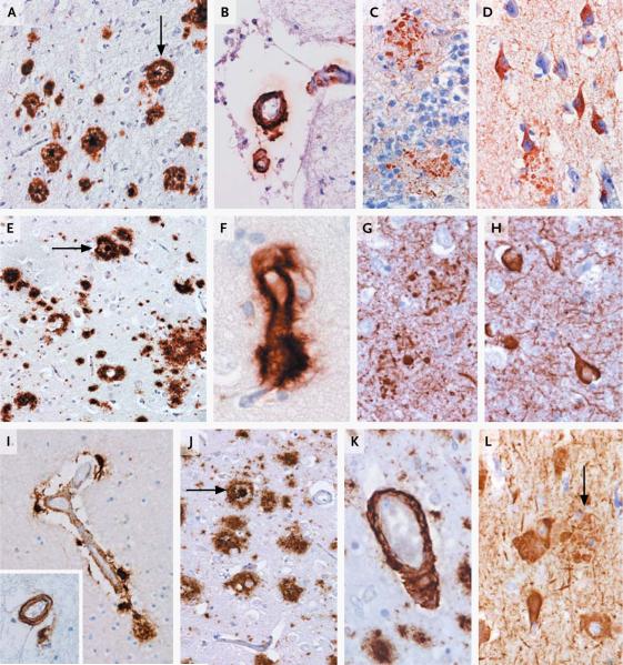
Pathological examination of five brains with variants that have possibly pathogenic effects (Q33X, R47H, and D87N) revealed that all were Braak stage 6 (fully developed Alzheimer's disease) and had typical findings with no distinguishing features. Two of the samples showed some mild Lewy-body disease, and one had documented TAR DNA-binding protein 43 (TDP-43) disease (Fig. 1). In two cases, some mild white-matter abnormalities were observed, but these were not outside the range one would expect for a typical case of Alzheimer's disease.
Figure 1. Pathological Hallmarks in Carriers of TREM2 Variants.
Shown are pathological findings in samples containing variant D87N (Panels A through D) and variant R47H (Panels E through L) obtained from patients with Alzheimer's disease. A carrier of the D87N variant had mature plaques (Panel A, arrow), along with more diffuse plaques and a moderate degree of cerebral amyloid angiopathy (Panel B). Beta amyloid was found deposited in leptomeningeal blood vessels. Tau immunohistochemical analysis revealed neuritic plaques (Panel C) in the hippocampus and numerous neurofibrillary tangles throughout the cortical regions (Panel D). Two carriers of the R47H variant also showed pathological features of Alzheimer's disease (with sections obtained from one patient in Panels E through H and from the other in Panels I through L) in the form of mature and diffuse plaques (Panels E and J, arrow). Severe cerebral amyloid angiopathy was evident with the presence of parenchymal capillary involvement (Panel F) and circumferential deposition of beta amyloid (Panel K). Also present were neuritic plaques (Panels G and L, arrow in Panel L), neurofibrillary tangles (Panels H and L), and abundant neuropil threads. However, one carrier of the R47H variant showed only moderate cerebral amyloid angiopathy with no other pathological abnormalities (Panel I, inset). Immunohistochemical analysis was carried out with the use of antibodies against beta amyloid (DAKO; 1:100) to identify the beta amyloid protein in both plaques and cerebral amyloid angiopathy, and AT8 antibody (Autogen Bioclear; 1:600) to identify tau.
In human control brain, TREM2 was expressed widely, at high levels in white matter and substantial abundance in hippocampus and neocortex but at low levels in the cerebellum (Fig. S2 in the Supplementary Appendix). This expression pattern is partly consistent with the pathological features observed in both Nasu–Hakola disease and Alzheimer's disease.
We assayed Trem2 expression in the brain tissue from TgCRND8 mice.23 To capture changes reflecting the very earliest responses to APP mis-processing and accumulation of beta amyloid, rather than responses to late-stage tissue injury, we focused on time points at which the Alzheimer's disease–like phenotype was largely a biochemical accumulation of beta amyloid without obvious morphological changes (i.e., at 70 to 80 days). We also analyzed Trem2 expression during the phase of emerging disease and functional deficits (150 days).23,24 At the earlier time point, we observed 85 genes to be differentially expressed in the brains of mutant mice and controls (Table S3 in the Supplementary Appendix). These genes comprised seven clusters. Clusters 1, 3, and 7 contained genes related to cholesterol or lipid metabolism, whose expression was coordinately altered from early time points (≤70 days). Cluster 5 included genes encoding complement factors, cytokines, and proinflammatory proteins, with changes in the expression of these factors at slightly later time points (>80 days). Genes encoding proteins that mediate vesicular trafficking genes did not appear to be differentially expressed (Tables S3 and S4 in the Supplementary Appendix).
To determine whether mRNA changes were reflected in changes in protein expression and to document the cell types involved, we assayed proteins in brain tissue from TgCRND8 mice, together with age- and sex-matched littermate controls, by means of Western blotting and immunohistochemical analysis. Suitable antibodies were not available for many proteins. Nevertheless, for proteins that could be assessed, the results were consistent with the mRNA data.
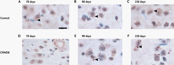
The expression of Trem2 mRNA was increased in TgCRND8 mice, as compared with non-TgCRND8 littermates (Table S4 in the Supplementary Appendix). This change, which is in agreement with a similar observation in another mouse transgenic model of Alzheimer's disease,25 was confirmed by means of Western blotting. We observed increased expression of Trem2 in large activated microglia (Fig. 2). In wild-type mice, Trem2 is expressed in small cytoplasmic granules in the cytoplasm of most neurons in the cortex and other brain regions. The number and size of these granules in control mice were relatively constant during aging (mean [±SD] at 70 days, 6.7±0.5 granules per cell; at 90 days, 7.2±0.4; and at 150 days, 5.8±0.8). In contrast, in TgCRND8 mice, the average number of granules increased significantly as the amyloid disease emerged (at 70 days, 8.2±0.3 granules per cell; at 90 days, 22.4±0.6; and at 150 days, 15.0±0.3) (P = 0.01 for all comparisons). There was also a progressive increase in the diameter of granules (at 70 days, 0.4 μm; at 90 days, 0.6 μm; and at 120 days, 2.1 μm) (P = 0.01 for all comparisons). We observed Trem2 to be expressed in microglia surrounding the outer border of amyloid plaques, as reported previously in the APP23 mouse model25 (Fig. 2). Perhaps these changes represent a response to rising levels of beta amyloid in the TgCRND8 and APP23 mice, which, like all mouse models, develop prominent pathological findings associated with beta amyloid but no neurode-generation.
Figure 2. Immunohistochemical Analyses of Trem2 in TgCRND8 Mice.

Slides of samples from TgCRND8 mice (a transgenic mouse model of Alzheimer's disease) and age-matched littermates (controls) at different ages show that Trem2 is expressed as small granules in the cytoplasm of neurons (arrowheads) in both the transgenic mice and controls. In a slide of a sample from a transgenic mouse, the amyloid plaques are also surrounded by Trem2-positive granules (Panel D, arrow), and the Trem2 granules increase in number and size, as compared with the controls. The scale bar represents 30 mm in Panels A, B, C, E, and F and 60 mm in Panel D.
DISCUSSION
TREM2 encodes a single-pass type I membrane protein that forms a receptor-signaling complex with the TYRO protein tyrosine kinase-binding protein (TYROBP) and thereby triggers the activation of immune responses in macrophages and dendritic cells.10 As we have shown, TREM2 is expressed throughout the central nervous system and at particularly high concentrations in white matter. Homozygous loss-of-function mutations in TREM2 cause Nasu–Hakola disease (Online Mendelian Inheritance in Man number, 605086). Here we show that heterozygous rare variants, including those that cause Nasu–Hakola disease in the homozygous state, predispose to Alzheimer's disease. Thus, we suggest that the reduced function of TREM2 is key to the pathogenic effect of these risk variants associated with Alzheimer's disease.
Trem2 is localized to microglia around plaques and neurons in the brains of TgCRND8 mice. In microglia, TREM2 is known to control two streams of signaling to regulate the reactive phenotype. One of these streams regulates phagocytosis. Increased expression of TREM2 on microglia is coupled to enhanced phagocytic pathways (which could be relevant to the removal of cell debris and the clearance of amyloid proteins in Alzheimer's disease) and promotion of the alternative activation state of microglia, which is thought to be protective.26–28 The other signaling stream suppresses inflammatory reactivity and involves the repression of cytokine production and secretion.28 TREM2 control of constitutive cytokine signaling may promote survival by prompting the secretion of tumor necrosis factor at levels that potentiate survival and repair pathways through tumor necrosis factor receptor 2 (TNFR2).
Consistent with the results described in a previous study,25 we have shown that the expression of TREM2 rises in parallel with a rise in cortical levels of beta amyloid. However, the dysregulation of expression that is induced by beta amyloid is relatively specific to TREM2. Other components of the same cascade, such as TYROBP, are not dysregulated. These data are consistent with a role for TREM2 as a gateway for controlling microglial responses. Compromised function of TREM2 is likely to have ramifications for the clearance of cell debris and possibly the removal of beta amyloid in Alzheimer's disease. At the same time, relief of the TREM2 lock on cytokine levels, potentially effected by the TREM2 variants described here, may fuel inflammatory cascades, leading to a systemic inflammatory response and the incidental death of neurons. In an animal model of multiple sclerosis, blocking of Trem2 exacerbated disease symptoms,27 whereas boosting of Trem2 signaling ameliorated them.29
Variants of the gene CR1 have been implicated as risk factors for Alzheimer's disease.30 We have recently described functional data that support the control of microglial reactivity and the production of tumor necrosis factor by CR1 protein, suggesting parallel or convergent signaling streams with respect to those initiated by TREM2.31,32 Thus, if there are variants with reduced function in either of these pathways, the subsequent functional defects in microglial responses may show similar effects.
The approach we have taken shows that whole-exome sequencing is capable of identifying not only very rare mendelian causes of disease but also low-frequency variability with medium-effect sizes modulating disease development. Our data add to a growing list of disease associations32 in which two diseases — one that is severe, has an early onset, and is caused by homozygous loss-of-function mutations and the other that has a late onset with susceptibility caused by heterozygous loss-of-function variants — had previously been thought to be unrelated.32 We and others have predicted that heterozygous loss-of-function variants may represent a substantial component of risk for common late-onset diseases.32,33 Our findings support this hypothesis, and we believe additional loss-of-function variants will be identified as risk factors for Alzheimer's disease and other late-onset complex disorders.
Supplementary Material
Supplement1
Acknowledgments
Supported by the Alzheimer's Research UK, the Medical Research Council (MRC), the Wellcome Trust, the Wellcome Trust/MRC Joint Call in Neurodegeneration Award (WT089698) to the UK Parkinson's Disease Consortium (whose members are from the University College London Institute of Neurology, the University of Sheffield, and the MRC Protein Phosphorylation Unit at the University of Dundee), grants (P50 AG016574, U01 AG006786, and R01 AG18023) from the Robert and Clarice Smith and Abigail Van Buren Alzheimer's Disease Research Program (which support the Mayo Alzheimer's Disease Research Center), grants (2P50 AG005681-27, 5P30 NS069329-02, NIRG-11-200110, and 1R01 AG041797-01) from the Washington University School of Medicine Alzheimer's Disease Research Center, the National Institute for Health Research Biomedical Research Unit in Dementia at University College London Hospitals, University College London; grants from the Ontario Research Fund, Canadian Institutes of Health Research (to Drs. Rogaeva and Hyslop); grants from the National Institutes of Health Research, the Wellcome Trust Medical Research Council, an anonymous charitable foundation, the Fondation pour la Recherche sur Alzheimer, and the Alzheimer Society of Ontario (to Dr. Hyslop); the Big Lottery (to Dr. Morgan); a fellowship from Alzheimer's Research UK (to Dr. Guerreiro); and the Intramural Research Programs of the National Institute on Aging and the National Institute of Neurological Disease and Stroke, National Institutes of Health (Department of Health and Human Services Project number, ZO1 AG000950-10). Additional funders of individual investigators and institutions are provided in the Supplementary Appendix.
We thank the controls, the patients, and their families; the National Cell Repository for Alzheimer's Disease and the Human Genetics Resource Center DNA and Cell Line Repository; the National Heart, Lung, and Blood Institute Grand Opportunity (GO) Exome Sequencing Project and its ongoing studies, which produced and provided exome variant calls for comparison; the 1000 Genomes Project; and the MRC London Neurodegenerative Diseases Brain Bank and the Manchester Brain Bank from Brains for Dementia Research for providing samples and pathological diagnosis.
Footnotes
Ms. Wojtas and Drs. Bras, Carrasquillo, and Rogaeva contributed equally to this article.
Disclosure forms provided by the authors are available with the full text of this article at NEJM.org.
References
- 1.Goate A, Chartier-Harlin MC, Mullan M, et al. Segregation of a missense mutation in the amyloid precursor protein gene with familial Alzheimer's disease. Nature. 1991;349:704–6. doi: 10.1038/349704a0. [DOI] [PubMed] [Google Scholar]
- 2.Rogaev EI, Sherrington R, Rogaeva EA, et al. Familial Alzheimer's disease in kindreds with missense mutations in a gene on chromosome 1 related to the Alzheimer's disease type 3 gene. Nature. 1995;376:775–8. doi: 10.1038/376775a0. [DOI] [PubMed] [Google Scholar]
- 3.Sherrington R, Rogaev EI, Liang Y, et al. Cloning of a gene bearing missense mutations in early-onset familial Alzheimer's disease. Nature. 1995;375:754–60. doi: 10.1038/375754a0. [DOI] [PubMed] [Google Scholar]
- 4.Strittmatter WJ, Saunders AM, Schmechel D, et al. Apolipoprotein E: high-avidity binding to beta-amyloid and increased frequency of type 4 allele in late-onset familial Alzheimer disease. Proc Natl Acad Sci U S A. 1993;90:1977–81. doi: 10.1073/pnas.90.5.1977. [DOI] [PMC free article] [PubMed] [Google Scholar]
- 5.Majounie E, Abramzon Y, Renton AE, et al. Repeat expansion in C9ORF72 in Alzheimer's disease. N Engl J Med. 2012;366:283–4. doi: 10.1056/NEJMc1113592. [DOI] [PMC free article] [PubMed] [Google Scholar]
- 6.Naj AC, Jun G, Beecham GW, et al. Common variants at MS4A4/MS4A6E, CD2AP, CD33 and EPHA1 are associated with late-onset alzheimer's disease. Nat Genet. 2011;43:436–41. doi: 10.1038/ng.801. [DOI] [PMC free article] [PubMed] [Google Scholar]
- 7.Hollingworth P, Harold D, Sims R, et al. Common variants at ABCA7, MS4A6A/MS4A4E, EPHA1, CD33 and CD2AP are associated with Alzheimer's disease. Nat Genet. 2011;43:429–35. doi: 10.1038/ng.803. [DOI] [PMC free article] [PubMed] [Google Scholar]
- 8.Lambert JC, Heath S, Even G, et al. Genome-wide association study identifies variants at CLU and CR1 associated with Alzheimer's disease. Nat Genet. 2009;41:1094–9. doi: 10.1038/ng.439. [DOI] [PubMed] [Google Scholar]
- 9.Harold D, Abraham R, Hollingworth P, et al. Genome-wide association study identifies variants at CLU and PICALM associated with Alzheimer's disease. Nat Genet. 2009;41:1088–93. doi: 10.1038/ng.440. [DOI] [PMC free article] [PubMed] [Google Scholar]
- 10.Paloneva J, Manninen T, Christman G, et al. Mutations in two genes encoding different subunits of a receptor signaling complex result in an identical disease phenotype. Am J Hum Genet. 2002;71:656–62. doi: 10.1086/342259. [Erratum, Am J Hum Genet 2003;72:225.] [DOI] [PMC free article] [PubMed] [Google Scholar]
- 11.Guerreiro RJ, Lohmann E, Brás J, et al. Using exome sequencing to reveal mutations in TREM2 presenting as a frontotemporal dementia-like syndrome without bone involvement. Arch Neurol. 2012 Oct 8; doi: 10.1001/jamaneurol.2013.579. Epub ahead of print. [DOI] [PMC free article] [PubMed] [Google Scholar]
- 12.Butler AW, Ng MY, Hamshere ML, et al. Meta-analysis of linkage studies for Alzheimer's disease — a web resource. Neurobiol Aging. 2009;30:1037–47. doi: 10.1016/j.neurobiolaging.2009.03.013. [DOI] [PubMed] [Google Scholar]
- 13.Chishti MA, Yang DS, Janus C, et al. Early-onset amyloid deposition and cognitive deficits in transgenic mice expressing a double mutant form of amyloid precursor protein 695. J Biol Chem. 2001;276:21562–70. doi: 10.1074/jbc.M100710200. [DOI] [PubMed] [Google Scholar]
- 14.Li H, Durbin R. Fast and accurate short read alignment with Burrows-Wheeler transform. Bioinformatics. 2009;25:1754–60. doi: 10.1093/bioinformatics/btp324. [DOI] [PMC free article] [PubMed] [Google Scholar]
- 15.DePristo MA, Banks E, Poplin R, et al. A framework for variation discovery and genotyping using next-generation DNA sequencing data. Nat Genet. 2011;43:491–8. doi: 10.1038/ng.806. [DOI] [PMC free article] [PubMed] [Google Scholar]
- 16.McKenna A, Hanna M, Banks E, et al. The Genome Analysis Toolkit: a MapReduce framework for analyzing next-generation DNA sequencing data. Genome Res. 2010;20:1297–303. doi: 10.1101/gr.107524.110. [DOI] [PMC free article] [PubMed] [Google Scholar]
- 17.Wang K, Li M, Hakonarson H. ANNOVAR: functional annotation of genetic variants from high-throughput sequencing data. Nucleic Acids Res. 2010;38(16):e164. doi: 10.1093/nar/gkq603. [DOI] [PMC free article] [PubMed] [Google Scholar]
- 18.Adzhubei IA, Schmidt S, Peshkin L, et al. A method and server for predicting damaging missense mutations. Nat Methods. 2010;7:248–9. doi: 10.1038/nmeth0410-248. [DOI] [PMC free article] [PubMed] [Google Scholar]
- 19.Breitner JC, Wyse BW, Anthony JC, et al. APOE-epsilon4 count predicts age when prevalence of AD increases, then declines: the Cache County Study. Neurology. 1999;53:321–31. doi: 10.1212/wnl.53.2.321. [Erratum, Neurology 2000;55: 161–2.] [DOI] [PubMed] [Google Scholar]
- 20.Bentley DR, Balasubramanian S, Swerdlow HP, et al. Accurate whole human genome sequencing using reversible terminator chemistry. Nature. 2008;456:53–9. doi: 10.1038/nature07517. [DOI] [PMC free article] [PubMed] [Google Scholar]
- 21.Li H, Handsaker B, Wysoker A, et al. The Sequence Alignment/Map format and SAMtools. Bioinformatics. 2009;25:2078–9. doi: 10.1093/bioinformatics/btp352. [DOI] [PMC free article] [PubMed] [Google Scholar]
- 22.Howie BN, Donnelly P, Marchini J. A flexible and accurate genotype imputation method for the next generation of genome-wide association studies. PLoS Genet. 2009;5(6):e1000529. doi: 10.1371/journal.pgen.1000529. [DOI] [PMC free article] [PubMed] [Google Scholar]
- 23.Janus C, Pearson J, McLaurin J, et al. A beta peptide immunization reduces behavioural impairment and plaques in a model of Alzheimer's disease. Nature. 2000;408:979–82. doi: 10.1038/35050110. [DOI] [PubMed] [Google Scholar]
- 24.Melchior B, Puntambekar SS, Carson MJ. Microglia and the control of autoreactive T cell responses. Neurochem Int. 2006;49:145–53. doi: 10.1016/j.neuint.2006.04.002. [DOI] [PMC free article] [PubMed] [Google Scholar]
- 25.Neumann H, Takahashi K. Essential role of the microglial triggering receptor expressed on myeloid cells-2 (TREM2) for central nervous tissue immune homeostasis. J Neuroimmunol. 2007;184:92–9. doi: 10.1016/j.jneuroim.2006.11.032. [DOI] [PubMed] [Google Scholar]
- 26.Takahashi K, Prinz M, Stagi M, Chechneva O, Neumann H. TREM2-transduced myeloid precursors mediate nervous tissue debris clearance and facilitate recovery in an animal model of multiple sclerosis. PLoS Med. 2007;4(4):e124. doi: 10.1371/journal.pmed.0040124. [DOI] [PMC free article] [PubMed] [Google Scholar]
- 27.Frank S, Burbach GJ, Bonin M, et al. TREM2 is upregulated in amyloid plaque-associated microglia in aged APP23 transgenic mice. Glia. 2008;56:1438–47. doi: 10.1002/glia.20710. [DOI] [PubMed] [Google Scholar]
- 28.Piccio L, Buonsanti C, Mariani M, et al. Blockade of TREM-2 exacerbates experimental autoimmune encephalomyelitis. Eur J Immunol. 2007;37:1290–301. doi: 10.1002/eji.200636837. [DOI] [PubMed] [Google Scholar]
- 29.Hazrati LN, Van Cauwenberghe C, Brooks PL, et al. Genetic association of CR1 with Alzheimer's disease: a tentative disease mechanism. Neurobiol Aging. 2012;33:2949.e5–2949.e12. doi: 10.1016/j.neurobiolaging.2012.07.001. [DOI] [PubMed] [Google Scholar]
- 30.Crehan H, Holton P, Wray S, Pocock J, Guerreiro R, Hardy J. Complement receptor 1 (CR1) and Alzheimer's disease. Immunobiology. 2012;217:244–50. doi: 10.1016/j.imbio.2011.07.017. [DOI] [PubMed] [Google Scholar]
- 31.Crehan H, Hardy J, Pocock J. Microglia, Alzheimer's disease, and complement. Int J Alzheimers Dis. 2012;2012:983640. doi: 10.1155/2012/983640. [DOI] [PMC free article] [PubMed] [Google Scholar]
- 32.Singleton AB, Hardy J, Traynor BJ, Houlden H. Towards a complete resolution of the genetic architecture of disease. Trends Genet. 2010;26:438–42. doi: 10.1016/j.tig.2010.07.004. [DOI] [PMC free article] [PubMed] [Google Scholar]
- 33.MacArthur DG, Balasubramanian S, Frankish A, et al. A systematic survey of loss-of-function variants in human protein-coding genes. Science. 2012;335:823–8. doi: 10.1126/science.1215040. [DOI] [PMC free article] [PubMed] [Google Scholar]
Associated Data
This section collects any data citations, data availability statements, or supplementary materials included in this article.
Supplementary Materials
Supplement1
