Formation of Human Myocardium in the Rat Heart from Human Embryonic Stem Cells (original) (raw)
Abstract
Human embryonic stem cells (hESCs) offer the opportunity to replenish cells lost in the postinfarct heart. We explored whether human myocardium could be generated in rat hearts by injecting differentiated cardiac-enriched hESC progeny into the left ventricular wall of athymic rats. Although initial grafts were predominantly epithelial, noncardiac elements were lost over time, and grafts consisted predominantly of cardiomyocytes by 4 weeks. No teratomatous elements were observed. Engrafted cardiomyocytes were glycogen-rich and expressed expected cardiac markers including β-myosin heavy chain, myosin light chain 2v, and atrial natriuretic factor. Heat-shock treatment improved graft size approximately threefold. The cardiac implants exhibited substantial angiogenesis, both recipient and graft derived. Importantly, there was greater proliferation in human cardiomyocytes than previously seen in rodent-derived cardiomyocytes: 14.4% of graft cardiomyocytes expressed the proliferation marker Ki-67, and 2.7% incorporated the thymidine analog BrdU 4 weeks after transplantation. This proliferation was associated with a sevenfold increase in graft size over the 4-week interval. Thus, hESCs can form human myocardium in the rat heart, permitting studies of human myocardial development and physiology and supporting the feasibility of their use in myocardial repair.
Because the adult human heart has little regenerative capacity, irreversible injury to the myocardium, such as by infarction, typically results in the formation of a noncontractile scar and often initiates progressive heart failure. Given the limited supply of transplant hearts in the face of a large population of patients with end-stage heart disease, much attention has recently been directed at cell transplantation strategies as an alternative strategy to ameliorate cardiac injury.1–3 As currently envisioned for human therapeutic application, a suitable myogenic cell type from either an autologous or appropriately matched allogeneic source is delivered to the infarcted zone in an attempt to repopulate the lost myocardium. A number of different cell types have been considered for such therapies, including skeletal myoblasts,4–6 bone marrow-derived hematopoietic stem cells,7–10 mesenchymal stem cells,11–14 intrinsic cardiac stem cells,15,16 and embryonic stem cells (ESCs).17–22
ESCs, pluripotent cells derived from the inner cell mass of pre-implantation-stage embryos, have a number of potential advantages over some of these other cell types for cardiac repair applications. First, there are well-defined protocols for the isolation and maintenance of ESCs, and they have a tremendous capacity for in vitro expansion, making them likely scalable for human applications.23 Second, ESCs have an unquestioned ability to differentiate into functional cardiomyocytes in vitro,17,18,21,24,25 which stands in contrast to a number of other candidate cell sources for which this capacity remains controversial.8,26 Furthermore, human ESC (hESC)-derived cardiomyocytes possess the cellular elements required for electromechanical coupling with the host myocardium (eg, gap and adherens junctions17,18), and it is therefore expected that, when transplanted, these cells could electrically integrate and contribute to systolic function. This property represents a significant advantage over other cell types, such as skeletal muscle, which might yield meaningful function improvement but should do so predominantly through modulation of diastolic function (eg, by passively ameliorating postinfarct ventricular dilatation5,27,28).
In the current study, we sought to form human myocardium by implanting cardiomyocytes from hESCs into the hearts of immunodeficient, “nude” rats. We report that this strategy resulted in the formation of substantial human myocardial tissue, and furthermore, that these cells proliferated at high rates for at least 1 month after implantation. Grafts showed associated angiogenesis, and the size of the resultant graft was enhanced by preceding heat-shock treatment of the implanted cells.
Materials and Methods
Cell Culture
Undifferentiated H1 (male) and H7 female hESCs29 were maintained under feeder-free conditions as previously described30 and detailed in the supplemental material (http://ajp.amjpathol.org). These were differentiated in vitro as embryoid bodies for 3 weeks and then enriched to ∼15% cardiomyocytes, using Percoll (Amersham Biosciences, Piscataway, NJ) gradient centrifugation18 (supplemental material at http://ajp.amjpathol.org). Unless otherwise stated, embryoid body outgrowths were heat-shocked 24 hours before implantation to improve survival (via 30-minute incubation at 43°C).
Cell Implantation and Histological Analysis
All studies were approved by the University of Wash-ington Animal Care and Use Committee and were conducted in accordance with federal guidelines. Using surgical techniques previously reported by our group6,28,31–33 and further detailed in the supplemental materials (http://ajp.amjpathol.org), 0.5 to 10 million Percoll-enriched hESC-derived cardiomyocytes were directly injected into the uninjured left ventricular walls of 200- to 300-g male nude rats (Harlan, Indianapolis, IN). At 1 to 28 days after engraftment, rats received a 1-hour pulse of 5-bromodeoxyuridine (BrdU, 10 mg), followed by euthanasia with pentobarbital. Engrafted hearts were fixed and vibratome-sectioned at 500-μm thickness to ensure equivalent sampling. These uniform transverse sections were routinely processed and paraffin-embedded for histology. Sections were analyzed histologically, phenotyping the implanted cells with the histochemical and immunohistochemical markers detailed in the supplemental materials (http://ajp.amjpathol.org). Graft lineage was traced using in situ hybridization with human-specific probes (including Y-chromosome, Alu repeat, and pan-centromeric targets), as is also detailed in the supplemental materials (http://ajp.amjpathol.org).
Statistics
Values are expressed as mean ± SEM, and statistical testing was performed using _t_-test with α = 0.05 for significance (Excel Data Analysis Toolpak; Microsoft, Redmond, WA).
Results
Time Course of Graft Composition
An early and important goal was to examine the time course of differentiation by the hESC-derived myocardial grafts after implantation in the heart of the nude rat recipient. One day after transient heat shock to improve survivability, human ESC-derived cardiomyocytes were harvested and enriched by Percoll centrifugation. Parallel in vitro experiments indicated that, before implantation, approximately 10 to 15% of the used cells were positive for cardiac markers such as sarcomeric myosin heavy chain and/or actin. For this time course study, 5 × 10⁶ cells were then implanted into the uninjured left ventricular myocardium of 10 nude rats. Recipient animals were sacrificed at 1, 7, 14, and 28 days after engraftment (n = 2–4/group), and hearts underwent histological analysis.
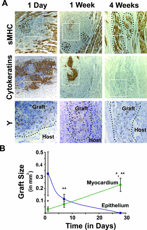
Representative images from this series are contained in Figure 1, and, for this study, in situ hybridization with a human-specific Y chromosome sequence was used to follow the lineage of the (male H1-derived) graft cells. One day after implantation, the grafts consisted of confluent masses of generally poorly differentiated cells, interspersed with strands of host myocardium. Graft cells had high nuclear-to-cytoplasmic ratios, and a relatively small population of cells with a clear cytoplasm was noted. There was extensive cell death in the grafts, as evidenced by the intense granulocytic infiltration, nuclear condensation, and Y-positive karyorrhectic debris noted among graft cells. Not surprisingly, evaluation of DNA fragmentation using terminal deoxynucleotidyl transferase biotin-dUTP nick end labeling staining confirmed the extensive cell death at the 1-day time point (not shown). In occasional areas, the adjacent host myocardium had undergone contraction band necrosis, likely from the trauma of injection. These findings were similar to the appearance of early grafts with other cell types in the heart, including neonatal rat cardiomyocytes.31
Figure 1.
Time course study of human ESC cardiomyocyte grafts. A: The three top panels show immunostaining (brown deposit) for sarcomeric myosin heavy chain (sMHC) at 1 day, 1 week, and 4 weeks after transplantation and identify host and graft cardiomyocytes. The three middle panels show immunostaining for epithelial cells (pan-cytokeratins) on adjacent sections. Note that, over time, the tightly clustered human myocardial implants (enclosed within dotted lines) appear to have expanded, whereas epithelial elements decrease in extent and are totally absent by 4 weeks. The lineage of the engrafted human cells is confirmed in the three bottom panels, in which adjacent sections have undergone in situ hybridization with a human-specific probe against the Y-chromosome (punctate intranuclear dots), these from fields corresponding to the boxes in the above panels. Scale bar = 50 μm; counterstain is hematoxylin. Y in situ images are magnified an additional 2.5-fold. B: The plot shows the changing composition of the graft versus time (n = 4 to 10 hearts per marker per time point). Whereas the mean total epithelial (ie, pan-cytokeratin+; blue line) graft cross-sectional area declines with time and is totally absent by 4 weeks, the mean total cardiac graft area shows a sevenfold increase from the 1-day to 4-week time points. Note that the mean cardiac graft area at 4 weeks shows a statistically significant difference from that at both the 1-day (*) and 1-week (**) time points by Student’s _t_-test (P < 0.05).
Immunohistochemistry on the 1-day-old grafts revealed them to be polymorphous in composition: the vast majority of cells were epithelial (pan-cytokeratin+), but there were small discrete islands of cardiac (sarcomeric myosin heavy chain) and endodermal (α-fetoprotein+; not shown) cells also present.
As illustrated in Figure 1, the composition of the hESC-derived grafts changed importantly over time. In contrast to the initial grafts, both 7 and 14 days after transplantation, there were only comparatively rare cytokeratin+ or α-fetoprotein+ elements, and the clustered sarcomeric myosin heavy chain+ cardiac cells appeared expanded. (Note that, as would be expected, the graft cells at all later time points were present on a highly fibrotic background.) Most remarkably, after 28 days, grafts were found to be composed predominantly of sarcomeric myosin heavy chain-positive human myocardium; all other cytokeratin+ or α-fetoprotein+ elements had been eliminated from the heart. (As is quantified in Figure 1B, there was a more than sevenfold increase in the cross-sectional area of the human cardiac implants between 1 and 28 days after transplantation.) A small number of graft-derived endothelial cells were retained at 4 weeks (as is detailed subsequently), but otherwise the human-specific Y chromosome in situ hybridization identified only graft-derived cardiomyocytes or extraordinarily rare fibroblastic cells. No heterologous or teratomatous elements were found after 28 days despite exhaustive searching (n = 10 rats receiving H1-derived cells).
Dose-Response Studies and Analysis of Graft Differentiation
Multiple additional grafting experiments have been performed, variably using cells derived from the H1 or H7 ESC line and evaluating grafts at time points up to 4 weeks (Supplemental Table 1 at http://ajp.amjpathol.org). Dose-response studies were performed to identify an optimal number of cells to implant into the heart. Rats (n = 4–6/group) received doses of 0.5, 1, 5, or 10 million hESC-derived cardiomyocytes, and their hearts were examined histologically 4 weeks after implantation. The resultant graft size varied considerably, even with a constant dose of cells. This variability likely is due in part to varyingly successful cell delivery and survival33,34 and could also be influenced by differential immunorecognition by the nude rat (which has been maintained on an outbred genetic background). No grafts were detected in rats receiving 0.5 million cells, and only a single small graft was observed in the four rats receiving 1 million cells. In contrast, grafts were present in the vast majority of hearts receiving 5 or 10 million cells, but no clear increase in graft size was seen at the highest dose. Therefore, for all described studies, unless stated otherwise, a “standard” dose of 5 million cells was implanted.
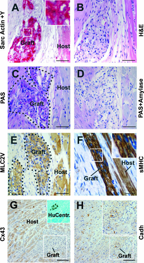
Extensive additional immunophenotyping has been performed on the numerous hESC-derived myocardial grafts (Table 1). No obvious differences have been observed between grafts derived from undifferentiated cultures that have varied in their number of passages in vitro or in their parental line of origin (ie, H1 or H7). The hESC-derived cardiac implants expressed numerous cardiac markers at all time points, including sarcomeric actin (Figure 2A) and myosin (Figure 2F), smooth muscle α-actin, myosin light chain 2v (Figure 2E), and atrial natriuretic peptide (Supplemental Figure 1 at http://ajp.amjpathol.org). As a general trend, grafts that were implanted for increasing lengths of time showed increasing maturation, such that 4-week-old transplants typically exhibited clear sarcomeric banding, generally in alignment with adjacent host cardiomyocytes (Figure 2F). The predominant isoform of sarcomeric myosin heavy chain in developing and adult human ventricular myocardium is the β-isoform,35,36 and, consistent with this, the human cardiac grafts showed immunoreactivity for the β-isoform and not for the α-isoform (Figures 3A; 4, B and C; and 5;, A and B). (In contrast, as expected for the mature rat heart,37 the host myocardium immunostained strongly for the α-myosin heavy chain and did not express the β-isoform, thereby serving as another means to identify the also morphologically distinct hESC-derived cardiac implants.)
Table 1.
Changing Composition and Immunophenotype of Human ESC-Derived Intracardiac Grafts versus Time
| 1 day | 1 week | 2 weeks | 4 weeks | |
|---|---|---|---|---|
| Sarcomeric actin | +/+++ | ++/+++ | ++/+++ | +++/+++ |
| β-Myosin heavy chain | +/+++ | ++/+++ | ++/+++ | +++/+++ |
| α-Myosin heavy chain | − | − | − | − |
| Smooth muscle α-actin | +/++ | ++/+++ | ++/++ | +++/++ |
| N-cadherin | +/+ | ++/+++ | ++/+++ | +++/+++ |
| Atrial natriuretic peptide | +/+ | ++/++ | ++/++ | +++/++ |
| Myosin light chain 2v | NE | ++/++ | ++/++ | +++/+++ |
| Connexin-43 | − | − | − | − |
| Pan-cytokeratins | +++/+++ | +/+++ | +/+++ | − |
| α-Fetoprotein | +/+++ | − | − | − |
| βIII-Tubulin | − | − | − | − |
| S-100 protein | − | − | − | − |
| Fast skeletal myosin heavy chain | − | − | − | − |
Figure 2.
Additional phenotyping of human ESC cardiomyocyte grafts. All images depict 4-week-old grafts. A: Clustered human ESC-derived cardiomyocyte graft cells are positive for sarcomeric actin (sarc Actin, immunostain indicated by red deposit) and are identified by their reactivity for the human-specific Y-chromosome probe by in situ hybridization (Y, punctate brown intranuclear signal, best seen in inset magnified an additional threefold). B: The peculiar appearance of the engrafted cardiac cells by routine H&E stain in a serial section, by which they show a distinctly vacuolated appearance. This pattern is likely accounted for by the results depicted C, wherein engrafted cardiomyocytes were found to be intensely PAS reactive. This PAS reaction is completely removed by preceding amylase digestion (PAS+Amylase), as depicted in D, indicating engrafted cardiomyocytes are heavily glycogen-laden. E: The cardiomyocyte graft cells stained positive for the ventricular-specific marker myosin light chain 2v (MLC2V). As depicted by sarcomeric myosin heavy chain (sMHC) immunostain in F, the engrafted cardiomyocytes assume sarcomeric banding and alignment with host fibers (inset magnified an additional twofold). G and H: Adjacent sections immunostained with antibodies against connexin-43 (Cx43) and pan-cadherins (Cadh), respectively. The inset in G shows in situ hybridization with the human-specific pan-centromeric probe (on adjacent section), thus identifying a clump of human ESC-derived cardiomyocytes residing within the indicated box. This clump of cells showed the expected strong reactivity with β-myosin heavy chain (not shown). Note the total absence of connexin-43 reactivity, despite the expected intercalated disk staining pattern on the surrounding host myocardium. In contrast, this same field does show definite diffuse membranous staining of the human cardiac implant with anti-pan-cadherins. Scale bars = 50 μm.
Figure 3.
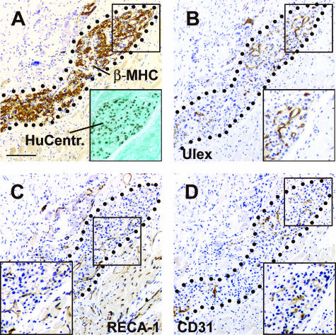
Human ESC-derived cardiac grafts show both recipient- and graft-derived angiogenesis. A: A typical cardiac implant 4 weeks after transplantation; the graft myocardium is readily distinguished from that of the host by its strong immunoreactivity for β-myosin heavy chain (β-MHC) and by in situ hybridization on an adjacent section with a human-specific pan-centromeric probe (HC, within inset). The ingrowth of both rat-derived and human-derived (ie, graft) vessels into this cardiac implant is demonstrated in B through D, which contain adjacent tissue sections stained with species-specific endothelial markers. B and D: Human endothelial cells, as detected by staining with U. europaeus agglutinin I and a human-specific anti-CD31 antibody, respectively. C: Rat endothelial cells, as defined by the rat-specific pan-endothelial antibody RECA-1, which marks the microvasculature of both the cardiac implant and the surrounding host myocardium. Scale bar = 100 μm; insets magnified an additional 1.5-fold. Sections are counterstained with hematoxylin, except for the inset of A, which is counterstained with Fast Green.
A surprising result was that, despite the preceding observation of immunoreactivity for the gap junction protein connexin-43 by the hESC-derived cardiomyocytes in vitro17,18 (and their clear-cut synchronous spontaneous contraction in culture), the engrafted human cardiomyocytes were found consistently negative for connexin-43 (not shown). At present, it is uncertain whether this failure to immunostain for connexin-43 reflects an in vivo switch to another connexin isoform or merely results from a level of expression below the threshold of immunohistochemical detection. (We have attempted immunohistochemistry for other connexin isoforms, including connexin-40 and -45, but have not identified antibodies with sufficient specificity on control sections.) It should be noted that the engrafted cardiomyocytes did show strong immunoreactivity, with a diffuse membranous pattern, with a pan-cadherin antibody (Figure 2H; Supplemental Figure 1, A and B, at http://ajp.amjpathol.org). Because N-cadherin is an important structural element of the intercalated disk, present in both the developing and adult heart,38 this staining pattern suggests that these cells may be at an early stage in the formation of these sites of specialized electromechanical cell contact. Similar membranous staining for pan-cadherin and absent staining for connexin-43 was observed in early-stage rat cardiomyocyte grafts, although the rat cell had formed mature intercalated disks by 4 weeks.31
Also as expected for embryonic human myocardium,39 the cardiac grafts were found to be heavily glycogen laden, as evidenced by strong reactivity for the histochemical periodic acid-Schiff reaction (PAS stain; Figure 2, C and D) that was entirely amylase digestible. Of note, despite fairly extensive searching by immunohistochemistry, grafts were negative at all time points for markers of skeletal muscle (fast skeletal myosin heavy chain), neuronal (βIII-tubulin), and glial (S-100 protein) cell types.
Recipient- and Graft-Derived Angiogenesis of Human Cardiac Implants
We tested whether the human myocardial grafts elicited an angiogenic response from either host- or donor-derived cells, using rat- and human-specific endothelial probes. The human cardiac implants showed a substantial degree of rat vessel ingrowth, this determined by immunohistochemistry for rat endothelial antigen-1 (RECA-1). In separate studies (not shown), we confirmed previously reported results that this antibody reacts uniformly and exclusively with rat (and not human) endothelial cells.40 Morphometric quantification of the RECA-1+, rat-derived graft angiogenesis was then undertaken, using adjacent sections immunostained for β-myosin heavy chain to identify the human cardiac implants of interest. In so doing, we observed a mean vascular density of 305.0 ± 46.2 rat vessels/mm2, representing 6.0 ± 1.3% of the cardiac graft cross-sectional area (n = 10 engrafted hearts) (Figure 3, A and C).
An unexpected finding was the roughly comparable contribution of human graft-derived angiogenesis within the cardiac implants. Two human-specific markers were used to identify human endothelial cells within the cardiac grafts, namely a human-specific antibody against CD31/PECAM and Ulex europaeus agglutinin I.41 Again, both markers were shown to react uniquely with human but not rodent endothelium in separate studies. Remarkably, Ulex identified a substantial number of human vessels within the β-myosin heavy chain+ cardiac implants: a mean density of 176.5 ± 42.4 human vessels/mm2, occupying a mean of 4.6 ± 1.2% of the cardiac graft cross-sectional area (n = 9 engrafted hearts). Qualitatively similar results were obtained with human-specific CD31/PECAM immunostaining (Figure 3, A, B, and D). Interestingly, when summed together, the density of host- and graft-derived vessels is reasonably close to that of the distant native myocardium, which we quantified as 1476.3 ± 187.3 vessels/mm2 or a mean of 15.0 ± 0.7% of the myocardial cross-sectional area (n = 11 hearts).
Improved Cardiac Graft Size with Preceding Heat-Shock Treatment
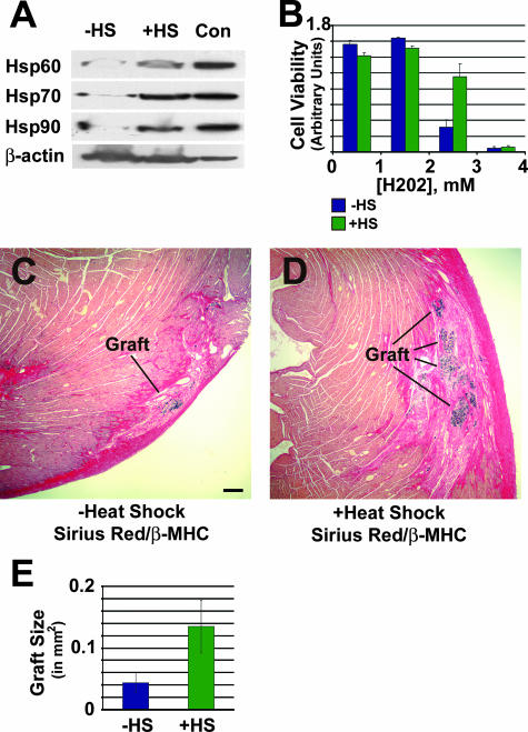
Our group had previously shown that heat shock significantly reduces death of neonatal cardiomyocytes after grafting.33 We therefore examined the effect of heat shock (by incubation at 43°C for 30 minutes) on hESC-derived cardiomyocytes. Heat-shocked cells showed a marked induction of Hsp-60, Hsp-70, and Hsp-90 (Figure 4A), and these cells showed significant protection against hydrogen peroxide induced injury in vitro (Figure 4B). Heat shock had no effect on the proliferation of hESC-derived cardiomyocytes in vitro (supplemental materials at http://ajp.amjpathol.org). We then compared graft size using heat-shocked or naïve hESC-derived cardiomyocytes in rats sacrificed 1 week after implanting 5 million cells. Figure 4, C and D, shows representative fields with islands of human myocardium on a fibrous background, taken from the median-sized grafts from the recipients of control and heat-shocked cells, respectively. Overall, heat-shock treatment produced an approximately threefold increase in cardiac graft cross-sectional area, with the mean total graft area increasing to 0.135 ± 0.043 mm2 for the heat-shocked cells versus 0.044 ± 0.016 mm2 for the control cells (P < 0.05). In contrast, no difference in total graft-associated fibrosis was observed between the two sets (4.9 ± 1.2 versus 4.7 ± 0.5 mm2 for heat shocked and non-heat shocked, respectively).
Figure 4.
Heat-shock pretreatment is cytoprotective and yields large grafts. A: The expression of heat-shock proteins in H7 hESC-derived, Percoll-fractionated cardiomyocytes, as determined by Western blot analysis with antibodies against Hsp60, Hsp70, and Hsp90. Note the induction of all three isoforms in heat-shocked cells (lane +HS) over untreated controls (lane −HS). Heat-shocked HeLa cell lysate (lane Con) was included as positive control. Expression of β-actin was measured to control for protein loading. B: The protective effect of heat shock on hESC-derived cardiomyocytes is examined by quantifying (by MTS assay) the survival of control (blue) and heat-shocked (green) Percoll-fractionated cells in the face of varying concentrations of hydrogen peroxide (n = 2 preparations per condition). Note the enhanced cell survival in response to heat shock at the intermediate peroxide concentration. C and D: Human ESC-derived cardiac implants and associated fibrosis in hearts receiving control and heat-shocked cells, respectively. Fields contain representative islands of β-myosin heavy chain+ human cardiomyocytes (blue/black deposit), each taken from the heart with the median-sized graft from its corresponding experimental group. As was typical, cardiac grafts were present on a background of Sirius Red-positive scar tissue (intense red staining, with staining specificity subsequently confirmed under polarized light). E: Heat-shock treatment (+HS) yielded a statistically significant, ≅3-fold difference in cardiac graft volume over non-heat-shocked cells (−HS). Scale bar = 200 μm.
Proliferation of Human ESC-Derived Cardiac Implants
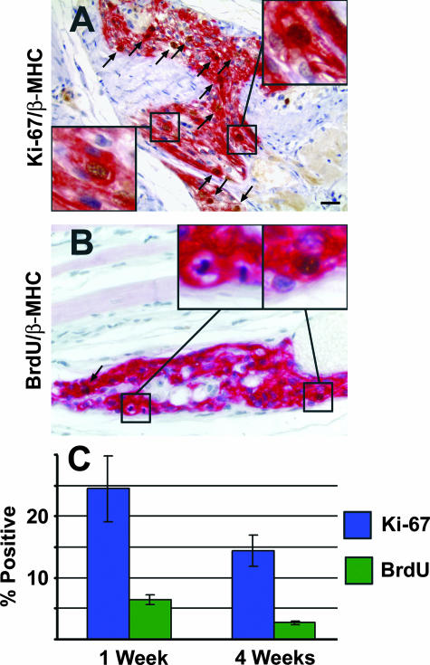
The progressive increase in cardiomyocyte graft size over time suggested that these cells might have significant in vivo cell cycle activity. To test this, rats received an intraperitoneal bolus of the thymidine analog BrdU 1 hour before euthanasia. The incorporation of BrdU, which marks cells in the S phase of the cell cycle, was detected immunohistochemically in sections from 1- and 4-week grafts. These sections were also immunostained with an antibody against β-myosin heavy chain to facilitate identification of the human cardiac implants, and the fraction of BrdU-positive cardiac graft cells was quantitated in a blinded fashion. This analysis revealed a remarkably high degree of proliferative activity, with 6.4 ± 0.8% (n = 5) of cardiac graft nuclei double-positive 1 week after transplantation and 2.7 ± 0.3% (n = 7) after 4 weeks (Figure 5). Analogous results were obtained by immunohistochemical studies with a human-specific monoclonal antibody recognizing the nuclear proliferative marker Ki-67, known to identify cells in all active phases of the cell cycle (eg, all cells outside of G0).42 In this instance, 24.5 ± 5.4% (n = 4) of cardiac graft nuclei after 1 week and 14.4 ± 2.6% (n = 6) after 4 weeks were double positive for Ki-67 and β-myosin heavy chain. Further evidence of the substantial proliferative capacity of the implanted human cardiomyocytes included the observation of infrequent but readily detectable mitotic figures (Figure 5B). The high proliferative rates of these human cardiomyocytes stand in stark contrast with experience with rodent embryonic, fetal, or neonatal cardiomyocytes, all of which have been found to rapidly exit the cell cycle on grafting, if not in their preceding in vitro preparation.31,43–45
Figure 5.
Human ESC-derived cardiac grafts are highly proliferative. A: A 4-week-old cardiac implant is double-immunostained for β-myosin heavy chain (red deposit), the predominant myosin isoform in the human cardiomyocytes, and a human-specific antibody against the proliferative marker Ki-67 (brown intranuclear staining). Note that the host myocardium is β-myosin heavy chain negative. Representative Ki-67+ cardiomyocyte nuclei are indicated by the insets (magnified an additional threefold) and arrows. B: Another 4-week-old implant is double-immunostained for β-myosin heavy chain (red deposit) and anti-BrdU (brown intranuclear staining). Two clear-cut BrdU+ and β-myosin heavy chain+ human cardiomyocytes are indicated by the arrow and right inset. In addition, occasional mitotic figures were noted within the human cardiomyocytes, as illustrated by the left inset. Magnification, ×400 (insets magnified an additional threefold). C: 1 week after transplantation, 28.3 ± 5.4% of β-myosin heavy chain-positive graft cells were Ki-67+ and 6.4 ± 0.8% were BrdU+. As long as 4 weeks after transplantation, a still robust 15.8 ± 3.4% of graft cardiomyocytes were Ki-67+ and 2.7 ± 0.3% were BrdU+. Scale bar = 100 μm.
Discussion
In the present study, we successfully demonstrated the capacity of the differentiated progeny of human ESCs to form stable grafts of human myocardium within the rat heart. In so doing, we have shown that these cells form reasonably sized and expanding grafts that, at least by 4 weeks, are composed predominantly of cardiomyocytes. The grafts expressed many cardiac markers (Figures 1 to 5; Table 1), including sarcomeric actins and myosins, ventricular myosin light chain, atrial natriuretic peptide, and others. Interestingly, the human cells expressed β-myosin heavy chain, the major motor protein of slow heart rate species (including humans), whereas the surrounding rat myocardium expressed the α-isoform. This indicates that the rat heart environment does not override the normal human pattern of myosin expression. The human cardiac implants clearly matured over time and responded to the host environment, as evidenced by the formation of graft fiber strands aligned in parallel with the host myocardium, as well as appropriately aligned sarcomeres (Figure 2F).
At 1 day after implantation, the grafts were composed predominantly of epithelial cells, with only much smaller populations of cardiomyocytes and α-fetoprotein+ endodermal cells. By 4 weeks, however, these heterologous elements were no longer present, such that the grafts were almost exclusively cardiac, otherwise containing only endothelial cells or very rare fibroblastic cells (total n = 36 hearts, including recipients of H1 and H7 cells, both heat shocked and untreated). The mechanisms by which the implanted noncardiac cells are cleared from the rat heart are presently unknown but are the subject of ongoing investigation by our group. Importantly, although we are encouraged by this eventual clearance of noncardiac elements by the nude rat heart and by the absence of teratomatous and/or heterologous elements at 28 days (despite exhaustive histological evaluation of each heart), we recognize the definite need for additional preclinical safety studies to exclude such tumor formation. Because of the obvious unacceptability of even rare or small teratomata within the heart, such studies will need to examine longer time points, as well as distant organs.
Furthermore, the present study demonstrates that the total volume of cardiac graft produced with implantation of hESC-derived cardiomyocytes can be significantly increased by preceding heat-shock treatment (Figure 4). In preceding cell grafting studies by our group with rat neonatal cardiomycytes, we showed that cell death at early time points was greatly reduced by heat-shock pretreatment.33 We speculate that a similar cytoprotective effect is occurring with the present cell type. (We also compared proliferation—obviously another means to a larger graft—in heat-shocked and untreated hESC-derived cardiomyocytes and found it to have a negligible effect.) Although the precise molecular events underlying this cytoprotection remain incompletely elucidated, heat-shock treatment of hESC-derived cardiomyocytes produced the expected induction in heat-shock proteins (including Hsp60, Hsp70, and Hsp90). The cardioprotective action of Hsp70 has been particularly well-demonstrated by numerous groups,46–49 and this and other heat-shock-induced effectors may limit cell death by acting as “molecular chaperones” for damaged proteins,50 inhibiting pro-apoptotic signaling,51 or opposing oxidative stress.52
The presence of both host (rat) and graft (human) vessels within the hESC-derived cardiac implants is also intriguing. Although the present histological study cannot address whether the human-derived vessels are in fact interconnected with the rest of the rodent vasculature, their extensive ingrowth invites speculation that they may be expanding to support the growth of the cardiac implants and that deliberate “doping” of the implanted hESC-derived cardiomyocytes with hESC-derived endothelial cells might further promote graft survival.
A final unexpected and important finding in the present study was the tremendous proliferative capacity of the engrafted hESC-derived cardiomyocytes. BrdU marking studies showed that 6.4% of these cells were synthesizing DNA at 1 week after transplantation, as well as a still-respectable 2.7% after 4 weeks (Figure 5). Because S phase is only a fraction of the cell cycle, the total percentage of cycling graft cells is likely severalfold higher. Consistent with this, the proliferation marker Ki-67, which identifies cells in all active phases of the cell cycle (G1, S, G2, and mitosis), identified 24.5% of engrafted cardiac cells after 1 week and 14.4% after 4 weeks. The persistent proliferative activity of this cell type after transplantation distinguishes them from rodent embryonic,43–45 fetal, and neonatal cardiomyocytes,31 each of which has been shown to very rapidly exit the cell cycle on culture or engraftment. We speculate that this species difference may reflect the 13-fold greater gestation period of humans relative to mice or rats. In any case, this substantial increase in cardiomyocyte proliferation may offer significant benefits for cardiac repair.
Although we have taken pains to carefully follow the lineage of the graft cells using human-specific in situ hybridization probes (as well as human-specific sarcomeric and proliferation markers), it is worth noting that recent studies have demonstrated the occurrence of rare but definite, spontaneous cell fusion events both in vitro53 and in vivo.15,54,55 Although very infrequent, cell fusion can lead to misattribution in the phenotype of implanted cells, because it can result in a single cell exhibiting both donor and host markers. With regard to the present studies, although we are open to the possibility of rare human-rat fusion events, this phenomenon seems highly unlikely to account for the substantial quantities of human myocardium formed in the present studies. (Cell fusion events have typically involved ≪1% of input cells in related experiments, even when using cells with a “fusiogenic” phenotype.55) Moreover, it is worth reemphasizing that we have here used a previously differentiated and cardiac-enriched cell preparation with a well-established cardiac phenotype18 and that the proportion and characteristics of these cells histologically at initial time points are entirely reflective of the input population. We thus conclude that hESC-derived cardiomyocytes can be used to form substantial and proliferating implants of developing human myocardium within the uninjured rat heart.
While this manuscript was under review, two groups also reported the successful formation of stable graft of hESC-derived cardiomyocytes in pharmacologically immunosuppressed pigs56 and guinea pigs.57 Importantly, these independent studies by the Gepstein and Li groups unambiguously demonstrated electromechanical integration of the cardiac implants with the host myocardium, and they provide exciting proof-of-concept for this cell type for applications as a biological pacemaker. Their cell preparation differed from our own in that they physically dissected spontaneous beating foci from differentiated embryoid body outgrowths, an approach not readily scalable to human cell therapeutic applications. On the other hand, because an important contribution of the present study is that hESC-derived cardiomyocytes have sustained proliferative capacity after implantation, input cell number requirements may be less limiting than intuitively expected. Given the gradual in situ expansion of the graft, one may not have to implant the full quantity of cells required for a therapeutic effect, and angiogenesis (perhaps both host- and graft-derived) may be able to match its increasing metabolic requirements. Finally, in addition to having implications for potential cell transplantation therapies, we expect this model to permit unique, previously inaccessible in vivo studies into the developmental biology of early human myocardium, in essence, using the rat heart as a “bioreactor” for the differentiating human cardiomyocytes.
Supplementary Material
Supplemental Material
Acknowledgments
We thank Drs. Jane Lebkowski and Melissa Carpenter for invaluable discussion and for facilitation of the research and Ms. Sarah Dupras and Ms. Brittany Hopkins for assistance with immunohistochemistry and in situ hybridization.
Footnotes
Address reprint requests to Charles E. Murry, MD, PhD, Center for Cardiovascular Biology and Regenerative Medicine, Department of Pathology, 815 Mercer Street, Room 453, University of Washington, Seattle, WA 98109. E-mail: murry@u.washington.edu.
Supported in part by a sponsored research grant from Geron and in part by National Institutes of Health grants HL61553, HL64387, and HL03174 to C.E.M. M.A.L. was supported by a National Research Service Award postdoctoral fellowship (HL07828-06).
References
- Hassink RJ, Brutel de, la Riviere A, Mummery CL, Doevendans PA. Transplantation of cells for cardiac repair. J Am Coll Cardiol. 2003;41:711–717. doi: 10.1016/s0735-1097(02)02933-9. [DOI] [PubMed] [Google Scholar]
- Hassink RJ, Dowell JD, Brutel de, la Riviere A, Doevendans PA, Field LJ. Stem cell therapy for ischemic heart disease. Trends Mol Med. 2003;9:436–441. doi: 10.1016/j.molmed.2003.08.002. [DOI] [PubMed] [Google Scholar]
- Murry CE, Whitney ML, Laflamme MA, Reinecke H, Field LJ. Cellular therapies for myocardial infarct repair. Cold Spring Harb Symp Quant Biol. 2002;67:519–526. doi: 10.1101/sqb.2002.67.519. [DOI] [PubMed] [Google Scholar]
- Jain M, DerSimonian H, Brenner DA, Ngoy S, Teller P, Edge AS, Zawadzka A, Wetzel K, Sawyer DB, Colucci WS, Apstein CS, Liao R. Cell therapy attenuates deleterious ventricular remodeling and improves cardiac performance after myocardial infarction. Circulation. 2001;103:1920–1927. doi: 10.1161/01.cir.103.14.1920. [DOI] [PubMed] [Google Scholar]
- Leobon B, Garcin I, Menasche P, Vilquin JT, Audinat E, Charpak S. Myoblasts transplanted into rat infarcted myocardium are functionally isolated from their host. Proc Natl Acad Sci USA. 2003;100:7808–7811. doi: 10.1073/pnas.1232447100. [DOI] [PMC free article] [PubMed] [Google Scholar]
- Murry CE, Wiseman RW, Schwartz SM, Hauschka SD. Skeletal myoblast transplantation for repair of myocardial necrosis. J Clin Invest. 1996;98:2512–2523. doi: 10.1172/JCI119070. [DOI] [PMC free article] [PubMed] [Google Scholar]
- Orlic D, Kajstura J, Chimenti S, Jakoniuk I, Anderson SM, Li B, Pickel J, McKay R, Nadal-Ginard B, Bodine DM, Leri A, Anversa P. Bone marrow cells regenerate infarcted myocardium. Nature. 2001;410:701–705. doi: 10.1038/35070587. [DOI] [PubMed] [Google Scholar]
- Murry CE, Soonpaa MH, Reinecke H, Nakajima H, Nakajima HO, Rubart M, Pasumarthi KB, Ismail Virag J, Bartelmez SH, Poppa V, Bradford G, Dowell JD, Williams DA, Field LJ. Haematopoietic stem cells do not transdifferentiate into cardiac myocytes in myocardial infarcts. Nature. 2004;428:664–668. doi: 10.1038/nature02446. [DOI] [PubMed] [Google Scholar]
- Penn MS, Francis GS, Ellis SG, Young JB, McCarthy PM, Topol EJ. Autologous cell transplantation for the treatment of damaged myocardium. Prog Cardiovasc Dis. 2002;45:21–32. doi: 10.1053/pcad.2002.123466. [DOI] [PubMed] [Google Scholar]
- Strauer BE, Brehm M, Zeus T, Kostering M, Hernandez A, Sorg RV, Kogler G, Wernet P. Repair of infarcted myocardium by autologous intracoronary mononuclear bone marrow cell transplantation in humans. Circulation. 2002;106:1913–1918. doi: 10.1161/01.cir.0000034046.87607.1c. [DOI] [PubMed] [Google Scholar]
- Shake JG, Gruber PJ, Baumgartner WA, Senechal G, Meyers J, Redmond JM, Pittenger MF, Martin BJ. Mesenchymal stem cell implantation in a swine myocardial infarct model: engraftment and functional effects. Ann Thorac Surg. 2002;73:1919–1925. doi: 10.1016/s0003-4975(02)03517-8. discussion, 1926. [DOI] [PubMed] [Google Scholar]
- Toma C, Pittenger MF, Cahill KS, Byrne BJ, Kessler PD. Human mesenchymal stem cells differentiate to a cardiomyocyte phenotype in the adult murine heart. Circulation. 2002;105:93–98. doi: 10.1161/hc0102.101442. [DOI] [PubMed] [Google Scholar]
- Min JY, Sullivan MF, Yang Y, Zhang JP, Converso KL, Morgan JP, Xiao YF. Significant improvement of heart function by cotransplantation of human mesenchymal stem cells and fetal cardiomyocytes in postinfarcted pigs. Ann Thorac Surg. 2002;74:1568–1575. doi: 10.1016/s0003-4975(02)03952-8. [DOI] [PubMed] [Google Scholar]
- Mangi AA, Noiseux N, Kong D, He H, Rezvani M, Ingwall JS, Dzau VJ. Mesenchymal stem cells modified with Akt prevent remodeling and restore performance of infarcted hearts. Nat Med. 2003;9:1195–1201. doi: 10.1038/nm912. [DOI] [PubMed] [Google Scholar]
- Oh H, Bradfute SB, Gallardo TD, Nakamura T, Gaussin V, Mishina Y, Pocius J, Michael LH, Behringer RR, Garry DJ, Entman ML, Schneider MD. Cardiac progenitor cells from adult myocardium: homing, differentiation, and fusion after infarction. Proc Natl Acad Sci USA. 2003;100:12313–12318. doi: 10.1073/pnas.2132126100. [DOI] [PMC free article] [PubMed] [Google Scholar]
- Beltrami AP, Barlucchi L, Torella D, Baker M, Limana F, Chimenti S, Kasahara H, Rota M, Musso E, Urbanek K, Leri A, Kajstura J, Nadal-Ginard B, Anversa P. Adult cardiac stem cells are multipotent and support myocardial regeneration. Cell. 2003;114:763–776. doi: 10.1016/s0092-8674(03)00687-1. [DOI] [PubMed] [Google Scholar]
- Mummery C, Ward-van Oostwaard D, Doevendans P, Spijker R, van den Brink S, Hassink R, van der Heyden M, Opthof T, Pera M, de la Riviere AB, Passier R, Tertoolen L. Differentiation of human embryonic stem cells to cardiomyocytes: role of coculture with visceral endoderm-like cells. Circulation. 2003;107:2733–2740. doi: 10.1161/01.CIR.0000068356.38592.68. [DOI] [PubMed] [Google Scholar]
- Xu C, Police S, Rao N, Carpenter MK. Characterization and enrichment of cardiomyocytes derived from human embryonic stem cells. Circ Res. 2002;91:501–508. doi: 10.1161/01.res.0000035254.80718.91. [DOI] [PubMed] [Google Scholar]
- Klug MG, Soonpaa MH, Koh GY, Field LJ. Genetically selected cardiomyocytes from differentiating embryonic stem cells form stable intracardiac grafts. J Clin Invest. 1996;98:216–224. doi: 10.1172/JCI118769. [DOI] [PMC free article] [PubMed] [Google Scholar]
- Behfar A, Zingman LV, Hodgson DM, Rauzier JM, Kane GC, Terzic A, Puceat M. Stem cell differentiation requires a paracrine pathway in the heart. FASEB J. 2002;16:1558–1566. doi: 10.1096/fj.02-0072com. [DOI] [PubMed] [Google Scholar]
- Kehat I, Kenyagin-Karsenti D, Snir M, Segev H, Amit M, Gepstein A, Livne E, Binah O, Itskovitz-Eldor J, Gepstein L. Human embryonic stem cells can differentiate into myocytes with structural and functional properties of cardiomyocytes. J Clin Invest. 2001;108:407–414. doi: 10.1172/JCI12131. [DOI] [PMC free article] [PubMed] [Google Scholar]
- Etzion S, Battler A, Barbash IM, Cagnano E, Zarin P, Granot Y, Kedes LH, Kloner RA, Leor J. Influence of embryonic cardiomyocyte transplantation on the progression of heart failure in a rat model of extensive myocardial infarction. J Mol Cell Cardiol. 2001;33:1321–1330. doi: 10.1006/jmcc.2000.1391. [DOI] [PubMed] [Google Scholar]
- Zandstra PW, Bauwens C, Yin T, Liu Q, Schiller H, Zweigerdt R, Pasumarthi KB, Field LJ. Scalable production of embryonic stem cell-derived cardiomyocytes. Tissue Eng. 2003;9:767–778. doi: 10.1089/107632703768247449. [DOI] [PubMed] [Google Scholar]
- Mummery C, Ward D, van den Brink CE, Bird SD, Doevendans PA, Opthof T, Brutel de, la Riviere A, Tertoolen L, van der Heyden M, Pera M. Cardiomyocyte differentiation of mouse and human embryonic stem cells. J Anat. 2002;200:233–242. doi: 10.1046/j.1469-7580.2002.00031.x. [DOI] [PMC free article] [PubMed] [Google Scholar]
- He JQ, Ma Y, Lee Y, Thomson JA, Kamp TJ. Human embryonic stem cells develop into multiple types of cardiac myocytes: action potential characterization. Circ Res. 2003;93:32–39. doi: 10.1161/01.RES.0000080317.92718.99. [DOI] [PubMed] [Google Scholar]
- Balsam LB, Wagers AJ, Christensen JL, Kofidis T, Weissman IL, Robbins RC. Haematopoietic stem cells adopt mature haematopoietic fates in ischaemic myocardium. Nature. 2004;428:668–673. doi: 10.1038/nature02460. [DOI] [PubMed] [Google Scholar]
- Reinecke H, MacDonald GH, Hauschka SD, Murry CE. Electromechanical coupling between skeletal and cardiac muscle: implications for infarct repair. J Cell Biol. 2000;149:731–740. doi: 10.1083/jcb.149.3.731. [DOI] [PMC free article] [PubMed] [Google Scholar]
- Reinecke H, Poppa V, Murry CE. Skeletal muscle stem cells do not transdifferentiate into cardiomyocytes after cardiac grafting. J Mol Cell Cardiol. 2002;34:241–249. doi: 10.1006/jmcc.2001.1507. [DOI] [PubMed] [Google Scholar]
- Thomson JA, Itskovitz-Eldor J, Shapiro SS, Waknitz MA, Swiergiel JJ, Marshall VS, Jones JM. Embryonic stem cell lines derived from human blastocysts. Science. 1998;282:1145–1147. doi: 10.1126/science.282.5391.1145. [DOI] [PubMed] [Google Scholar]
- Xu C, Inokuma MS, Denham J, Golds K, Kundu P, Gold JD, Carpenter MK. Feeder-free growth of undifferentiated human embryonic stem cells. Nat Biotechnol. 2001;19:971–974. doi: 10.1038/nbt1001-971. [DOI] [PubMed] [Google Scholar]
- Reinecke H, Zhang M, Bartosek T, Murry CE. Survival, integration, and differentiation of cardiomyocyte grafts: a study in normal and injured rat hearts. Circulation. 1999;100:193–202. doi: 10.1161/01.cir.100.2.193. [DOI] [PubMed] [Google Scholar]
- Reinecke H, Murry CE. Transmural replacement of myocardium after skeletal myoblast grafting into the heart: too much of a good thing? Cardiovasc Pathol. 2000;9:337–344. doi: 10.1016/s1054-8807(00)00055-7. [DOI] [PubMed] [Google Scholar]
- Zhang M, Methot D, Poppa V, Fujio Y, Walsh K, Murry CE. Cardiomyocyte grafting for cardiac repair: graft cell death and anti-death strategies. J Mol Cell Cardiol. 2001;33:907–921. doi: 10.1006/jmcc.2001.1367. [DOI] [PubMed] [Google Scholar]
- Muller-Ehmsen J, Whittaker P, Kloner RA, Dow JS, Sakoda T, Long TI, Laird PW, Kedes L. Survival and development of neonatal rat cardiomyocytes transplanted into adult myocardium. J Mol Cell Cardiol. 2002;34:107–116. doi: 10.1006/jmcc.2001.1491. [DOI] [PubMed] [Google Scholar]
- Bouvagnet P, Neveu S, Montoya M, Leger JJ. Development changes in the human cardiac isomyosin distribution: an immunohistochemical study using monoclonal antibodies. Circ Res. 1987;61:329–336. doi: 10.1161/01.res.61.3.329. [DOI] [PubMed] [Google Scholar]
- Bouvagnet P, Mairhofer H, Leger JO, Puech P, Leger JJ. Distribution pattern of alpha and beta myosin in normal and diseased human ventricular myocardium. Basic Res Cardiol. 1989;84:91–102. doi: 10.1007/BF01907006. [DOI] [PubMed] [Google Scholar]
- Dechesne CA, Leger JO, Leger JJ. Distribution of alpha- and beta-myosin heavy chains in the ventricular fibers of the postnatal developing rat. Dev Biol. 1987;123:169–178. doi: 10.1016/0012-1606(87)90439-8. [DOI] [PubMed] [Google Scholar]
- Angst BD, Khan LU, Severs NJ, Whitely K, Rothery S, Thompson RP, Magee AI, Gourdie RG. Dissociated spatial patterning of gap junctions and cell adhesion junctions during postnatal differentiation of ventricular myocardium. Circ Res. 1997;80:88–94. doi: 10.1161/01.res.80.1.88. [DOI] [PubMed] [Google Scholar]
- Tuganowski W, Samek D, Glenc F. Glycogen content in human embryonic heart. Recent Adv Stud Cardiac Struct Metab. 1975;8:179–180. [PubMed] [Google Scholar]
- Duijvestijn AM, van Goor H, Klatter F, Majoor GD, van Bussel E, van Breda Vriesman PJ. Antibodies defining rat endothelial cells: rECA-1, a pan-endothelial cell-specific monoclonal antibody. Lab Invest. 1992;66:459–466. [PubMed] [Google Scholar]
- Kalka C, Masuda H, Takahashi T, Kalka-Moll WM, Silver M, Kearney M, Li T, Isner JM, Asahara T. Transplantation of ex vivo expanded endothelial progenitor cells for therapeutic neovascularization. Proc Natl Acad Sci USA. 2000;97:3422–3427. doi: 10.1073/pnas.070046397. [DOI] [PMC free article] [PubMed] [Google Scholar]
- Scholzen T, Gerdes J. The Ki-67 protein: from the known and the unknown. J Cell Physiol. 2000;182:311–322. doi: 10.1002/(SICI)1097-4652(200003)182:3<311::AID-JCP1>3.0.CO;2-9. [DOI] [PubMed] [Google Scholar]
- Dowell JD, Rubart M, Pasumarthi KB, Soonpaa MH, Field LJ. Myocyte and myogenic stem cell transplantation in the heart. Cardiovasc Res. 2003;58:336–350. doi: 10.1016/s0008-6363(03)00254-2. [DOI] [PubMed] [Google Scholar]
- Soonpaa MH, Koh GY, Klug MG, Field LJ. Formation of nascent intercalated disks between grafted fetal cardiomyocytes and host myocardium. Science. 1994;264:98–101. doi: 10.1126/science.8140423. [DOI] [PubMed] [Google Scholar]
- Klug MG, Soonpaa MH, Field LJ. DNA synthesis and multinucleation in embryonic stem cell-derived cardiomyocytes. Am J Physiol. 1995;269:H1913–H1921. doi: 10.1152/ajpheart.1995.269.6.H1913. [DOI] [PubMed] [Google Scholar]
- Marber MS, Mestril R, Chi SH, Sayen MR, Yellon DM, Dillmann WH. Overexpression of the rat inducible 70-kD heat stress protein in a transgenic mouse increases the resistance of the heart to ischemic injury. J Clin Invest. 1995;95:1446–1456. doi: 10.1172/JCI117815. [DOI] [PMC free article] [PubMed] [Google Scholar]
- Radford NB, Fina M, Benjamin IJ, Moreadith RW, Graves KH, Zhao P, Gavva S, Wiethoff A, Sherry AD, Malloy CR, Williams RS. Cardioprotective effects of 70-kDa heat shock protein in transgenic mice. Proc Natl Acad Sci USA. 1996;93:2339–2342. doi: 10.1073/pnas.93.6.2339. [DOI] [PMC free article] [PubMed] [Google Scholar]
- Jayakumar J, Suzuki K, Sammut IA, Smolenski RT, Khan M, Latif N, Abunasra H, Murtuza B, Amrani M, Yacoub MH. Heat shock protein 70 gene transfection protects mitochondrial and ventricular function against ischemia-reperfusion injury. Circulation. 2001;104:I303–I307. doi: 10.1161/hc37t1.094932. [DOI] [PubMed] [Google Scholar]
- Williams RS, Thomas JA, Fina M, German Z, Benjamin IJ. Human heat shock protein 70 (hsp70) protects murine cells from injury during metabolic stress. J Clin Invest. 1993;92:503–508. doi: 10.1172/JCI116594. [DOI] [PMC free article] [PubMed] [Google Scholar]
- Benjamin IJ, McMillan DR. Stress (heat shock) proteins: molecular chaperones in cardiovascular biology and disease. Circ Res. 1998;83:117–132. doi: 10.1161/01.res.83.2.117. [DOI] [PubMed] [Google Scholar]
- Mosser DD, Caron AW, Bourget L, Denis-Larose C, Massie B. Role of the human heat shock protein hsp70 in protection against stress-induced apoptosis. Mol Cell Biol. 1997;17:5317–5327. doi: 10.1128/mcb.17.9.5317. [DOI] [PMC free article] [PubMed] [Google Scholar]
- Su CY, Chong KY, Owen OE, Dillmann WH, Chang C, Lai CC. Constitutive and inducible hsp70s are involved in oxidative resistance evoked by heat shock or ethanol. J Mol Cell Cardiol. 1998;30:587–598. doi: 10.1006/jmcc.1997.0622. [DOI] [PubMed] [Google Scholar]
- Terada N, Hamazaki T, Oka M, Hoki M, Mastalerz DM, Nakano Y, Meyer EM, Morel L, Petersen BE, Scott EW. Bone marrow cells adopt the phenotype of other cells by spontaneous cell fusion. Nature. 2002;416:542–545. doi: 10.1038/nature730. [DOI] [PubMed] [Google Scholar]
- Alvarez-Dolado M, Pardal R, Garcia-Verdugo JM, Fike JR, Lee HO, Pfeffer K, Lois C, Morrison SJ, Alvarez-Buylla A. Fusion of bone-marrow-derived cells with Purkinje neurons, cardiomyocytes and hepatocytes. Nature. 2003;425:968–973. doi: 10.1038/nature02069. [DOI] [PubMed] [Google Scholar]
- Reinecke H, Minami E, Poppa V, Murry CE. Evidence for fusion between cardiac and skeletal muscle cells. Circ Res. 2004;94:e56–e60. doi: 10.1161/01.RES.0000125294.04612.81. [DOI] [PubMed] [Google Scholar]
- Kehat I, Khimovich L, Caspi O, Gepstein A, Shofti R, Arbel G, Huber I, Satin J, Itskovitz-Eldor J, Gepstein L. Electromechanical integration of cardiomyocytes derived from human embryonic stem cells. Nat Biotechnol. 2004;22:1282–1289. doi: 10.1038/nbt1014. [DOI] [PubMed] [Google Scholar]
- Xue T, Cho HC, Akar FG, Tsang SY, Jones SP, Marban E, Tomaselli GF, Li RA. Functional integration of electrically active cardiac derivatives from genetically engineered human embryonic stem cells with quiescent recipient ventricular cardiomyocytes: insights into the development of cell-based pacemakers. Circulation. 2004;111:11–20. doi: 10.1161/01.CIR.0000151313.18547.A2. [DOI] [PubMed] [Google Scholar]
Associated Data
This section collects any data citations, data availability statements, or supplementary materials included in this article.
Supplementary Materials
Supplemental Material
