Molecular Regulation of H3K4 Trimethylation by Wdr82, a Component of Human Set1/COMPASS (original) (raw)
Abstract
In yeast, the macromolecular complex Set1/COMPASS is capable of methylating H3K4, a posttranslational modification associated with actively transcribed genes. There is only one Set1 in yeast; yet in mammalian cells there are multiple H3K4 methylases, including Set1A/B, forming human COMPASS complexes, and MLL1-4, forming human COMPASS-like complexes. We have shown that Wdr82, which associates with chromatin in a histone H2B ubiquitination-dependent manner, is a specific component of Set1 complexes but not that of MLL1-4 complexes. RNA interference-mediated knockdown of Wdr82 results in a reduction in the H3K4 trimethylation levels, although these cells still possess active MLL complexes. Comprehensive in vitro enzymatic studies with Set1 and MLL complexes demonstrated that the Set1 complex is a more robust H3K4 trimethylase in vitro than the MLL complexes. Given our in vivo and in vitro observations, it appears that the human Set1 complex plays a more widespread role in H3K4 trimethylation than do the MLL complexes in mammalian cells.
The MLL gene located on chromosome 11q23 is found in a variety of chromosomal translocations possessing unique clinical and biological characteristics (11, 23, 31). The MLL rearrangements are found in more than 70% of infant leukemia, with phenotypes more consistent with acute lymphoid leukemia and/or acute myeloid leukemia (23). Such translocations have also been reported in ca. 10% of adults with a diagnosis of acute myeloid leukemia related to the treatment of other malignancies with the use of topoisomerase II inhibitors.
For more than 20 years, little was known about the molecular function(s) of MLL until its yeast homologue, Set1, was identified in a macromolecular complex named COMPASS (for complex of proteins associated with Set1) (18). COMPASS is capable of mono-, di-, and trimethylating histone H3 on lysine 4 (H3K4) (12, 18, 20, 22, 26). We now know that human MLL is also found in a COMPASS-like complex capable of methylating H3K4 (7, 35). In addition to MLL, there are three MLL homologues (MLL2 to MLL4) (3, 7) and two Set1-related proteins (Set1A and Set1B) (13, 15) in humans; all are found in COMPASS-like complexes capable of methylating H3K4 (26, 27). We do not understand why there are several H3K4 methylases in mammalian cells capable of methylating H3K4. However, it is clear that the H3K4 methylase activities in mammals are not redundant. This is evident by the observation that the deletion of many individual MLL genes result in embryonic lethality (6, 36). Why do mammals need so many different H3K4 methylases? Perhaps mammals need to control the H3K27 methylation mark, which is associated with polycomb and transcriptional silencing at different genomic loci in different cellular contexts, while single-celled eukaryotes do not (1). It is possible that mammals have a built-in intricate network of H3K4 methylases that perform the ancient functions, as well as oppose and perhaps reverse H3K27. All of the H3K4 methylase-containing complexes in mammals have COMPASS-like conserved subunits, as well as complex-specific subunits (27). The MLL1 and MLL2 complexes both contain the tumor suppressor gene menin (7, 35). The MLL3 and MLL4 complexes are associated with PTIP, PA1, NCOA6, and an H3 lysine 27 demethylase, UTX (3, 17, 27). The Set1A and Set1B complexes in human are much more similar to the yeast COMPASS, and almost all of the yeast components have corresponding mammalian partners (13, 15, 27).
Most studies defining the H3K4 methylase activities in mammals have taken advantage of reagents (antibodies and RNA interference [RNAi], etc.) created toward common subunits of these complexes, such as Ash2, WDR5, and Rbbp5. These studies have demonstrated that human homologues of the components of yeast COMPASS function similarly. For example, the Cps40 and Cps60 components of COMPASS were shown to be required for proper H3K4 trimethylation by COMPASS (19, 24). The human protein related to these two proteins, ASH2L, is also required for proper H3K4 trimethylation (4, 29). Furthermore, yeast Cps30 was shown to be required for proper COMPASS assembly (29). The human homologue of Cps30, the Wdr5 protein, also functions in proper MLL complex formation and is required for the mono-, di-, and trimethylation of H3K4 (4, 29).
H3K4 trimethylation is highly correlated with active transcription, and this modification requires the presence of histone H2B ubiquitination, a process known as histone cross talk (5, 26, 30). Many aspects of cross talk between H2B ubiquitination and H3K4 methylation appear to be highly conserved from yeasts to humans (26). Recently, yeast Cps35, the only essential component of COMPASS, which is required for H3K4 di- and trimethylation, was shown to interact with chromatin in an H2B ubiquitination-dependent manner but in a Set1-independent manner (16). It has been proposed that the interaction of Set1 with Cps35 on chromatin can result in the assembly of the trimethylation-competent COMPASS (16). We show in this study that human Wdr82, the human homologue of yeast Cps35, also interacts with chromatin in a H2B ubiquitination-dependent manner. Wdr82 is found to be associated with the Set1A/B complexes (13), but its function in histone methylation is not well understood. We show here that Wdr82 is only associated with the Set1A/B complexes and not the other MLL1-4 complexes. Surprisingly, a reduction of the Wdr82 levels by RNAi results in a decrease in the total pattern of H3K4 trimethylation, even though the cells still carry functional MLL1-4 complexes. Comprehensive in vitro enzymological studies demonstrated that the Set1 complexes are much more robust H3K4 trimethylases than the MLL1-4 complexes in vitro, demonstrating a functional difference between these H3K4 methylases.
MATERIALS AND METHODS
Antibody, cell lines, and siRNA.
The histone H3 lysine 4 mono-, di-, and trimethylation antibodies were purchased from Upstate and Abcam. For Western blotting, the H3K4 trimethylation antibodies were used at a 1:5,000 dilution (Upstate 07-473 [discontinued] and 04-745) or a 1:30,000 dilution (Abcam). Antibodies for Flag (M2; Sigma), Set1A (A300-289A; Bethyl), MLL2 (A300-113A; Bethyl), MLL1 (A300-086A; Bethyl), Rbbp5 (A300-109A; Bethyl), Ash2 (A300-112A; Bethyl), WDR5 (07-706; Upstate), H3 (ab1791-100; Abcam), H4(ab31827; Abcam), and α-tubulin (Santa Cruz) were purchased from various sources as indicated. Antiserum for hBre1 was made against peptide. Antiserum and full-length cDNA of Wdr82 were gifts from David G. Skalnik (Indiana University School of Medicine). HeLa and U2OS cell lines were purchased from the American Type Culture Collection. The 293 and 293FRT cell lines were gifts from Joan and Ron Conaway (Stowers Institute). The MLL1+/+ and MLL1−/− mouse embryonic fibroblast (MEF) cell lines were gifts from Jay L. Hess (University of Michigan Medical School). The Flag-PTIP HeLa S3 stable cell line is a gift from Kai Ge (National Institutes of Health). All of the cell lines were cultured in Dulbecco modified Eagle medium plus 10% fetal bovine serum for adherent growth. The 293 and 293FRT cells were grown in suspension with Cd293 medium (Invitrogen) as described by the manufacturer. The Wdr82 small interfering RNAs (siRNAs), either SMARTpool (L-016629-01) or customized (AGAGAACCCUGUACAGUAAUU), were obtained from Dharmacon. All of the other siRNAs were purchased from Dharmacon SMARTpool.
Purification of COMPASS-like complexes from Flag-tagged stable cell lines.
The cDNAs of human WDR5, Rbbp5, Ash2, and Wdr82 genes were cloned into either pCMV-TAG2B or pCDNA5/FRT vectors with N-terminal Flag tag. The plasmids were then transfected into 293 or 293FRT cell lines and selected by neomycin or hygromycin. The single clones were picked and cultured up to 3 liters. Nuclear extracts were prepared and subjected to anti-Flag agarose immunoaffinity chromatography.
In vitro methyltransferase assay.
Approximately 107 cells for each assay were collected, washed with phosphate-buffered saline once, and lysed in high-salt lysis buffer (20 mM HEPES [pH 7.4], 10% glycerol, 0.35 M NaCl, 1 mM MgCl2, 0.5% Triton X-100, 1 mM dithiothreitol) containing proteinase inhibitors (Sigma). After incubation at 4°C for 30 min, the lysate was centrifuged thoroughly at 4°C twice. The balance buffer (20 mM HEPES [pH 7.4], 1 mM MgCl2, 10 mM KCl) was added to the resulting supernatant to make the final NaCl concentration 300 mM. The lysate was then mixed with antibodies and protein A beads or with anti-Flag agarose (Sigma). After incubation at 4°C for 4 h, the beads were spun down and washed three times with wash buffer (10 mM HEPES [pH 7.4], 1 mM MgCl2, 300 mM NaCl, 10 mM KCl, 0.2% Triton X-100) and once with 1× MAB buffer (50 mM Tris [pH 8.5], 20 mM KCl, 10 mM MgCl2, 10 mM β-mercaptoethanol, 250 mM sucrose). The residual buffer was completely moved by aspiration; the beads were mixed with 1 μg of histone H3, 1 μl of 2 mM S-adenosyl methionine, and 0.1 μg of bovine serum albumin/μl, and 1× MAB buffer was added to bring the total volume to 25 μl. For experiments with multiple assays, large numbers of cells were grown, and the resulting extracts were purified as described above. The purified histone methyltransferase (HMTase) on the beads was then divided equally among the tubes, and HMTase assays were performed as described above. After incubation at 37°C for 3 h or overnight, sodium dodecyl sulfate loading buffer was added to stop each reaction mixture, and the methylation of histone H3 was determined by analysis of the reaction mixture by sodium dodecyl sulfate-polyacrylamide gel electrophoresis and Western blotting.
MudPIT analysis.
Trichloroacetic acid-precipitated protein mixtures from purifications were digested with endoproteinase Lys-C and trypsin (Roche) as previously described (24). Peptide mixtures were loaded onto triphasic 100-mm fused silica microcapillary columns as described previously. Loaded microcapillary columns were placed in-line with a Quaternary Agilent 1100 series high-pressure liquid chromatography pump and a Deca-XP ion trap mass spectrometer (Thermo Fisher) equipped with a nano-LC electrospray ionization source. Fully automated multidimensional protein identification technology (MudPIT) runs were carried out on the electrosprayed peptides. Tandem mass spectra were interpreted by using SEQUEST against a database containing Homo sapiens protein sequences downloaded from the National Center for Biotechnology Information. In addition to estimate false discovery rates, each sequence was randomized (keeping the same amino acid composition and length), and the resulting “shuffled” sequences were added to the “normal” human database and searched at the same time. Peptide/spectrum matches were sorted and selected using DTASelect, keeping false discovery rates at 2% or less, and peptide hits from multiple runs were compared using CONTRAST. To estimate protein levels, spectral counts of nonredundant proteins were normalized by using the in-house-developed script NSAF7.
ChIP assay.
Cells were fixed, lysed, and sonicated. Sonicated lysates equivalent to 4 × 10⁶ cells were subjected to chromatin immunoprecipitation (ChIP) analysis. ChIP products were analyzed by quantitative (real-time) PCR using Sybr green real-time PCR with a Bio-Rad iCycler. The comparative CT method was used to determine relative expression compared to input or total histone H3, which was then averaged over three independent experiments.
RESULTS AND DISCUSSION
Wdr82 is specifically associated with Set1 but not any of the MLL complexes.
We searched the NCBI database with the full amino acid sequence of Saccharomyces cerevisiae Cps35. In Drosophila melanogaster, we found the closest gene to be CG17293. In Homo sapiens, the homologue yeast CPS35 is called a WD repeat domain 82 or a transmembrane protein 113 (WDR82 or TMEM113), which is located at chromosome 3q21.1. Interestingly, we have also identified another predicted sequence, termed the WD repeat domain 82 pseudogene 1 (WDR82p1) in H. sapiens. The two human versions of Wdr82 are 93% identical in the amino acid sequence and 95% identical in the DNA sequence. Moreover, Wdr82p1 does not have any nonsense mutation in the coding region; however, we cannot find any sequences in the expressed sequence tag database that are 100% identical. Whether Wdr82p1 is an expressed gene needs to be investigated. The alignment of the amino acid sequences of the genes described above suggests that Wdr82 is conserved from S. cerevisiae to H. sapiens. In addition to the above sequences, there is one more predicted sequence in the human genome, Wdr82p2, which has several nonsense mutations and is unlikely to encode a functional protein.
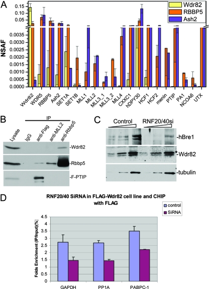
Wdr82 interacts with the Set1 complexes and not with the MLL1 complexes (14). However, due to the lack of antibodies recognizing MLLs 2 to 4, the association of Wdr82 with these complexes was not determined. In order to define the composition of the Wdr82 complexes, we established stable cell lines expressing N-terminal Flag-tagged Wdr82 under a cytomegalovirus promoter. We purified the Flag-Wdr82 complexes via Flag affinity purification and analyzed the purified complexes by silver staining (data not shown), as well as by using MudPIT (32) (Fig. 1A). These studies were performed in triplicate. The proteins were reproducibly detected in all three analyses: the Wdr82 affinity-purified complexes included Wdr82, Wdr5, Rbbp5, Ash2, SET1A, and SET1B, as well as CXXC1, hDPY30, and HCF1 (Fig. 1A). We only identified background levels of MLLs in the Flag-WDR82 preparations. On the other hand, proteins purified through the Flag-RBBP5 or Flag-Ash2 subunits shared between the human Set1 complexes and MLL complexes reproducibly (two out of two runs) included high levels of all four MLLs (i.e., MLL1 to MLL4). HCF2, menin, PTIP, PA1, NCOA6, and UTX were detected in these preparations as well, but hardly any were detected in the Flag-Wdr82 affinity-purified complexes, further supporting the view that Wdr82 is a Set1 complex (preferentially Set1A)-specific subunit. We also tested for proteins associated with Wdr82 by Western blot analysis after immunoprecipitation in a Flag-PTIP stable HeLa cell line (Fig. 1B). Wdr82 was only coimmunoprecipitated with anti-RbbP5 (a common subunit of the MLLs and Set1 complexes) and not with MLL2 or PTIP (a component of the MLL3/4 complexes) (Fig. 1B).
FIG. 1.
Wdr82 is a component of the Set1 complex that associates with chromatin in an H2B monoubiquitination-dependent manner. (A) Mammalian H3K4 trimethylase complexes were affinity purified through Flag-Wdr82, Flag-RBBP5, or Flag-Ash2. Relative protein levels were estimated by their dNSAF values (calculated based on unique spectral counts and shared spectral counts distributed among isoforms). dNSAF values were averaged across three (Wdr82) or two (RBBP5 and Ash2) independent runs. (B) Extracts from the Flag-tagged PTIP HeLa stable cells were immunoprecipitated with rabbit immunoglobulin G (IgG), anit-Flag, anti-MLL2, anti-Rbbp5 antibodies, followed by Western blotting with anti-Rbbp5, anti-Wdr82, and anti-Flag antibodies. (C) Human Bre1 (RNF20/40) were co-knocked down in Flag-Wdr82 stable cell lines via RNAi. Increasing amounts of cell extract were analyzed by Western analysis. (D) ChIP assays were carried out with anti-Flag antibody with the same samples as in panel C. The 3′-untranslated region of hemoglobin gene was used as a nonexpressing control gene internal control. The recruitment of Flag-Wdr82 to GAPDH, PP1A, and PABPC-1 promoters was decreased in the H2B ubiquitination-deficient cells. The asterisk in panel C indicates the presence of the nonspecific bands in the Western analysis.
In yeast, histone H2B ubiquitination by Rad6/Bre1 is required for proper H3K4 methylation (5, 8, 26, 30, 33). The Wdr82 homologue in yeast, Cps35, interacts with chromatin in an H2B ubiquitination-dependent manner (16). We investigated whether the Wdr82 interaction with chromatin also requires histone H2B ubiquitination. Human Bre1 is required for H2B ubiquitination (9, 21, 37). Partial knockdown of its levels through RNAi of RNF20 and RNF40 together in 293 cells expressing Flag-Wdr82, resulted in a partial reduction in the localization pattern of Wdr82 on Set1A/B-regulated genes (Fig. 1C and D). Partial knockdown of human Bre1 levels resulted in a decrease (∼50%) in the recruitment of Wdr82 to the promoters of the genes tested (Fig. 1D), indicating that the mechanism of the regulation of H3K4 trimethylation through H2B ubiquitination could be conserved from yeasts to humans.
Wdr82 regulates H3K4 trimethylation pattern in mammalian cells.
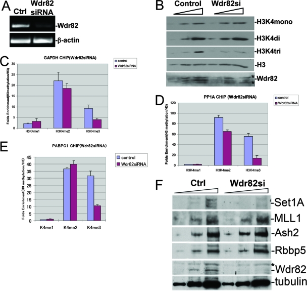
In S. cerevisiae, Cps35 is a critical regulator for H3K4 di- and trimethylation (2, 16). We investigated the effect of Wdr82 on H3K4 by reducing Wdr82 levels via RNAi in mammalian cells. To be certain about the results of the present study, we initially characterized our antibodies for H3K4 di- and trimethylation by using yeast Cps60 and Cps40-null extracts as the markers for H3K4 di- and trimethylation, respectively (data not shown). After Wdr82 knockdown, the global level of H3K4 trimethylation decreased, with little to no effect on the mono- or dimethylation levels of H3K4 (Fig. 2A and B). To confirm these observations, we tested two different siRNAs (data not shown). We also analyzed Wdr82 knockdown in several cell types, including U2OS, 293, HeLa, and MEF cells. In all experiments we observed a specific loss of H3K4 trimethylation. This observation was unexpected since the cells depleted of Wdr82 still contained functional MLL1-4 complexes. Our observations suggest that Wdr82-containing complexes could play a major role in the global pattern of H3K4 trimethylation in mammalian cells.
FIG. 2.
Reduction of Wdr82 levels results in the loss of H3K4 trimethylation and Set1A. (A) RNAi knockdown of Wdr82 in HeLa cells was confirmed by reverse transcription-PCR. (B) RNAi knockdown of Wdr82 in HeLa cells results in a reduction in H3K4 trimethylation levels but not a reduction in H3K4 mono- and dimethylation. WDR82 mRNA was knocked down using RNAi. Cell extracts from mock RNAi and WDR82 RNAi were tested by Western analysis with antibodies specific to mono-, di-, and trimethylated H3K4. (C to E) Wdr82 mRNA levels were knocked down in HeLa cells with siRNA, and CHIP assays were performed with H3K4 mono-, di-, and trimethylation antibodies on the promoters of GAPDH (C), PP1A (D), and PABPC1 (E) genes. The H3K4 trimethylation of all three genes decreased approximately two- to threefold, while the dimethylation was relatively unchanged for GAPDH and PP1A, and no changes were observed for PABPC1. (F) RNAi knockdown of Wdr82 results in the reduction in Set1A levels with no effect on MLL1, Ash2, or Rbbp5 levels. Reductions in the levels of WDR82, result in destabilization of Set1 complexes. The asterisks in panels B and F indicate the presence of nonspecific bands in the Western analysis.
To confirm these observations obtained by Western blot studies, we performed ChIP in cells expressing Flag-Wdr82 and found that Wdr82 is present on the promoter regions of several housekeeping genes (data not shown). After Wdr82 knockdown in these cells via RNAi, the levels of H3K4 mono-, di-, and trimethylation were measured via ChIP (Fig. 2C to E). We observed a marginal reduction in mono- and dimethylation levels; however, H3K4 trimethylation levels for all of the three genes tested via ChIP were reduced approximately two- to threefold when the Wdr82 levels were reduced by RNAi (Fig. 2C to E).
Set1A, but not MLL1, protein levels are reduced after Wdr82 knockdown.
Since Wdr82 is one of the components in the Set1 complexes, we investigated the stability of these proteins following Wdr82 knockdown. We found that the levels of Rbbp5, Ash2, and MLL1 remain unchanged as a result of Wdr82 reduction. As previously reported, the Set1A levels were reduced significantly in the absence of Wdr82 (Fig. 2F) (14). The loss of Wdr82 possibly affects the stability of the entire Set1A complex. This is very similar to the role of Cps35 in COMPASS's stability in yeast. We do not have specific antibodies toward Set1B proteins. However, our MudPIT studies demonstrated that the Set1B interaction with Wdr82 in Wdr82-Flag purification is marginal (Fig. 1A). Since Wdr82 does not appear to be a major component of the MLL complexes, the stability of the MLL complexes is not altered in the absence of Wdr82. Together, our data suggest that the reduction of the total H3K4 trimethylation level might be due to the reduction of the Set1A protein and its complex and not the MLL complexes.
The loss of Wdr82 significantly reduces H3K4 trimethylation activity in vitro.
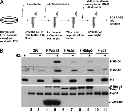
Our results at this point suggest that in vivo the Set1 complexes could play a major role as H3K4 trimethylases in mammalian cells. We set out to define the in vitro kinetic properties of the Set1 and MLL complexes. Therefore, we set up a new system for an in vitro H3K4 HMTase assay, which can be performed on a small scale (Fig. 3A). We purified mammalian H3K4 methylase complexes by immunoprecipitation via either Flag-Wdr82, Flag-Ash2, or Flag-Rbbp5 from stable cell lines and performed in vitro HMTase assays. Purified Set1 complexes (Flag-Wdr82) demonstrated very strong H3K4 di- and trimethylase activities (Fig. 3B, lanes 4 to 5). Purified H3K4 methylase complexes through either Flag-Rbbp5 or Flag-Ash2 also demonstrated H3K4 di- and trimethylase activities (Fig. 3B, lanes 7 to 9). The complexes purified through Rbbp5 or Ash2 (common subunits of all of the H3K4 methylases in human) contain a mixture of the MLLs and Set1 complexes (Fig. 1A). As an internal negative control for our assay, we used the purified Flag-p53 complex (Fig. 3B, lanes 10 to 11). We demonstrate here that H3K4 HMTase purified either through Flag-Wdr82 (Set1 specific) or Ash2 and Rbbp5 (a mixture of Set1 and MLL complexes) are active in vitro.
FIG. 3.
In vitro H3K4 trimethylase activities. (A) Schematic flowchart of the in vitro histone H3K4 methyltransferase assay with either human MLL- or Set1-containing complexes. (B) In vitro HMTase assays were performed with the complexes purified by anti-Flag antibody. HMTase was immunoprecipitated from either Flag-Wdr82, Flag-Ash2, Flag-Rbbp5, or Flag-p53 cell lines and tested for H3K4 di- and trimethylase activities.
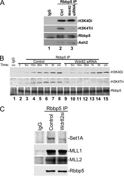
To determine whether Wdr82 knockdown via RNAi alters the HMTase activities of these complexes, we purified the HMTase through Rbbp5 (a common subunit of the Set1 and MLL1-4 complexes) (Fig. 1A) in the presence or absence of Wdr82. The resulting complexes were then tested for H3K4 di- and trimethylase activity in vitro (Fig. 4A). The loss of Wdr82 did not significantly alter the H3K4 dimethylation activity (Fig. 4A, lanes 2 and 3). However, the H3K4 trimethylase activity of the complexes was reduced (Fig. 4A, lanes 2 and 3). We also checked the kinetic properties of the purified HMTase in di- and trimethylation as a function of time in the presence or absence of Wdr82 (Fig. 4B). Clearly, the dimethylase activity of the Rbbp5 purified complexes is slowed down in the absence of Wdr82; however, it reaches levels similar to that of wild type after ∼3 h (Fig. 4B, compare lanes 1 to 8 to lanes 9 to 15). However, the trimethylation activity of the complexes is reduced in the enzyme preparations lacking Wdr82 (Fig. 4B, compare lanes 1 to 8 to lanes 9 to 15). Purified complexes lacking Wdr82 are devoid of Set1A but not of MLL or MLL2 (Fig. 4C). We do not have antibodies toward MLL3 and MLL4, so their presence cannot be tested at this time. This indicates that in these HMTase preparations, we are comparing the H3K4 trimethylase activity of the MLL complexes (Wdr82 RNAi) (Fig. 4B, lanes 9 to 15) to that of the Set1 and MLL complexes (control) (Fig. 4B, lanes 1 to 8). In other words, the complexes tested in Fig. 4B lanes 9 to 15, lack Set1 and contain MLLs but have reduced H3K4 trimethylase activity. This observation is consistent with our in vivo studies in which the specific loss of Wdr82 results in a decrease in H3K4 trimethylation levels (Fig. 2).
FIG. 4.
Role of Wdr82 in H3K4 trimethylation in vitro. (A) The loss of Wdr82 via RNAi results in the purification of HMTase complexes with low H3K4 trimethylase activity. Wdr82 was knocked down in HeLa cells bearing Flag-Rbbp5, which is a common subunit of the MLLs and Set1-containing complexes. After a Flag immunoprecipitation, the effect of Wdr82 reduction on H3K4 trimethylation activities was tested. In the absence of Wdr82, the H3K4 trimethylation activities of Rbbp5-containing complexes were reduced. (B) Kinetics of H3K4 di- and trimethylation in the presence or absence of Wdr82. HMTase assays with Rbbp5 purified complexes as in panel A were performed at different time points. The dimethylation activity was retarded in the Wdr82 knockdown samples, but both the control and the knockdown samples reached the similar maximum levels after 3 h. However, the trimethylation activities were decreased substantially after Wdr82 knockdown. (C) To demonstrate the effect of Wdr82 RNAi on Set1, MLL, and MLL2, and their complex stabilities, Rbbp5 purified complexes from Wdr82 knockdown were tested by Western analysis with antibodies to Set1, MLL1, and MLL2 and a shared member of their complexes RBbp5. This panel demonstrates a decrease in Set1A levels but not in MLL1 and MLL2 complexes as a result of Wdr82 RNAi.
Bulk levels of H3K4 trimethylation reduced with RNAi of Set1A/B but not MLL1.
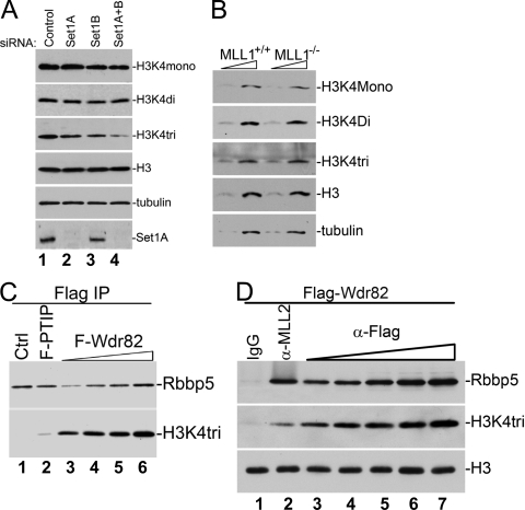
Since Wdr82 is a specific component of the Set1 complexes and its deficiency causes a reduction of global H3K4 trimethylation levels, the loss of Set1A and Set1B methyltransferases should phenocopy this effect. Therefore, we knocked down Set1A and Set1B by siRNAs, either individually or together, and investigated their effect on bulk H3K4 methylation levels. Consistent with the results for Wdr82 RNAi, the loss of Set1A or Set1B results in a reduction of H3K4 trimethylation (Fig. 5A). However, the RNAi of both Set1A and Set1B results in a similar decrease in bulk H3K4 trimethylation levels as Wdr82 (Fig. 5A, lane 4). Meanwhile, we investigated the consequence of MLL1 deficiency on the pattern of H3K4 methylation. We used the MLL1 knockout MEF cell (Fig. 5B). Surprisingly, the loss of MLL1 did not alter bulk H3K4 methylation. Therefore, our data suggest that the Set1 complexes contribute significantly to the global H3K4 trimethylation in mammalian cells. We also investigated the methylation on the HOXC8 and HOXA9 promoters in the MLL1 knockout cell, which was reported to be the target gene of MLL1, and found that H3K4 trimethylation decreased (data not shown), suggesting that MLL's role in H3K4 trimethylation is rather specific to certain regions of chromosomes and that the Set1 complexes may have broader H3K4 trimethylase function than in mammalian cells.
FIG. 5.
Comparative analyses of the HMTase activities of the Set1 and MLL complexes in vivo and in vitro. (A) The levels of Set1A and Set1B were knocked down in HeLa cells by siRNA either alone or together. H3K4 mono-, di-, and trimethylation was tested in the resulting cell extracts. Compared to the control, H3K4 trimethylation decreased in all of the knockdown samples, with most in double knockdown, while the dimethylation levels did not change. (B) The total histone H3K4 methylation status was tested by Western blot analysis in MLL1+/+ and MLL1−/− MEF cells. The total levels of mono-, di-, and trimethylation were unchanged in the MLL1 knockout cell line. (C) MLL3/4 trimethylase activity in comparison to the Set1 complexes. Flag immunoprecipitation was performed in Flag-PTIP and Flag-Wdr82 stable cell lines to purify the MLL3/4 and Set1 complexes, respectively. In vitro HMTase assays were performed with each purified complex. The level of Rbbp5 was used for normalization of the purified HMTase complexes. With the same amount of Rbbp5, the Set1 complexes demonstrated a much more robust H3K4 trimethylase activity than did the MLL3/4 complexes. (D) MLL2 trimethylase activity in comparison to Set1 complexes. After their purification, the MLL2 and Set1 complexes were tested for HMT activities. With the same amount of Rbbp5, the Set1 complexes demonstrate a much more robust H3K4 trimethylase activity than did the MLL2 complex.
The in vitro specific activity of H3K4 trimethylation of the Set1 complexes is higher than that of the MLL complexes.
To investigate whether Set1 or the MLLs have similar specific activities in H3K4 trimethylation, we purified the MLL3/4 complexes from a Flag-PTIP stable cell line and the Set1 complexes from a Flag-Wdr82 stable cell line by anti-Flag antibody and then performed an in vitro HMTase assay. RbBP5, Ash2L, and WDR5 form a structural entity independently of the catalytic subunit of either of the MLLs or the Set1 proteins (4, 29). Since the interaction of the RbBP5, Ash2L, and WDR5 trimeric complex with the MLLs and the Set1 proteins is required for proper catalytic activity (4, 29) and since these subunits are common among all of the human H3K4 HMTase, we have made the assumption that their levels within each complex are also similar. Therefore, we normalized the levels of the purified MLL3/4 and Set1 complexes by Rbbp5 levels (Fig. 5C). The Set1 complexes purified via Flag-Wdr82 seem to have much more robust activity toward trimethylation than the purified MLL3/4 complexes (Fig. 5C). Comparing lanes 2 and 3 of Fig. 5C, although lower amounts of the Wdr82 complex were used in the assay, as determined by RbBp5 levels (Fig. 5C, lane 3), the purified Set1 complex has a much more robust trimethylation activity than the MLL3/4 complex (Fig. 5C, lane 2). The Set1 complex in Fig. 5C, lane 5, has levels of RbBp5 similar to those for the Flag-PTIP purified MLL3/4 complexes in lane 2. However, the catalytic activity of Set1 in H3K4 trimethylation appears to be much higher than that of the MLL3/4 complex. We have also compared the activities of MLL2 and the Set1 complexes. We purified the Set1 complexes as described above and the MLL2 complex using an MLL2-specific antibody from the same cells and tested their activities toward H3K4 trimethylation (Fig. 5D). When the same amounts of Set1 and MLL2 complexes were used, as determined by the RbBp5 levels (Fig. 5D, lanes 2 and 5), the Set1 complex demonstrated a much more robust H3K4 trimethylation activity than that of the purified MLL2 complex.
Methylation of histones on H3K4 is a posttranslational modification that is exclusively associated with actively transcribed genes (10, 25, 26, 28). The first reported H3K4 methylase complex, COMPASS, was identified in the yeast S. cerevisiae and consists of Set1/KMT2 and seven other polypeptides (named Cps60-Cps15) (18). Set1/KMT2 alone is not enzymatically active but functions within COMPASS and is capable of mono-, di-, and trimethylating H3K4 (12, 18, 20, 22, 24, 26, 34). After the identification of Set1/COMPASS as an H3K4 methylase, it was demonstrated that its mammalian homologues, the MLL proteins, MLL1-4 and hSet1A and hSet1B, are found in COMPASS-like complexes capable of methylating the fourth lysine of histone H3 (7, 26, 31).
Although there are several H3K4 methylases in mammals, their HMTase activities do not appear to be redundant as the deletion of many of these enzymes, such as MLL1 or MLL2, results in embryonic lethality. All H3K4 methylases have conserved COMPASS-like subunits, and these subunits function similarly from yeasts to humans. Many of the studies performed to date in the mammalian cells in regard to H3K4 methylation take advantage of the reagents developed to these shared subunits, such as Ash2L, WDR5, and Rbbp5. We have identified here Wdr82 to be Set1 complex specific. The reduction in Wdr82 levels via RNAi results in a substantial decrease in the H3K4 trimethylation levels of bulk histone and on the chromatin of actively transcribed genes. We have also compared the in vitro catalytic properties of the human Set1 complex to that of the MLL complexes. The present findings indicate that human Set1 complexes have a much more robust H3K4 trimethylase activity in vitro compared to that of purified MLL complexes. Altogether, our data suggest that the Set1-containing complexes in mammalian cells play a more widespread role in H3K4 trimethylation. Therefore, future studies analyzing properties of the H3K4 methylation pattern and function in mammalian cells should also consider the role of Wdr82 and the Set1 complexes in this process.
Acknowledgments
We thank Edwin Smith for critical reading of the manuscript and helpful conversations throughout the study and Laura Shilatifard for editorial assistance.
This study in A.S.'s laboratory was supported by grant CA089455 from the National Institutes of Health to A.S.
Footnotes
▿
Published ahead of print on 6 October 2008.
REFERENCES
- 1.Bernstein, B. E., A. Meissner, and E. S. Lander. 2007. The mammalian epigenome. Cell 128669-681. [DOI] [PubMed] [Google Scholar]
- 2.Cheng, H., X. He, and C. Moore. 2004. The essential WD repeat protein Swd2 has dual functions in RNA polymerase II transcription termination and lysine 4 methylation of histone H3. Mol. Cell. Biol. 242932-2943. [DOI] [PMC free article] [PubMed] [Google Scholar]
- 3.Cho, Y. W., T. Hong, S. Hong, H. Guo, H. Yu, D. Kim, T. Guszczynski, G. R. Dressler, T. D. Copeland, M. Kalkum, and K. Ge. 2007. PTIP associates with MLL3- and MLL4-containing histone H3 lysine 4 methyltransferase complex. J. Biol. Chem. 28220395-20406. [DOI] [PMC free article] [PubMed] [Google Scholar]
- 4.Dou, Y., T. A. Milne, A. J. Ruthenburg, S. Lee, J. W. Lee, G. L. Verdine, C. D. Allis, and R. G. Roeder. 2006. Regulation of MLL1 H3K4 methyltransferase activity by its core components. Nat. Struct. Mol. Biol. 13713-719. [DOI] [PubMed] [Google Scholar]
- 5.Dover, J., J. Schneider, M. A. Tawiah-Boateng, A. Wood, K. Dean, M. Johnston, and A. Shilatifard. 2002. Methylation of histone H3 by COMPASS requires ubiquitination of histone H2B by Rad6. J. Biol. Chem. 27728368-28371. [DOI] [PubMed] [Google Scholar]
- 6.Glaser, S., J. Schaft, S. Lubitz, K. Vintersten, F. van der Hoeven, K. R. Tufteland, R. Aasland, K. Anastassiadis, S. L. Ang, and A. F. Stewart. 2006. Multiple epigenetic maintenance factors implicated by the loss of MLL2 in mouse development. Development 1331423-1432. [DOI] [PubMed] [Google Scholar]
- 7.Hughes, C. M., O. Rozenblatt-Rosen, T. A. Milne, T. D. Copeland, S. S. Levine, J. C. Lee, D. N. Hayes, K. S. Shanmugam, A. Bhattacharjee, C. A. Biondi, G. F. Kay, N. K. Hayward, J. L. Hess, and M. Meyerson. 2004. Menin associates with a trithorax family histone methyltransferase complex and with the hoxc8 locus. Mol. Cell 13587-597. [DOI] [PubMed] [Google Scholar]
- 8.Hwang, W. W., S. Venkatasubrahmanyam, A. G. Ianculescu, A. Tong, C. Boone, and H. D. Madhani. 2003. A conserved RING finger protein required for histone H2B monoubiquitination and cell size control. Mol. Cell 11261-266. [DOI] [PubMed] [Google Scholar]
- 9.Kim, J., S. B. Hake, and R. G. Roeder. 2005. The human homolog of yeast BRE1 functions as a transcriptional coactivator through direct activator interactions. Mol. Cell 20759-770. [DOI] [PubMed] [Google Scholar]
- 10.Kouzarides, T. 2007. Chromatin modifications and their function. Cell 128693-705. [DOI] [PubMed] [Google Scholar]
- 11.Krivtsov, A. V., and S. A. Armstrong. 2007. MLL translocations, histone modifications and leukaemia stem-cell development. Nat. Rev. Cancer 7823-833. [DOI] [PubMed] [Google Scholar]
- 12.Krogan, N. J., J. Dover, S. Khorrami, J. F. Greenblatt, J. Schneider, M. Johnston, and A. Shilatifard. 2002. COMPASS, a histone H3 (lysine 4) methyltransferase required for telomeric silencing of gene expression. J. Biol. Chem. 27710753-10755. [DOI] [PubMed] [Google Scholar]
- 13.Lee, J. H., and D. G. Skalnik. 2005. CpG-binding protein (CXXC finger protein 1) is a component of the mammalian Set1 histone H3-Lys4 methyltransferase complex, the analogue of the yeast Set1/COMPASS complex. J. Biol. Chem. 28041725-41731. [DOI] [PubMed] [Google Scholar]
- 14.Lee, J. H., and D. G. Skalnik. 2008. Wdr82 is a C-terminal domain-binding protein that recruits the Setd1A histone H3-Lys4 methyltransferase complex to transcription start sites of transcribed human genes. Mol. Cell. Biol. 28609-618. [DOI] [PMC free article] [PubMed] [Google Scholar]
- 15.Lee, J. H., C. M. Tate, J. S. You, and D. G. Skalnik. 2007. Identification and characterization of the human Set1B histone H3-Lys4 methyltransferase complex. J. Biol. Chem. 28213419-13428. [DOI] [PubMed] [Google Scholar]
- 16.Lee, J. S., A. Shukla, J. Schneider, S. K. Swanson, M. P. Washburn, L. Florens, S. R. Bhaumik, and A. Shilatifard. 2007. Histone crosstalk between H2B monoubiquitination and H3 methylation mediated by COMPASS. Cell 1311084-1096. [DOI] [PubMed] [Google Scholar]
- 17.Lee, M. G., R. Villa, P. Trojer, J. Norman, K. P. Yan, D. Reinberg, L. Di Croce, and R. Shiekhattar. 2007. Demethylation of H3K27 regulates polycomb recruitment and H2A ubiquitination. Science 318447-450. [DOI] [PubMed] [Google Scholar]
- 18.Miller, T., N. J. Krogan, J. Dover, H. Erdjument-Bromage, P. Tempst, M. Johnston, J. F. Greenblatt, and A. Shilatifard. 2001. COMPASS: a complex of proteins associated with a trithorax-related SET domain protein. Proc. Natl. Acad. Sci. USA 9812902-12907. [DOI] [PMC free article] [PubMed] [Google Scholar]
- 19.Morillon, A., N. Karabetsou, A. Nair, and J. Mellor. 2005. Dynamic lysine methylation on histone H3 defines the regulatory phase of gene transcription. Mol. Cell 18723-734. [DOI] [PubMed] [Google Scholar]
- 20.Nagy, P. L., J. Griesenbeck, R. D. Kornberg, and M. L. Cleary. 2002. A trithorax-group complex purified from Saccharomyces cerevisiae is required for methylation of histone H3. Proc. Natl. Acad. Sci. USA 9990-94. [DOI] [PMC free article] [PubMed] [Google Scholar]
- 21.Pavri, R., B. Zhu, G. Li, P. Trojer, S. Mandal, A. Shilatifard, and D. Reinberg. 2006. Histone H2B monoubiquitination functions cooperatively with FACT to regulate elongation by RNA polymerase II. Cell 125703-717. [DOI] [PubMed] [Google Scholar]
- 22.Roguev, A., D. Schaft, A. Shevchenko, W. W. Pijnappel, M. Wilm, R. Aasland, and A. F. Stewart. 2001. The Saccharomyces cerevisiae Set1 complex includes an Ash2 homologue and methylates histone 3 lysine 4. EMBO J. 207137-7148. [DOI] [PMC free article] [PubMed] [Google Scholar]
- 23.Rowley, J. D. 1998. The critical role of chromosome translocations in human leukemias. Annu. Rev. Genet. 32495-519. [DOI] [PubMed] [Google Scholar]
- 24.Schneider, J., A. Wood, J. S. Lee, R. Schuster, J. Dueker, C. Maguire, S. K. Swanson, L. Florens, M. P. Washburn, and A. Shilatifard. 2005. Molecular regulation of histone H3 trimethylation by COMPASS and the regulation of gene expression. Mol. Cell 19849-856. [DOI] [PubMed] [Google Scholar]
- 25.Seward, D. J., G. Cubberley, S. Kim, M. Schonewald, L. Zhang, B. Tripet, and D. L. Bentley. 2007. Demethylation of trimethylated histone H3 Lys4 in vivo by JARID1 JmjC proteins. Nat. Struct. Mol. Biol. 14240-242. [DOI] [PubMed] [Google Scholar]
- 26.Shilatifard, A. 2006. Chromatin modifications by methylation and ubiquitination: implications in the regulation of gene expression. Annu. Rev. Biochem. 75243-269. [DOI] [PubMed] [Google Scholar]
- 27.Shilatifard, A. 2008. Molecular implementation and physiological roles for histone H3 lysine 4 (H3K4) methylation. Curr. Opin. Cell Biol. 20341-348. [DOI] [PMC free article] [PubMed] [Google Scholar]
- 28.Sims, R. J., III, and D. Reinberg. 2006. Histone H3 Lys 4 methylation: caught in a bind? Genes Dev. 202779-2786. [DOI] [PubMed] [Google Scholar]
- 29.Steward, M. M., J. S. Lee, A. O'Donovan, M. Wyatt, B. E. Bernstein, and A. Shilatifard. 2006. Molecular regulation of H3K4 trimethylation by ASH2L, a shared subunit of MLL complexes. Nat. Struct. Mol. Biol. 13852-854. [DOI] [PubMed] [Google Scholar]
- 30.Sun, Z. W., and C. D. Allis. 2002. Ubiquitination of histone H2B regulates H3 methylation and gene silencing in yeast. Nature 418104-108. [DOI] [PubMed] [Google Scholar]
- 31.Tenney, K., and A. Shilatifard. 2005. A COMPASS in the voyage of defining the role of trithorax/MLL-containing complexes: linking leukemogenesis to covalent modifications of chromatin. J. Cell Biochem. 95429-436. [DOI] [PubMed] [Google Scholar]
- 32.Washburn, M. P., D. Wolters, and J. R. Yates III. 2001. Large-scale analysis of the yeast proteome by multidimensional protein identification technology. Nat. Biotechnol. 19242-247. [DOI] [PubMed] [Google Scholar]
- 33.Wood, A., N. J. Krogan, J. Dover, J. Schneider, J. Heidt, M. A. Boateng, K. Dean, A. Golshani, Y. Zhang, J. F. Greenblatt, M. Johnston, and A. Shilatifard. 2003. Bre1, an E3 ubiquitin ligase required for recruitment and substrate selection of Rad6 at a promoter. Mol. Cell 11267-274. [DOI] [PubMed] [Google Scholar]
- 34.Wood, A., A. Shukla, J. Schneider, J. S. Lee, J. D. Stanton, T. Dzuiba, S. K. Swanson, L. Florens, M. P. Washburn, J. Wyrick, S. R. Bhaumik, and A. Shilatifard. 2007. Ctk complex-mediated regulation of histone methylation by COMPASS. Mol. Cell. Biol. 27709-720. [DOI] [PMC free article] [PubMed] [Google Scholar]
- 35.Yokoyama, A., Z. Wang, J. Wysocka, M. Sanyal, D. J. Aufiero, I. Kitabayashi, W. Herr, and M. L. Cleary. 2004. Leukemia proto-oncoprotein MLL forms a SET1-like histone methyltransferase complex with menin to regulate Hox gene expression. Mol. Cell. Biol. 245639-5649. [DOI] [PMC free article] [PubMed] [Google Scholar]
- 36.Yu, B. D., J. L. Hess, S. E. Horning, G. A. Brown, and S. J. Korsmeyer. 1995. Altered Hox expression and segmental identity in MLL-mutant mice. Nature 378505-508. [DOI] [PubMed] [Google Scholar]
- 37.Zhu, B., Y. Zheng, A. D. Pham, S. S. Mandal, H. Erdjument-Bromage, P. Tempst, and D. Reinberg. 2005. Monoubiquitination of human histone H2B: the factors involved and their roles in HOX gene regulation. Mol. Cell 20601-611. [DOI] [PubMed] [Google Scholar]
