Thymic development beyond β-selection requires phosphatidylinositol 3-kinase activation by CXCR4 (original) (raw)
Abstract
T cell development requires phosphatidylinositol 3-kinase (PI3K) signaling with contributions from both the class IA, p110δ, and class IB, p110γ catalytic subunits. However, the receptors on immature T cells by which each of these PI3Ks are activated have not been identified, nor has the mechanism behind their functional redundancy in the thymus. Here, we show that PI3K signaling from the preTCR requires p110δ, but not p110γ. Mice deficient for the class IB regulatory subunit p101 demonstrated the requirement for p101 in T cell development, implicating G protein–coupled receptor signaling in β-selection. We found evidence of a role for CXCR4 using small molecule antagonists in an in vitro model of β-selection and demonstrated a requirement for CXCR4 during thymic development in CXCR4-deficient embryos. Finally, we demonstrate that CXCL12, the ligand for CXCR4, allows for Notch-dependent differentiation of DN3 thymocytes in the absence of supporting stromal cells. These findings establish a role for CXCR4-mediated PI3K signaling that, together with signals from Notch and the preTCR, contributes to continued T cell development beyond β-selection.
T lymphocytes develop in the thymus from a multistep differentiation program characterized by the sequential VDJ rearrangement of the tcrb and tcra genes in combination with stringent quality control checkpoints. The most immature T lymphocytes are CD4 and CD8 double-negative (DN), and this population can be further subdivided based upon their expression of CD44 and CD25 (Godfrey et al., 1993). At the CD25+CD44lo DN3 stage, thymocytes undergo the β-selection checkpoint. DN3 cells are tested for the successful expression of a TCRβ polypeptide in the context of the invariant pTα subunit and CD3, which together form the preTCR (Yamasaki and Saito, 2007). DN3 cells that have successfully rearranged TCRβ undergo proliferative expansion, further differentiation, and allelic exclusion of the TCRβ locus. This occurs as DN3 cells lose expression of CD25 to become DN4. Subsequently, thymocytes express CD8 and CD4, which together define the double-positive (DP) population, where tcra rearrangement occurs and the mature TCR is expressed. The completion of β-selection is contingent upon Notch signaling, which is necessary for survival, metabolic fitness, and proliferation after TCRβ rearrangement (Ciofani and Zuñiga-Pflucker, 2005; Maillard et al., 2006). Together, Notch and preTCR signaling are thought to constitute the minimal requirements for continued differentiation beyond β-selection; however, our understanding of how the signaling events downstream of these receptors are integrated is limited.
The class I phosphatidylinositol 3-kinase (PI3K) family of enzymes mediates the phosphorylation of phosphatidylinositol-4,5-trisphosphate (PIP2) to generate phosphatidylinositol-3,4,5-trisphosphate (PIP3). This lipid binds the pleckstrin homology domains of effector molecules, which in turn regulate cellular processes including survival, proliferation, metabolism, differentiation, and movement (Fruman and Bismuth, 2009). The class I PI3Ks are comprised of two subgroups designated class IA and IB. The class IA subgroup, whose members are activated by tyrosine kinase–associated receptors, consists of three ∼110-kD catalytic subunits termed α, β, and δ that pair with regulatory subunits p50α, 55α, 85α, p85β, and p55γ. The class IB subgroup, activated by G protein–coupled receptors, consists of p110γ that interacts with p101 or p84 regulatory subunits.
Functional redundancy between PI3K isoforms for T cell development is revealed in mice lacking both p110γ and p110δ in the germline. These mice have small thymi characterized by a marked reduction in DP thymocyte numbers (Webb et al., 2005; Swat et al., 2006; Ji et al., 2007), reflecting defective survival of DP thymocytes (Webb et al., 2005; Swat et al., 2006). The conditional deletion in DN T cells of phosphatase and tensin homologue, which opposes PI3K activity by converting PIP3 to PIP2, rescues T cell development in mice with defective IL-7 or preTCR signaling (Hagenbeek et al., 2004; Shiroki et al., 2007). Despite this, it remains unclear how P110γ and P110δ contribute to the earlier stages of T cell development and to which activating receptors these PI3K isoforms are coupled (Webb et al., 2005; Swat et al., 2006). Genetic and biochemical studies have revealed important roles for the PI3K-dependent protein kinases PDK1 and Akt in T cell development (Hinton et al., 2004; Fayard et al., 2007; Juntilla et al., 2007; Mao et al., 2007). DN4 cells of these mutant mice were small and failed to up-regulate nutrient receptors despite having normal expression of intracellular TCRβ, whereas expression of constitutively active Akt resulted in the rescue of the DN3 developmental block in Rag2-deficient mice (Mao et al., 2007).
Although the preTCR utilizes protein tyrosine kinases for signaling, a direct link between the preTCR and PI3K activation has yet to be shown. Similarly, the PI3K effector molecules PDK1 and AKT have been reported to be required for Notch-dependent metabolic changes in DN3 cells (Ciofani and Zuñiga-Pflucker, 2005; Kelly et al., 2007). However, the mechanistic basis for the interaction between Notch and PI3K remains to be clarified. Further, the implication of class IB PI3K in early T cell development raises the possibility that a GPCR may also be involved in this process, but such a receptor has not been identified.
Among GPCRs, chemokine receptors are important in directing thymocytes through the cortex and medulla during their development, thereby guiding thymocytes to the appropriate microenvironment for their specific developmental stage (Petrie and Zuñiga-Pflucker, 2007). However, it is not known if chemokines can directly contribute to the induction of thymocyte proliferation and differentiation. CXCL12, and its receptor CXCR4, was first identified as a growth factor for pre B cells (Nagasawa et al., 1994) and using mutant mice it was subsequently shown that CXCR4 is critical for B cell and myeloid hematopoiesis (Ma et al., 1998; Nagasawa et al., 1996; Tachibana et al., 1998; Zou et al., 1998). In the thymus, CXCR4 is highly expressed on thymocytes from the DN2 to the DP stage (Ara et al., 2003; Plotkin et al., 2003), but its role during T cell development is controversial. Both CXCR4 and CXCL12 are required for embryo viability, and some studies examining T cell lymphopoiesis at fetal stages of development have concluded that T cell development is normal in the absence of CXCR4 (Ma et al., 1998; Zou et al., 1998) Conversely, competitive chimera studies using donor fetal liver cells showed a reduced reconstitution of CXCR4-deficient thymocytes compared with WT (Ara et al., 2003). In addition, cre-mediated CXCR4 deletion in DN1 thymocytes resulted in complete developmental arrest of T cells (Plotkin et al., 2003) and human CD34+ cells seeded into mouse fetal thymic organ cultures treated with neutralizing antibodies to CXCL12 or CXCR4 showed impaired differentiation and reduced numbers of recovered T cells (Hernández-López et al., 2002).
We have revealed the role of P110γ and P110δ in T cell development using a combination of genetic and pharmacological approaches. Stimulation of different PI3K mutant DN4 cells through the preTCR showed that p110δ is essential for the initiation of PI3K signaling from this receptor, but we found no role for p110γ. Experiments with chimeric mice and in vitro cell differentiation cultures demonstrated the combined deletion of p110δ and the class IB regulatory subunit p101 recapitulate the phenotype of p110γ−/−δ−/− mice, suggesting a requirement for G-protein coupled receptor (GPCR)–dependent PI3K activation during T cell development. Further evidence for this was shown using small molecule inhibitors of CXCR4 to impede the proliferation and differentiation of DN3 thymocytes in vitro. Moreover, we found that stimulation of DN3 thymocytes with the chemokine CXCL12 elicits a PI3K-mediated signaling response which is predominantly p110γ-dependent, but also involves p110δ. Analysis of CXCR4-deficient embryos confirmed that signaling through CXCR4 is critical for normal thymic T cell development. Using these insights, we tested the requirement for CXCR4 signaling by culturing DN3 cells with Notch ligand in the presence or absence of CXCL12. Only in the presence of CXCL12 could DN3 cells differentiate to become DP. These insights permitted the differentiation and expansion of DN3 in the absence of supporting stromal cells and that the additional inclusion of a GSK3 inhibitor allowed PI3K-mediated accessory cell–free proliferation and differentiation to a similar extent as that seen upon co-culture of DN3 thymocytes with stromal cells.
RESULTS
p110δ, and not p110γ, is required for preTCR-mediated Akt phosphorylation
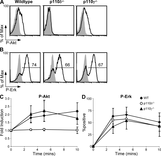
Although the PI3K pathway has been implicated in preTCR signaling, a direct link between the preTCR and individual PI3K catalytic subunits has yet to be shown. This is caused in part by the technical difficulties of studying signaling events initiated by the preTCR, as it is rapidly degraded and difficult to detect at the cell surface (Panigada et al., 2002). To determine whether p110γ and/or p110δ are required for preTCR-mediated Akt phosphorylation, we used the OP9-DL1 culture system. BM-derived hematopoietic stem cells (HSCs) from WT, p110δ−/−, or p110γ−/− were cultured in the continued presence of IL-7, which enabled differentiation to the DN3/DN4 stage of T cell development (Huang et al., 2005 and unpublished data). Under these culture conditions, we found that DN4 cells express detectable CD3ε at the cell surface (Fig. S1). DN4 cells from WT, p110δ−/−, and p110γ−/− cultures were similarly responsive to stimulation with anti-CD3ε antibody for the induction of Erk1/2 phosphorylation as measured by flow cytometry (Fig. 1). The staining profile for phospho-Erk appeared bi-modal and is thus similar to the Erk1/2 response in more mature TCR-stimulated thymocytes (Prasad et al., 2009). In contrast, the staining profile of Akt phosphorylation at residue serine 473 showed an overall shift in median fluorescence indicative of a graded analogue response. The increase in phospho-Akt in p110γ−/− DN4 cells upon anti-CD3ε stimulation was similar to the WT, but p110δ−/− cells failed to generate detectable p-Akt under identical conditions. These results suggested that the p110δ subunit is critical for PI3K signaling from the preTCR, whereas p110γ is dispensable. Moreover, preTCR activation of the Erk signaling pathway is independent of either p110γ or p110δ.
Figure 1.
PI3K p110δ is required for preTCR signaling in DN4 thymocytes. (A) Phosphorylated Akt (S473) and (B) phosphorylated Erk1/2 (T202/Y204) in WT and PI3K mutant DN4 T cells in response to preTCR signaling. Histograms show staining in either resting cells (gray area) or cells stimulated with 10 µg/ml of anti-CD3ε antibody 2C11 for 5 min (black line). DN4 cells were derived from long-term BM HSC cultures. Intracellular p-Akt and p-Erk1/2 were detected using flow cytometry in combination with cell surface markers to distinguish the DN4 population (defined as Thy1.2+ and [CD4/CD8/CD19/CD25]neg). (C and D) Time course of p-Akt (S473) and pErk induction in WT, p110δ−/−, and p110γ−/− cultured DN4 cells in response to stimulation with 2C11. Graphs show the mean and SD (n = 3–5) and are comprised of five independent experiments.
Impaired thymocyte development in mice deficient for p110δ in combination with p101
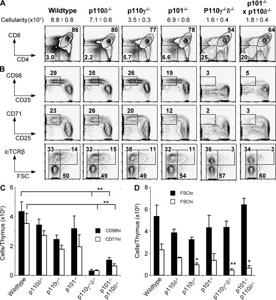
p110γ is recruited to the Gβγ subunits of GPCRs via its association with p101 or p84 regulatory subunits (Stephens et al., 1997; Suire et al., 2005). We found that p101 mRNA was expressed in DN3 and DN4 thymocytes, whereas p84 mRNA was undetectable (Fig. S2 A). Thymocytes from mice lacking p101 (Suire et al., 2006) had a CD4/CD8 FACS staining profile similar to the p110γ−/− single mutants (Fig. 2 A), and they had a reduced number of DP thymocytes compared with WT (P = 0.01; using the unpaired Student’s t test; not depicted). However, mice deficient for both p101 and p110δ showed thymic atrophy to a similar degree to the p110γ−/−δ−/− mice (Fig. 2 A). These mice had a marked increase in the proportion of thymocytes that were CD4–CD8– DN and a decrease in the proportion of CD4+CD8+ DP cells. DN4 cells (CD25–/CD44–) from p110γ−/−δ−/− and p101−/−/p110δ−/− mice failed to show increased expression of the CD98 heavy chain of amino acid transporters and CD71 (transferrin receptor), nor did they show increased forward angle light scatter indicative of cell growth (Fig. 2, B–D). This defect was not secondary to a failure to express a productively rearranged tcrb gene as the frequency of small DN3 cells expressing the β polypeptide identified by intracellular flow cytometry was not different from the WT (Fig. 2 D). Additionally, the amounts of p110γ protein were unchanged in the absence of p101 (Fig. S2 B). Thus, the phenotype observed in the p101−/−/p110δ−/− mice is not a consequence of reduced expression of the p110γ. As p101 is required to regulate p110γ function we conclude that, in its absence, p110γ is uncoupled from its activating receptor.
Figure 2.
Defective maturation of DN3 thymocytes in mice that lack p110δ in combination with either p110γ or p101. (A) Flow cytometric analysis of CD4 and CD8 expression on T lymphocytes in the thymus from WT and PI3K mutant mice. The percentages of CD4+CD8+ DP and CD4−CD8− DN cells are shown. The mean thymus cellularity + SEM for each genotype is also shown (n = 9–33). (B) Flow cytometric analysis of the DN3/DN4 thymic subsets in PI3K mutant mice (identified as Thy1+ and [CD4/CD8/CD44/B220/CD11b/ NK1.1/Gr1/Ter119/γδTCR]neg). The percentages of CD25loCD98hi, CD25loCD71hi, icTCRβ−, icTCRβ+FSClo, and icTCRβ+FSChi are shown. Enumeration of CD25loCD98hi and CD25loCD71hi DN3/DN4 cells (C) and numbers of icTCR+ FSClo and FSChi cells (D) for each genotype are shown. In C, graphs represent the mean and SD (n = 3–5). In D, graphs show the mean and SEM (n = 5–27).
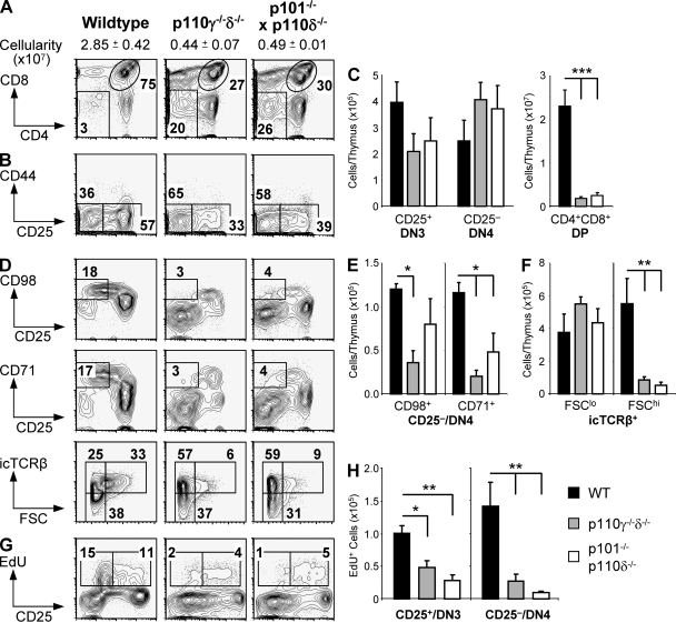
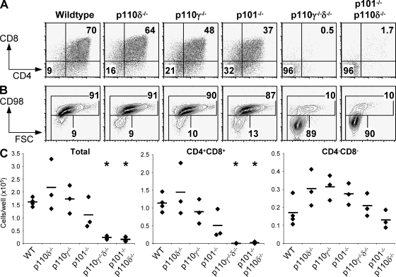
Previous studies have shown that when WT BM is transplanted into p110γδ−/− recipient mice, thymic development is impaired to the same extent as that seen in p110γ−/− mice, suggesting a non–cell autonomous role for p110γ in T cell development (Swat et al., 2006). Therefore, to determine whether there was a thymocyte-intrinsic requirement for p110γ/p110δ or p101/p110δ, we created chimeras by reconstituting recipient mice doubly deficient in Rag2 and the common cytokine receptor γ chain (_Rag2_−/− _Il2rg_−/−) with BM from WT, p110γ−/−δ−/−, or p101−/−/p110δ−/− mutant mice. Chimeric mice reconstituted with p110γ−/−δ−/− or p101−/−/p110δ−/− BM had a reduction in the number of DP thymocytes compared with control chimeras reconstituted with WT BM (Fig. 3 A). Although the numbers of DN3/DN4 cells were equivalent to WT chimeras, these cells failed to become CD98hi, CD71hi or to increase in size (Fig. 3, B–F). In addition, pulse-labeling with the thymidine analogue 5-ethynyl-2′-deoxyuridine (EdU) revealed that both p110γ−/−δ−/− and p101−/−/p110δ−/− DN3/4 thymocytes incorporated EdU poorly compared with WT (Fig. 3 G-H). Defective expansion and differentiation was recapitulated in vitro using sorted DN3 cells from WT and PI3K single- or double mutant mice cultured on OP9-DL1 stromal cells (Fig. 4). After 3 d of culture, the majority of WT and PI3K single-mutant DN3 cells had proliferated and differentiated to become large CD4+CD8+CD98hi cells. However, both p110γ−/−δ−/− and p101−/−/p110δ−/− DN3 cells remained undifferentiated, did not proliferate, and failed to increase in size. Thus, thymocytes have a cell autonomous requirement for p110γ/p110δ or p101/p110δ to progress beyond β-selection.
Figure 3.
T lymphocyte development in BM chimeras. (A) CD4 and CD8 expression on thymocytes with the percentages of DP and DN cells shown. The average thymus cellularity + SEM for each group is shown (n = 15–20). (B) Expression of CD25 and CD44 on DN thymocytes with the percentages of CD44−CD25+ and CD44−CD25− cells shown. (C) Enumeration of CD44−CD25+ and CD44−CD25− and CD4+CD8+ DP thymocytes in chimeras reconstituted with WT, p110γ−/−δ−/−, and p101−/−/p110δ−/− marrow. Graphs represent the mean and SEM. n = 15–19. (D) Flow cytometric analysis of the DN3/DN4 thymic subset. The percentages of CD25loCD98hi, CD25loCD71hi, icTCRβ−, icTCRβ+FSClo, and icTCRβ+FSChi are shown. Plots and percentages are representative of at least five individual mice. Enumeration of CD25loCD98hi and CD25loCD71hi cells (E) and icTCR+FSClo and FSChi cells (F) for each genotype are shown. (G and H) Analysis of DN3/DN4 cell proliferation by EdU incorporation. Graphs show the mean and SD from analysis of five independent mice.
Figure 4.
DN3 cells from p110γ−/−δ−/− and p101−/−/p110δ−/− mice fail to differentiate in vitro. 4 × 10⁴ DN3 cells from WT and PI3K mutant mice were cultured on OP9-DL1 stroma for 3 d. (A) The percentage of CD4+CD8+ cells. (B) The percentage of CD98+ cells. (C) The number of total CD4+CD8+ and CD4−CD8− cells recovered. Graphs show each biological sample (black diamond) as well as the mean (black bars) for each group. This experiment is representative of three independent replicates.
Evidence for the involvement of CXCR4 at the β-selection checkpoint
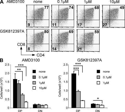
The in vivo and in vitro requirement for p101 at the β-selection checkpoint prompted us to seek evidence of a role for a GPCR in this process. We observed that CXCL12, the ligand for CXCR4, was secreted by the OP9-DL1 stromal line (Fig. S3 A). We examined whether CXCL12 was required for DN3 proliferation and differentiation by culturing sorted DN3 cells on OP9-DL1 in the presence of the CXCR4 antagonist AMD3100. This small molecule binds to CXCR4 with high specificity and inhibits chemotaxis of human PBMCs to CXCL12 with an IC50 of 0.16 µM (Hatse et al., 2002). Culturing DN3 cells in the presence of AMD3100 inhibited their ability to become CD4+CD8+ (Fig. 5). The numbers of DP cells recovered after 3 d of culture were decreased in a dose-dependent manner, indicating that in the absence of CXCR4 signaling DN3 cells had reduced proliferation and/or survival. This effect on DN3 cells was also seen with a chemically distinct small molecule inhibitor of CXCR4 termed GSK812397A, which inhibits the CXCL12-mediated chemotaxis of the U937 cell line with an IC50 of <1 nM (GlaxoSmithKline; personal communication). These results show that signaling through CXCR4 contributes to the development of DN3 cells to the DP developmental stage.
Figure 5.
Inhibition of CXCR4 signaling blocks DN3 cell differentiation. 4 × 10⁴ DN3 cells from WT mice were cultured on OP9-DL1 stromal cells for 3 d in the presence of increasing concentrations of AMD3100 or GSK812397A. Cells were harvested, analyzed for the expression of CD4 and CD8 (A), and absolute numbers of CD4+CD8+ DP and CD4−CD8− DN cells recovered (B). Graphs show the mean and SD. n = 3. This experiment is representative of three independent replicates.
CXCR4 signals through both PI3K p110γ and p110δ in DN3 thymocytes
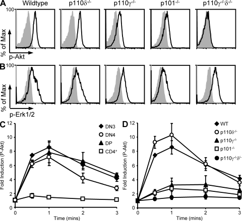
We sought evidence to determine whether CXCR4 activation of PI3K required p110γ and/or p110δ. Using intracellular flow cytometry, we measured p-Akt (S473) and p-Erk1/2 (T202/Y204) levels in freshly isolated thymocytes after in vitro stimulation with CXCL12. WT DN3, DN4, and DP, but not CD4+ SP, thymocytes showed increased p-Akt and p-Erk in response to CXCL12 stimulation (Fig. 6, A and B). DN3 cells lacking p110δ responded similarly to WT but the p-Akt response in CXCL12-stimulated p110γ−/− and p101−/− DN3 cells was reduced, although not eliminated. These results indicate that the PI3K signaling pathway is activated in DN3 cells in response to CXCL12 and that p110γ and p101 are required for this response. We also measured CXCL12-mediated Akt in p110γ−/−δ−/− DN3 cells and were unable to detect any increase in p-Akt (Fig. 6, A and D). Thus, in the absence of p110γ, p110δ also contributes to the CXCR4 activation of PI3K. Erk phosphorylation was found to be equivalent to WT in all PI3K mutant genotypes showing that p110δ, p110γ, and p101 were not required for CXCL12-mediated Erk phosphorylation and that some aspects of CXCR4 signaling are independent of these PI3Ks.
Figure 6.
PI3K p110γ and p110δ are required for CXCR4 signaling in DN3 thymocytes. (A) Phosphorylated Akt (S473) or (B) phosphorylated Erk1/2 (T202/Y204) in WT and PI3K mutant DN3 thymocytes after stimulation with CXCL12. Histograms show staining of resting cells or cells stimulated with 10 nM of CXCL12 for either 1 min (p-Akt) or 2 min (p-Erk1/2). Single-cell suspensions of whole thymus were used for stimulation. Intracellular p-Akt (S473) and p-Erk1/2 was detected using flow cytometry in combination with cell surface markers to distinguish the DN3 population. (C) Time course of p-Akt (S473) induction in WT DN3, DN4, DP, and CD4+SP thymocytes. (D) Time course of p-Akt (S473) induction in WT, p110δ−/−, p110γ−/−, p101−/−, and p110γ−/−δ−/− DN3 thymocytes. Data are presented as fold induction, which is calculated as the median fluorescence of phosphorylated protein in stimulated/unstimulated cells. Each data point shows the mean and SD of three to four biological replicates and is comprised from two independent experiments.
CXCR4 is required for PI3K activation in DN3 thymocytes
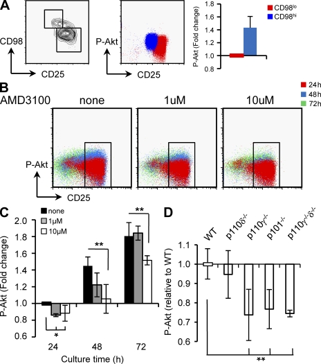
Our findings have identified the preTCR and CXCR4 as receptors on DN3 cells through which p110δ and p110γ are activated. However, it is unclear whether these receptors operate independently or cooperatively to induce activation of PI3K required for development beyond β-selection. To address this, we first compared PI3K activity before and after β-selection by measuring p-Akt (S473) in freshly isolated DN3 cells which were either CD98lo or CD98hi (Fig. 7 A) We have found that the expression of CD98 in DN3 cells correlates with expression of CD27 (data not shown), thus identifying DN3 cells with expression of TCRβ (Taghon et al., 2006). Our results show that CD98hi DN3 cells have an ∼1.4-fold increase in detectable p-Akt compared with CD98lo cells. This is consistent with increased PI3K activity upon β-selection.
Figure 7.
PI3K activity increases during β-selection. (A) CD98lo (red) and CD98hi (blue) DN3 thymocytes were sort purified from WT mice before fixation, permeabilization, and intracellular staining for p-Akt (S473). The fold change in p-Akt was calculated as the median fluorescence of p-Akt in CD98hi compared with CD98lo cells. (B and C) 4 × 10⁴ DN3 cells from WT mice were cultured on OP9-DL1 stromal cells for 3 d in the presence of increasing concentrations of AMD3100. At 24 (red), 48 (blue), and 72 h (green) cells were harvested and analyzed for the expression of p-Akt. The fold change in p-Akt levels was measured relative to cells harvested at 24 h that had been cultured in the absence of AMD3100. Results presented in A–C are from a single experiment of three biological replicates (with each biological sample comprising a pool of sorted cells from five mice). (D) p-Akt (S473) expression in DN3 cells isolated ex vivo from PI3K mutant mice. Data are presented as p-Akt expression relative to WT and 4–11 mice were analyzed for each genotype. All graphs show the mean and SD.
Although we have demonstrated that blocking the CXCR4 signaling pathway inhibited DN3 cell differentiation in vitro, we wanted to determine whether this also affected PI3K activity. To test this, CD98lo DN3 cells were cultured on the OP9-DL1 stromal line in the presence of increasing concentrations of AMD3100. Cells were harvested at 24-h intervals, and their p-Akt levels were measured. We observed an increase in the p-Akt levels of DN3 cells with time, which was diminished in the presence of AMD3100 (Fig. 7, B and C). Thus, signaling through CXCR4 contributes to PI3K activity. We also measured p-Akt levels in DN3 cells isolated ex vivo from PI3K mutant mice (Fig. 7 D). Despite the requirement of p110δ for preTCR-mediated Akt phosphorylation, p-Akt levels in the DN3 cells of p110δ−/− mice were similar to WT. In contrast, DN3 cells from p110γ−/−, p101−/−, and p110γ−/−δ−/− mice had 70–80% of WT p-Akt levels, demonstrating a role for the class IB subunits in the generation and/or maintenance of PI3K activity in DN3 thymocytes.
Impaired thymic development in CXCR4-deficient embryos
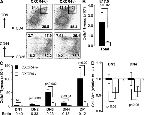
To examine the role of CXCR4 in thymocyte development, we analyzed thymi from CXCR4-deficient mice. CXCR4 is required for embryo viability (Ma et al., 1998; Tachibana et al., 1998; Zou et al., 1998), therefore we compared T cell development in E17.5 CXCR4+/− and CXCR4−/− embryos (Fig. 8). We found a decreased proportion of DP thymocytes and increased proportion of DN thymocytes in CXCR4−/− mice (Fig. 8 A). We also observed significant thymic atrophy in CXCR4−/− mice (Fig. 8 B), which corresponded to reduced numbers of all thymic subsets from DN2 to DP (Fig. 8 C). This phenotype may, in part, be attributed to the reduced number of T cell precursors colonizing the thymus. In addition, we noted a progressive decrease in the ratio of CXCR4−/− to CXCR4+/− cells at each successive developmental stage, indicating a continual requirement for CXCR4 as thymocytes progress through β-selection and become DP T cells. Finally, we compared the cell size of DN3 and DN4 thymocytes from CXCR4+/− and CXCR4−/− mice, as we had observed that DN4 cells from PI3K mutant mice were smaller compared with WT (Fig. 1 D). Unlike the adult thymus, we found that there was no increase in size between DN3 and DN4 thymocytes of the E17.5 embryo. However, both DN3 and DN4 cells were significantly smaller in CXCR4−/− thymi (Fig. 8 D). Collectively, these results suggest CXCR4 is required during T cell development.
Figure 8.
Embryonic thymic development is perturbed in CXCR4-deficient mice. Thymocyte composition fin E17.5 CXCR4+/− or CXCR4−/− embryos (A, top). Flow cytometric analysis of CD4 and CD8 expression. The percentages of CD4+CD8+ (DP) and CD4−CD8− (DN) cells are shown. (A, bottom) Flow cytometric analysis CD25 and CD44 expression on CD3ε/CD4/CD8neg thymocytes. The percentages of CD44hiCD25lo (DN1), CD44hiCD25hi (DN2), CD44loCD25hi (DN3), and CD44l°CD25lo (DN4) are shown. (B and C) The number of DN1, DN2, DN3, DN4, and DP thymocytes. For subset analysis, the ratio of CXCR4−/− to CXCR4+/− numbers is shown. (D) Comparison of CXCR4+/− and CXCR4−/− DN3 and DN4 cell size based on forward light scatter analysis. Size is represented relative to CXCR4+/−. All graphs represent the mean and SD (n = 3).
CXCL12, together with Notch ligand, allows for the differentiation of DN3 cells in vitro without supporting stroma
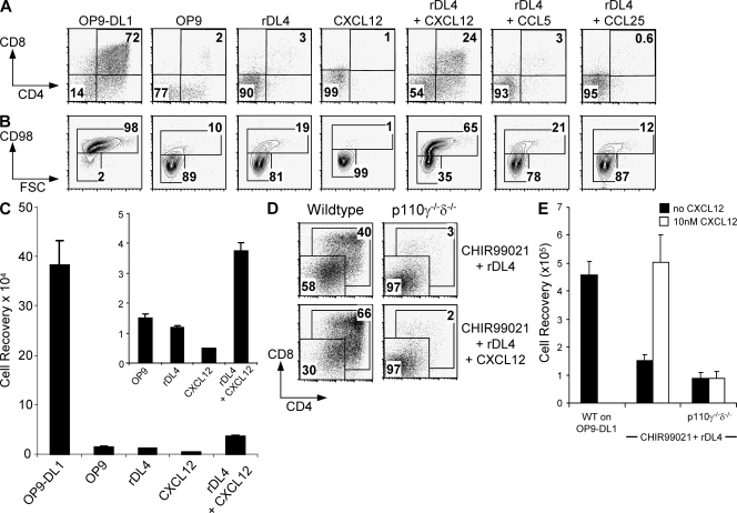
Our findings suggest that, in addition to the preTCR and Notch, DN3 cells also require signals through CXCR4 for their continued development. When sorted DN3 cells were cultured with the recombinant Notch ligand Delta-like 4 (rDL4), they failed to proliferate or differentiate and minimally increased expression of CD98, demonstrating the requirement for a factor(s) in addition to the preTCR and Notch (Fig. 9, A–C). In contrast, when DN3 cells were cultured in the presence of rDL4 with CXCL12, CD98 up-regulation occurred, the cells increased in size and DP cells were formed (Fig. 9, A–C). The effect of CXCL12 was found to be dose-dependent (Fig. S3, B-C). The absolute requirement for preTCR signaling in this culture system was also confirmed as Rag2−/− DN3 cells failed to differentiate (unpublished data). The effect of CXCL12 could not be substituted by two other chemokines. Culturing DN3 cells in the presence of a CCR5 ligand CCL5 did not induce DP differentiation (Fig. 9 A), nor did it have any additive effects in combination with CXCL12 (not depicted). Similarly, the addition of CCL25 neither reproduced nor potentiated the effect of CXCL12 (Fig. 9 A). Together, these results demonstrate that signaling through the preTCR, Notch, and CXCR4 combine to promote in vitro thymocyte differentiation beyond the DN3 stage.
Figure 9.
Establishment of an accessory cell–free culture system for the differentiation of immature thymocytes. (A–C) 105 DN3 cells from WT mice were cultured on OP9-DL1 or OP9 stromal cells, plate-bound recombinant Delta-like 4 (rDL4), CXCL12, or rDL4 and either CXCL12, CCL5, or CCL25. After 3 d, cells were harvested and analyzed for the percentage of CD4+CD8+, CD4−CD8− (A) cells and the percentage of CD98+ cells (B). (C) The total number of cells harvested under different culture conditions. The graph shows the mean and SEM of five biological replicates, except for cells cultured with CXCL12 alone, where n = 1. (D and E) 105 DN3 cells from WT or p110γ−/−δ−/− mice were cultured with rDL4 with 10 µM CHIR99021 in the presence or absence of 10nM CXCL12. After 3 d, cells were harvested and analyzed for the percentages of CD4+CD8+ and CD4−CD8− cells (D) and the total number of cells harvested (E). The graph shows the mean and SD of two biological replicates. Results from WT represent one of three experiments.
Inhibition of GSK3β augments in vitro thymocyte development
The combination of rDL4 and CXCL12 defined minimal requirements for a stromal cell–free in vitro system that promoted thymocyte differentiation. However, these conditions did not support DN3 cell proliferation or differentiation to the same extent as the OP9-DL1 stromal cell line, suggesting that additional factors were contributed by the OP9-DL1 cells. Glycogen synthase kinase 3β (GSK3β) has been implicated as a regulator of multiple signaling pathways, and we tested whether inhibition of GSK3β in DN3 cells could further augment the stromal-free in vitro culture. DN3 cells from WT and p110γ−/−/δ−/− mice were cultured on rDL4 with the GSK3β inhibitor CHIR99021 in the presence or absence of CXCL12 (Fig. 9, D and E). CHIR99021 enabled the differentiation of WT DN3 cells in the absence of CXCL12, but together CHIR99021 and CXCL12 increased the proportion of cells that became CD4+CD8+ and increased cell numbers to that achieved when DN3 cells were cultured on OP9-DL1. This result suggests a role for the GSK3β signaling pathway in DN3 cells and further demonstrates that CXCL12 is required for optimal differentiation and proliferation. In contrast to WT DN3 cells, p110γ−/−/δ−/− DN3 cells failed to differentiate when cultured with rDL4 and CHIR99021. Further, the addition of CXCL12 did not enhance the numbers of p110γ−/−/δ−/− cells recovered or support their differentiation. Thus, for DN3 cells cultured under in vitro conditions, p110γ and p110δ activation is absolutely required for CXCR4-dependent expansion and differentiation.
DISCUSSION
Until recently, T cell development in vitro was studied in the context of fetal thymic organ cultures. The creation of the OP9-DL1 stromal cell line allowed the differentiation of HSCs into functional T cells in a two-dimensional culture system (Schmitt and Zuñiga-Pflucker, 2002). Exploitation of this system has allowed T cell precursors to be genetically manipulated and subsequently used in the treatment of tumor models in mice (Zakrzewski et al., 2008). Accessory cell–free culture conditions for T cell precursors have been described previously, but these did not permit cells to differentiate beyond the DN2 stage (Dallas et al., 2007). This suggests that additional factors elaborated by the stromal cell line contribute to the proliferation and differentiation of thymocytes. However, the identity of these additional factors has remained unknown. Here, we have provided evidence that CXCR4 and its ligand CXCL12, which is produced by OP9-DL1, are important for the proliferation and differentiation of DN3 cells. CXCR4 was identified as being essential in the OP9-DL1 culture system through the use of highly selective receptor antagonists. CXCL12 promoted DN3 differentiation that was dependent on Notch and the preTCR. We suggest these three receptors are functionally nonredundant, with each receptor–system interacting to promote continued thymic development beyond β-selection. Our findings offer the possibility of studying thymocyte development in a stromal cell–free system in which the effects of extrinsic factors or genetic manipulation can be controlled.
The combination of rDL4 and CXCL12 alone did not support DN3 cell proliferation or differentiation to the same extent as the OP9-DL1 stromal cell line. This might have been caused by the suboptimal presentation of the Notch ligand, which varies in effectiveness depending on density (Dallas et al., 2005). Similarly, presentation of CXCL12, which binds to extracellular matrix (Amara et al., 1999), may not be optimal in our system. It is likely that an additional factor besides Notch, preTCR, and CXCL12 are required for the proliferation and differentiation of DN3 cells. Utilization of the GSK3β inhibitor CHIR99021, in conjunction with CXCL12, appeared to compensate for this and allowed the proliferation and differentiation of DN3 cells to a similar degree to that observed when cultured with OP9-DL1. CHIR99021 did not potentiate the differentiation of p110γ−/−δ−/− DN3 cells; however, indicating that it was not able to compensate for the lack of PI3K. It is possible that the GSK3β inhibitor may promote Wnt/β-catenin signaling that has been identified as important for T cell development at the β-selection checkpoint (Gounari et al., 2001; Xu et al., 2003). Alternatively, CHIR99021 may be potentiating the effects of Notch (McKenzie et al., 2006) or regulating Hedgehog signaling, which has also been shown to regulate DN thymocyte differentiation (Outram et al., 2000). These issues can be explored further using an accessory cell–free culture system.
Although we have shown here that T cell development is impaired in CXCR4 knockout mice, controversy exists regarding the role of CXCR4 in T cell development (Ma et al., 1998; Zou et al., 1998; Kawabata et al., 1999; Ara et al., 2003; Plotkin et al., 2003). These discrepancies may be caused by the differing requirements for T lymphopoiesis in the fetal and adult thymus. For example, fetal T cell development has been shown to be independent of the IL-7 receptor (Crompton et al., 1998) and α4 integrin (Arroyo et al., 1996), but these molecules are essential for postnatal T cell development. However, a complete block in adult T cell development upon the conditional deletion of CXCR4 in DN1 cells in chimeric mice suggests that CXCR4 is important for directing T cell progenitors beyond the corticomedullary junction in the adult thymus (Plotkin et al., 2003). To fully explore the role for CXCR4 at the later developmental stage of β-selection in the adult, a conditional model whereby CXCR4 is deleted upon the expression of TCRβ is required.
The identification of a critical role for CXCR4 signaling arose from an investigation into the functional redundancy between PI3K isoforms during T cell development. This study has identified receptors through which each of these catalytic isoforms is activated. PreTCR-mediated PI3K activation is p110δ dependent. In contrast, activation of PI3K by CXCR4 is predominantly mediated by p110γ in a p101-dependent manner. Activation of PI3K signaling through CXCR4 at the point of β-selection is critical, as in the presence of CXCR4 antagonists the phosphorylation of the PI3K effector Akt in DN3 cells is impaired and cells fail to differentiate. The relatively normal phenotypes of the p110δ−/−, p110γ−/−, and p101−/− DN thymocytes suggest that the loss of one catalytic isoform is compensated for by the presence of the other and that major defects in thymocyte development are only observed when class IA and IB PI3K subunits are inactivated simultaneously. Thus, p110δ−/− DN3 cells are unable to phosphorylate Akt downstream of the preTCR, but possess a robust p-Akt response to CXCR4 stimulation caused by the presence of p110γ. Conversely, the ability of p110γ−/− and p101−/− thymocytes to phosphorylate Akt upon CXCR4 stimulation is poor, but their PI3K response to preTCR signaling, mediated by p110δ, is intact. In each of these scenarios, PI3K activity in DN3 cells is sufficient for continued thymocyte development. The mechanistic rationale for having multiple cell surface receptors activate the same growth promoting pathway may be to promote proliferation in defined regions of the thymus. Thus, CXCL12 is concentrated at the subcapsular zone (Plotkin et al., 2003), which is where the most intensive proliferation of thymocytes is known to occur.
Although p110γ appears to be relatively more important for Akt phosphorylation after CXCR4 activation in DN3 cells, p110δ clearly plays an ancillary role as the p-Akt response of the p110γ−/−δ−/− or p101−/−/p110δ−/− double mutants was further reduced when compared with the p110γ−/− or p101−/− single mutants. The potential of class IA PI3K to contribute to CXCR4 signaling has been noted previously in studies using Jurkat cells (Curnock et al., 2003). In peripheral T cells, CXCR4 signaling leads to the phosphorylation of Lck and Zap-70 (Patrussi et al., 2007), possibly via direct interactions with the TCR (Kumar et al., 2006; Contento et al., 2008). It is not yet known whether these mechanisms are relevant to p110δ-mediated CXCR4 signaling in DN thymocytes; however, we have noted that pAkt responses to CXCL12 are intact in DN3 cells from Rag-2 mutant mice (unpublished data). Thus, TCRβ is dispensable for PI3K-mediated CXCR4 signaling in DN3 cells.
CXCL12 is predominantly considered as a chemokine but it has been recently shown to enhance the proliferation and activation of CD3-stimulated peripheral T cells (Molon et al., 2005). Indeed CXCL12 was originally identified through its preB cell growth factor properties (Nagasawa et al., 1994). Our results with stromal cell–free in vitro cultures suggest that CXCL12 also promotes the growth and differentiation of developing thymocytes. Presumably, in stromal cell–free culture, this function of CXCL12 is independent of any chemotactic activity. Notch has been documented as a regulator of DN3 metabolism (Ciofani and Zuñiga-Pflucker, 2005) but our results demonstrate that the provision of a Notch signal alone to DN3 thymocytes is not sufficient to support the metabolic changes associated with β-selection and that CXCL12 was additionally required. The contribution of CXCL12 to DN3 cell survival, proliferation, and differentiation have yet to be fully addressed, but further investigation using specific CXCR4 inhibitors in combination with mathematical modeling of time course data may provide further insight (Callard and Hodgkin, 2007).
Our findings may be relevant in the context of some T cell acute lymphoblastic leukemias (T-ALLs) as altered Notch and PI3K signaling frequently underpins the transformed phenotype of these cells, yet primary explants of these tumors are prone to apoptosis and difficult to propagate in vitro (Armstrong et al., 2009). Recently, some primary T-ALLs were propagated on stromal cell lines expressing recombinant Notch ligand (Armstrong et al., 2009). Our finding that CXCL12 promotes growth in concert with Notch and the preTCR may be relevant to the in vitro culture of T-ALL and prompts the hypothesis that CXCL12 may have an in vivo role in T-ALL. Examining the responses of T-ALL to AMD3100 alone or in combination with other drugs with efficacy against T-ALL may suggest a new therapeutic modality.
MATERIALS AND METHODS
Mice.
Mice with deletions of p110γ, 110δ, p101, and CXCR4 have been described previously (Tachibana et al., 1998; Webb et al., 2005; Suire et al., 2006). Mice were on 129/B6 mixed backgrounds and 5–13 wk of age, except CXCR4-deficient mice which were on a B6 background and analyzed at embryonic day 17.5. For chimera studies, irradiated Rag2−/−Il2rγ−/− mice were injected intravenously with 3 × 10⁶ BM cells from WT, p110γ−/−δ−/−, or p101−/−/p110δ−/− mice and analyzed 8–12 wk later. All mice husbandry and experimentation were in accordance with UK Home Office regulations and local ethical review (except CXCR4 embryo studies, which were approved by the animal ethics committee of the Institute for Frontier Medical Sciences, Kyoto University).
Cell isolation.
BM-derived HSCs were isolated from single-cell suspensions of BM by a two-step process using lineage depletion followed by ScaI+ enrichment by MACS (Miltenyi Biotec). Typically, the ScaI+-enriched population represented 0.1% of the starting BM preparation. DN3 and DN4 thymocytes were isolated by FACS. Single-cell thymocyte suspensions were first enriched for DN cells by incubation with biotinylated anti-CD8α antibody followed by streptavidin-coated MACS beads and magnetic depletion before staining for DN subsets. DN3 and DN4 cells were isolated as Thy1+, [CD4/CD8/CD44/B220/CD11b/NK1.1/Gr1/Ter119/γδTCR]neg and either CD25hiCD98lo (DN3) or CD25loCD98hi (DN4).
Cell culture.
OP9 and OP9-DL1 cells were maintained as previously described (Ciofani and Zuñiga-Pflucker, 2005). BM-derived HSCs were cultured in α-MEM supplemented with 20% FCS, 100 U/ml penicillin, 100 µg/ml streptomycin, 5 × 10⁻⁵M 2ME, 10 ng/ml recombinant IL-7, and 5 ng/ml recombinant Flt3L (PeproTech) at a density of 4 × 10³ cells/ml in 24-well tissue culture plates that had been seeded with 2 × 10⁴ OP9-DL1 cells. After 7 d, and at weekly intervals thereafter, cells were harvested, passed through a 40-µM filter, and recultured at 2 × 10⁵ cells/ml in 24-well tissue culture plates that had been seeded with 2 × 10⁴ OP9-DL1 cells in supplemented α-MEM complete with IL-7 and Flt3L. For assays using sorted DN3 thymocytes on OP9-DL1 cell lines, 4 × 10⁴ cells were cultured in supplemented α-MEM, without additional cytokines, in 96-well plates that had been previously seeded with 4,000 OP9-DL1 cells. AMD3100 (Sigma-Aldrich) and GSK812397A (GlaxoSmithKline) were added with the DN3 cells at the start of culture. For stromal cell–free cultures, 105 DN3 cells were cultures in 96-well plates that had been coated overnight with 10 µg/ml of recombinant mouse Delta-like ligand 4 (R&D Systems) supplemented with α-MEM in the presence or absence of recombinant CXCL12 (10 nM), CCL5 (10 nM), CCL25 (10 ng/ml; PeproTech), and/or CHIR99021 (10 µM; Axon MedChem). For controls, equivalent cell numbers were cultured on OP9 or OP9-DL1 stromal cells. Assays using sorted DN3 cells were harvested after 3 d of culture and analyzed by FACS. Cell numbers were determined by reference to the inclusion of a fixed number of microbeads (Spherotech).
Thymocyte population analysis by FACS.
Single cell suspensions from the thymus or cultured cells were stained using various combinations of antibodies conjugated to fluorochromes for analysis by FACS (LSRII; BD). DN3/DN4 thymocytes were identified as Thy1+ and [CD4/CD8/CD44/B220/CD11b/NK1.1/Gr1/Ter119/γδTCR]neg. For intracellular staining, cells were treated with BD Cytofix/Cytoperm solutions before staining for cell-surface markers in conjunction with PE-conjugated anti-mouse TCRβ (clone H57) or intracellular Notch1 (clone mN1A; BD).
EdU incorporation studies.
Mice were injected intraperitoneally with 100 µg of EdU in PBS. After 4 h, thymi were harvested and single-cell suspensions were prepared and stained for cell surface markers. Cells were then prepared for EdU detection as per the manufacturer’s instructions (Invitrogen; Salic and Mitchison, 2008).
Phosphospecific flow cytometry.
For preTCR stimulation studies, cultured cells were harvested, washed, and passed through a 40-µM filter before resuspension at 107 cells/ml in supplemented α-MEM. Cells were incubated at 37°C for 20 min before the addition of 10 µg/ml of the α-CD3ε monoclonal antibody 2C11. At various time intervals, 106 cells were fixed in 2% paraformaldehyde, washed in PBS, and resuspended in 90% ice-cold methanol at −20°C for at least 30 min. Cells were then washed three times in PBS with 2% FCS before incubation with antibodies to surface markers as well as to phosphorylated Akt (clone #4058; Cell Signaling Technology) or phosphorylated-Erk (clone #4370; Cell Signaling Technology) for 30 min at room temperature. Phosphospecific antibodies were detected by subsequent incubation with Cy5-conjugated donkey anti–rabbit antibody. Cells were then analyzed by FACS. CXCL12 stimulation studies were conducted as above, except that unfractionated thymocyte suspensions were resuspended at 107 cells/ml in DMEM with 0.1% BSA. Cells were incubated at 37°C for 20 min before the addition of 10 nM CXCL12. For data analysis, fold induction was calculated as the median fluorescence of stimulated sample/median fluorescence of unstimulated sample. Unstimulated samples showed no increase in phospho-Akt or phospho-Erk throughout the timecourse.
RT-PCR analysis.
RNA was extracted from 106 isolated cells using TRIzol (Invitrogen) and converted to cDNA. Controls without reverse transcription were included for all samples. p101 and p84 mRNA was quantified using a Taqman probe and normalized to the expression of β2 microglobulin (Applied Biosystems).
Western blots.
Lysates from 106 thymocytes were separated by SDS-PAGE and transferred to nitrocellulose. Blots were probed with rabbit anti-p110γ (Onxy Pharmaceuticals) and antibody binding was revealed using HRP-conjugated anti–rabbit antibody (Dako) followed by ECL (GE Healthcare). Blots were subsequently probed using mouse anti-β actin.
Statistics.
Data presented throughout represent the mean and either the SD or SEM (if n ≥ 5). For statistical analyses containing two groups, data were analyzed using unpaired Student’s t test and P values are shown in the figures. For statistical analyses containing more than two groups, data were analyzed by one-way analysis of variance and statistical significance represented as: *, P < 0.05; **, P < 0.01; and ***, P < 0.001 compared with WT, except for the data in Fig. 4 where statistical significance is shown compared with cells cultured without agonist.
Online supplemental material.
Fig. S1 presents the FACS profile of DN3 and DN4 cells derived from long-term BM HSC cultures. Fig. S2 presents relative RNA expression levels of the PI3K isoforms p101 and p84 in WT DN3 and DN4 cells, as well as p110γ protein levels in WT, p101−/− and p110γ−/− thymus lysates. Fig. S3 presents the secretion of CXCL12 by OP9-DL1 stromal cells as well as the cellular responses of WT DN3 cells cultured in the presence of increasing concentrations of CXCL12. Online supplemental material is available at http://www.jem.org/cgi/content/full/jem.20091430/DC1.
Acknowledgments
We thank A.M. Condliffe, L.R. Stephens, and P.T. Hawkins for the provision of the p101−/− mice and the anti-p110γ antibody, S. Kulkarni for providing isolated mouse neutrophils, J.C. Zúñiga-Pflücker for provision of the OP9-DL1 stromal cell line, G. Butcher, A. Corcoran, P. Hawkins, K. Okkenhaug, M. Sims, E. Vigorito, and L. Webb for critical discussion, K. Bates and SABU staff for technical support and G. Morgan for FACS expertise.
This work was supported by the Biotechnology and Biological Sciences Research Council and the Ministry of Education, Culture, Sports, Science and Technology of Japan. G.V. was supported by a Da Vinci scholarship (Italy) and M.T. by a fellowship from the Medical Research Council (U.K.).
For the course of this work K. Gudmundsson was an employee of GlaxoSmithKline. The authors have no further conflicting financial interests.
Footnotes
Abbreviations used:
DN
double-positive
DP
double-negative
EdU
5-ethynyl-2′-deoxyuridine
GPCR
G protein–coupled receptor
GSK3β
glycogen synthase kinase 3β
HSC
hematopoietic stem cell
PI3K
phosphatidylinositol 3-kinase
PIP
phosphatidylinositol-4,5-trisphosphate
rDL4
recombinant Delta-like 4
T-ALL
T cell acute lymphoblastic leukaemia
References
- Amara A., Lorthioir O., Valenzuela A., Magerus A., Thelen M., Montes M., Virelizier J.L., Delepierre M., Baleux F., Lortat-Jacob H., Arenzana-Seisdedos F. 1999. Stromal cell-derived factor-1alpha associates with heparan sulfates through the first beta-strand of the chemokine. J. Biol. Chem. 274:23916–23925 10.1074/jbc.274.34.23916 [DOI] [PubMed] [Google Scholar]
- Ara T., Itoi M., Kawabata K., Egawa T., Tokoyoda K., Sugiyama T., Fujii N., Amagai T., Nagasawa T. 2003. A role of CXC chemokine ligand 12/stromal cell-derived factor-1/pre-B cell growth stimulating factor and its receptor CXCR4 in fetal and adult T cell development in vivo. J. Immunol. 170:4649–4655 [DOI] [PubMed] [Google Scholar]
- Armstrong F., Brunet de la Grange P., Gerby B., Rouyez M.C., Calvo J., Fontenay M., Boissel N., Dombret H., Baruchel A., Landman-Parker J., et al. 2009. NOTCH is a key regulator of human T-cell acute leukemia initiating cell activity. Blood. 113:1730–1740 10.1182/blood-2008-02-138172 [DOI] [PubMed] [Google Scholar]
- Arroyo A.G., Yang J.T., Rayburn H., Hynes R.O. 1996. Differential requirements for alpha4 integrins during fetal and adult hematopoiesis. Cell. 85:997–1008 10.1016/S0092-8674(00)81301-X [DOI] [PubMed] [Google Scholar]
- Callard R., Hodgkin P. 2007. Modeling T- and B-cell growth and differentiation. Immunol. Rev. 216:119–129 [DOI] [PubMed] [Google Scholar]
- Ciofani M., Zúñiga-Pflücker J.C. 2005. Notch promotes survival of pre-T cells at the beta-selection checkpoint by regulating cellular metabolism. Nat. Immunol. 6:881–888 10.1038/ni1234 [DOI] [PubMed] [Google Scholar]
- Contento R.L., Molon B., Boularan C., Pozzan T., Manes S., Marullo S., Viola A. 2008. CXCR4-CCR5: a couple modulating T cell functions. Proc. Natl. Acad. Sci. USA. 105:10101–10106 10.1073/pnas.0804286105 [DOI] [PMC free article] [PubMed] [Google Scholar]
- Crompton T., Outram S.V., Buckland J., Owen M.J. 1998. Distinct roles of the interleukin-7 receptor alpha chain in fetal and adult thymocyte development revealed by analysis of interleukin-7 receptor alpha-deficient mice. Eur. J. Immunol. 28:1859–1866 [DOI] [PubMed] [Google Scholar]
- Curnock A.P., Sotsios Y., Wright K.L., Ward S.G. 2003. Optimal chemotactic responses of leukemic T cells to stromal cell-derived factor-1 requires the activation of both class IA and IB phosphoinositide 3-kinases. J. Immunol. 170:4021–4030 [DOI] [PubMed] [Google Scholar]
- Dallas M.H., Varnum-Finney B., Delaney C., Kato K., Bernstein I.D. 2005. Density of the Notch ligand Delta1 determines generation of B and T cell precursors from hematopoietic stem cells. J. Exp. Med. 201:1361–1366 10.1084/jem.20042450 [DOI] [PMC free article] [PubMed] [Google Scholar]
- Dallas M.H., Varnum-Finney B., Martin P.J., Bernstein I.D. 2007. Enhanced T-cell reconstitution by hematopoietic progenitors expanded ex vivo using the Notch ligand Delta1. Blood. 109:3579–3587 10.1182/blood-2006-08-039842 [DOI] [PMC free article] [PubMed] [Google Scholar]
- Fayard E., Gill J., Paolino M., Hynx D., Holländer G.A., Hemmings B.A. 2007. Deletion of PKBalpha/Akt1 affects thymic development. PLoS One. 2:e992 10.1371/journal.pone.0000992 [DOI] [PMC free article] [PubMed] [Google Scholar]
- Fruman D.A., Bismuth G. 2009. Fine tuning the immune response with PI3K. Immunol. Rev. 228:253–272 10.1111/j.1600-065X.2008.00750.x [DOI] [PubMed] [Google Scholar]
- Godfrey D.I., Kennedy J., Suda T., Zlotnik A. 1993. A developmental pathway involving four phenotypically and functionally distinct subsets of CD3-CD4-CD8- triple-negative adult mouse thymocytes defined by CD44 and CD25 expression. J. Immunol. 150:4244–4252 [PubMed] [Google Scholar]
- Gounari F., Aifantis I., Khazaie K., Hoeflinger S., Harada N., Taketo M.M., von Boehmer H. 2001. Somatic activation of beta-catenin bypasses pre-TCR signaling and TCR selection in thymocyte development. Nat. Immunol. 2:863–869 10.1038/ni0901-863 [DOI] [PubMed] [Google Scholar]
- Hagenbeek T.J., Naspetti M., Malergue F., Garçon F., Nunès J.A., Cleutjens K.B., Trapman J., Krimpenfort P., Spits H. 2004. The loss of PTEN allows TCR alphabeta lineage thymocytes to bypass IL-7 and Pre-TCR-mediated signaling. J. Exp. Med. 200:883–894 10.1084/jem.20040495 [DOI] [PMC free article] [PubMed] [Google Scholar]
- Hatse S., Princen K., Bridger G., De Clercq E., Schols D. 2002. Chemokine receptor inhibition by AMD3100 is strictly confined to CXCR4. FEBS Lett. 527:255–262 10.1016/S0014-5793(02)03143-5 [DOI] [PubMed] [Google Scholar]
- Hernández-López C., Varas A., Sacedón R., Jiménez E., Muñoz J.J., Zapata A.G., Vicente A. 2002. Stromal cell-derived factor 1/CXCR4 signaling is critical for early human T-cell development. Blood. 99:546–554 10.1182/blood.V99.2.546 [DOI] [PubMed] [Google Scholar]
- Hinton H.J., Alessi D.R., Cantrell D.A. 2004. The serine kinase phosphoinositide-dependent kinase 1 (PDK1) regulates T cell development. Nat. Immunol. 5:539–545 10.1038/ni1062 [DOI] [PubMed] [Google Scholar]
- Huang J., Garrett K.P., Pelayo R., Zúñiga-Pflücker J.C., Petrie H.T., Kincade P.W. 2005. Propensity of adult lymphoid progenitors to progress to DN2/3 stage thymocytes with Notch receptor ligation. J. Immunol. 175:4858–4865 [DOI] [PMC free article] [PubMed] [Google Scholar]
- Ji H., Rintelen F., Waltzinger C., Bertschy Meier D., Bilancio A., Pearce W., Hirsch E., Wymann M.P., Rückle T., Camps M., et al. 2007. Inactivation of PI3Kgamma and PI3Kdelta distorts T-cell development and causes multiple organ inflammation. Blood. 110:2940–2947 10.1182/blood-2007-04-086751 [DOI] [PubMed] [Google Scholar]
- Juntilla M.M., Wofford J.A., Birnbaum M.J., Rathmell J.C., Koretzky G.A. 2007. Akt1 and Akt2 are required for alphabeta thymocyte survival and differentiation. Proc. Natl. Acad. Sci. USA. 104:12105–12110 10.1073/pnas.0705285104 [DOI] [PMC free article] [PubMed] [Google Scholar]
- Kawabata K., Ujikawa M., Egawa T., Kawamoto H., Tachibana K., Iizasa H., Katsura Y., Kishimoto T., Nagasawa T. 1999. A cell-autonomous requirement for CXCR4 in long-term lymphoid and myeloid reconstitution. Proc. Natl. Acad. Sci. USA. 96:5663–5667 10.1073/pnas.96.10.5663 [DOI] [PMC free article] [PubMed] [Google Scholar]
- Kelly A.P., Finlay D.K., Hinton H.J., Clarke R.G., Fiorini E., Radtke F., Cantrell D.A. 2007. Notch-induced T cell development requires phosphoinositide-dependent kinase 1. EMBO J. 26:3441–3450 10.1038/sj.emboj.7601761 [DOI] [PMC free article] [PubMed] [Google Scholar]
- Kumar A., Humphreys T.D., Kremer K.N., Bramati P.S., Bradfield L., Edgar C.E., Hedin K.E. 2006. CXCR4 physically associates with the T cell receptor to signal in T cells. Immunity. 25:213–224 10.1016/j.immuni.2006.06.015 [DOI] [PubMed] [Google Scholar]
- Ma Q., Jones D., Borghesani P.R., Segal R.A., Nagasawa T., Kishimoto T., Bronson R.T., Springer T.A. 1998. Impaired B-lymphopoiesis, myelopoiesis, and derailed cerebellar neuron migration in CXCR4- and SDF-1-deficient mice. Proc. Natl. Acad. Sci. USA. 95:9448–9453 10.1073/pnas.95.16.9448 [DOI] [PMC free article] [PubMed] [Google Scholar]
- Maillard I., Tu L., Sambandam A., Yashiro-Ohtani Y., Millholland J., Keeshan K., Shestova O., Xu L., Bhandoola A., Pear W.S. 2006. The requirement for Notch signaling at the beta-selection checkpoint in vivo is absolute and independent of the pre-T cell receptor. J. Exp. Med. 203:2239–2245 10.1084/jem.20061020 [DOI] [PMC free article] [PubMed] [Google Scholar]
- Mao C., Tili E.G., Dose M., Haks M.C., Bear S.E., Maroulakou I., Horie K., Gaitanaris G.A., Fidanza V., Ludwig T., et al. 2007. Unequal contribution of Akt isoforms in the double-negative to double-positive thymocyte transition. J. Immunol. 178:5443–5453 [DOI] [PubMed] [Google Scholar]
- McKenzie G., Ward G., Stallwood Y., Briend E., Papadia S., Lennard A., Turner M., Champion B., Hardingham G.E. 2006. Cellular Notch responsiveness is defined by phosphoinositide 3-kinase-dependent signals. BMC Cell Biol. 7:10 10.1186/1471-2121-7-10 [DOI] [PMC free article] [PubMed] [Google Scholar]
- Molon B., Gri G., Bettella M., Gómez-Moutón C., Lanzavecchia A., Martínez-A C., Mañes S., Viola A. 2005. T cell costimulation by chemokine receptors. Nat. Immunol. 6:465–471 10.1038/ni1191 [DOI] [PubMed] [Google Scholar]
- Nagasawa T., Kikutani H., Kishimoto T. 1994. Molecular cloning and structure of a pre-B-cell growth-stimulating factor. Proc. Natl. Acad. Sci. USA. 91:2305–2309 10.1073/pnas.91.6.2305 [DOI] [PMC free article] [PubMed] [Google Scholar]
- Nagasawa T., Hirota S., Tachibana K., Takakura N., Nishikawa S., Kitamura Y., Yoshida N., Kikutani H., Kishimoto T. 1996. Defects of B-cell lymphopoiesis and bone-marrow myelopoiesis in mice lacking the CXC chemokine PBSF/SDF-1. Nature. 382:635–638 10.1038/382635a0 [DOI] [PubMed] [Google Scholar]
- Outram S.V., Varas A., Pepicelli C.V., Crompton T. 2000. Hedgehog signaling regulates differentiation from double-negative to double-positive thymocyte. Immunity. 13:187–197 10.1016/S1074-7613(00)00019-4 [DOI] [PubMed] [Google Scholar]
- Panigada M., Porcellini S., Barbier E., Hoeflinger S., Cazenave P.A., Gu H., Band H., von Boehmer H., Grassi F. 2002. Constitutive endocytosis and degradation of the pre-T cell receptor. J. Exp. Med. 195:1585–1597 10.1084/jem.20020047 [DOI] [PMC free article] [PubMed] [Google Scholar]
- Patrussi L., Ulivieri C., Lucherini O.M., Paccani S.R., Gamberucci A., Lanfrancone L., Pelicci P.G., Baldari C.T. 2007. p52Shc is required for CXCR4-dependent signaling and chemotaxis in T cells. Blood. 110:1730–1738 10.1182/blood-2007-01-068411 [DOI] [PubMed] [Google Scholar]
- Petrie H.T., Zúñiga-Pflücker J.C. 2007. Zoned out: functional mapping of stromal signaling microenvironments in the thymus. Annu. Rev. Immunol. 25:649–679 10.1146/annurev.immunol.23.021704.115715 [DOI] [PubMed] [Google Scholar]
- Plotkin J., Prockop S.E., Lepique A., Petrie H.T. 2003. Critical role for CXCR4 signaling in progenitor localization and T cell differentiation in the postnatal thymus. J. Immunol. 171:4521–4527 [DOI] [PubMed] [Google Scholar]
- Prasad A., Zikherman J., Das J., Roose J.P., Weiss A., Chakraborty A.K. 2009. Origin of the sharp boundary that discriminates positive and negative selection of thymocytes. Proc. Natl. Acad. Sci. USA. 106:528–533 10.1073/pnas.0805981105 [DOI] [PMC free article] [PubMed] [Google Scholar]
- Salic A., Mitchison T.J. 2008. A chemical method for fast and sensitive detection of DNA synthesis in vivo. Proc. Natl. Acad. Sci. USA. 105:2415–2420 10.1073/pnas.0712168105 [DOI] [PMC free article] [PubMed] [Google Scholar]
- Schmitt T.M., Zúñiga-Pflücker J.C. 2002. Induction of T cell development from hematopoietic progenitor cells by delta-like-1 in vitro. Immunity. 17:749–756 10.1016/S1074-7613(02)00474-0 [DOI] [PubMed] [Google Scholar]
- Shiroki F., Matsuda S., Doi T., Fujiwara M., Mochizuki Y., Kadowaki T., Suzuki H., Koyasu S. 2007. The p85alpha regulatory subunit of class IA phosphoinositide 3-kinase regulates beta-selection in thymocyte development. J. Immunol. 178:1349–1356 [DOI] [PubMed] [Google Scholar]
- Stephens L.R., Eguinoa A., Erdjument-Bromage H., Lui M., Cooke F., Coadwell J., Smrcka A.S., Thelen M., Cadwallader K., Tempst P., Hawkins P.T. 1997. The G β γ sensitivity of a PI3K is dependent upon a tightly associated adaptor, p101. Cell. 89:105–114 10.1016/S0092-8674(00)80187-7 [DOI] [PubMed] [Google Scholar]
- Suire S., Coadwell J., Ferguson G.J., Davidson K., Hawkins P., Stephens L. 2005. p84, a new Gbetagamma-activated regulatory subunit of the type IB phosphoinositide 3-kinase p110γ. Curr. Biol. 15:566–570 10.1016/j.cub.2005.02.020 [DOI] [PubMed] [Google Scholar]
- Suire S., Condliffe A.M., Ferguson G.J., Ellson C.D., Guillou H., Davidson K., Welch H., Coadwell J., Turner M., Chilvers E.R., et al. 2006. Gbetagammas and the Ras binding domain of p110γ are both important regulators of PI(3)Kgamma signalling in neutrophils. Nat. Cell Biol. 8:1303–1309 10.1038/ncb1494 [DOI] [PubMed] [Google Scholar]
- Swat W., Montgrain V., Doggett T.A., Douangpanya J., Puri K., Vermi W., Diacovo T.G. 2006. Essential role of PI3Kdelta and PI3Kgamma in thymocyte survival. Blood. 107:2415–2422 10.1182/blood-2005-08-3300 [DOI] [PMC free article] [PubMed] [Google Scholar]
- Tachibana K., Hirota S., Iizasa H., Yoshida H., Kawabata K., Kataoka Y., Kitamura Y., Matsushima K., Yoshida N., Nishikawa S., et al. 1998. The chemokine receptor CXCR4 is essential for vascularization of the gastrointestinal tract. Nature. 393:591–594 10.1038/31261 [DOI] [PubMed] [Google Scholar]
- Taghon T., Yui M.A., Pant R., Diamond R.A., Rothenberg E.V. 2006. Developmental and molecular characterization of emerging beta- and gammadelta-selected pre-T cells in the adult mouse thymus. Immunity. 24:53–64 10.1016/j.immuni.2005.11.012 [DOI] [PubMed] [Google Scholar]
- Webb L.M., Vigorito E., Wymann M.P., Hirsch E., Turner M. 2005. Cutting edge: T cell development requires the combined activities of the p110gamma and p110delta catalytic isoforms of phosphatidylinositol 3-kinase. J. Immunol. 175:2783–2787 [DOI] [PubMed] [Google Scholar]
- Xu Y., Banerjee D., Huelsken J., Birchmeier W., Sen J.M. 2003. Deletion of beta-catenin impairs T cell development. Nat. Immunol. 4:1177–1182 10.1038/ni1008 [DOI] [PubMed] [Google Scholar]
- Yamasaki S., Saito T. 2007. Molecular basis for pre-TCR-mediated autonomous signaling. Trends Immunol. 28:39–43 10.1016/j.it.2006.11.006 [DOI] [PubMed] [Google Scholar]
- Zakrzewski J.L., Suh D., Markley J.C., Smith O.M., King C., Goldberg G.L., Jenq R., Holland A.M., Grubin J., Cabrera-Perez J., et al. 2008. Tumor immunotherapy across MHC barriers using allogeneic T-cell precursors. Nat. Biotechnol. 26:453–461 10.1038/nbt1395 [DOI] [PMC free article] [PubMed] [Google Scholar]
- Zou Y.R., Kottmann A.H., Kuroda M., Taniuchi I., Littman D.R. 1998. Function of the chemokine receptor CXCR4 in haematopoiesis and in cerebellar development. Nature. 393:595–599 10.1038/31269 [DOI] [PubMed] [Google Scholar]
