A potential role for the nuclear factor of activated T cells family of transcriptional regulatory proteins in adipogenesis (original) (raw)
Abstract
NFAT (nuclear factor of activated T cells) is a family of transcription factors implicated in the control of cytokine and early immune response gene expression. Recent studies have pointed to a role for NFAT proteins in gene regulation outside of the immune system. Herein we demonstrate that NFAT proteins are present in 3T3-L1 adipocytes and, upon fat cell differentiation, bind to and transactivate the promoter of the adipocyte-specific gene aP2. Further, fat cell differentiation is inhibited by cyclosporin A, a drug shown to prevent NFAT nuclear localization and hence function. Thus, these data suggest a role for NFAT transcription factors in the regulation of the aP2 gene and in the process of adipocyte differentiation.
Nuclear factor of activated T cells (NFAT) is a family of transcriptional regulatory proteins that controls the expression of cytokine genes in T lymphocytes (1). NFAT was initially identified as a transcriptional complex in activated T cells that bound, in electrophoretic mobility shift assays (EMSAs) and by DNase I footprinting experiments, to two purine-rich sequences located at positions −290 and −35 upstream of the coding region of the interleukin 2 (IL-2) cytokine gene (2–4). The NFAT family has four members that are approximately 65% similar throughout a 290-amino acid domain-related to the DNA binding and dimerization domain of the Rel family of transcription factors (RHD) (5–8). The RHD is essential for the dimerization of NFAT with an inducible nuclear component composed of AP-1 family member proteins (9–13). The NFAT proteins reside in the cytoplasm in resting T and B lymphocytes and translocate to the nucleus upon activation through T and B cell receptors. This process is controlled by the dephosphorylation of cytoplasmic NFAT by the phosphatase calcineurin, which becomes activated upon the delivery of a signal that results in a sustained calcium flux (14–16). The inhibition of cytokine gene transcription by the immunosuppressive drugs cyclosporin A (CsA) and tacrolimus (FK506) can be explained by their interaction with their receptors, cyclophilin and FKBP12, thereby creating high-affinity binding sites for the phosphatase calcineurin. This interaction interferes with calcineurin activity and thereby inhibits NFAT translocation to the nucleus (17). The export of NFAT from the nucleus back into the cytosol has recently been shown to depend on its rephosphorylation by glycogen synthase kinase 3 (18).
The distribution of two of the four NFAT proteins, NFATc and NFAT4 (also called NFATc3/NFATx), is tightly restricted to the lymphoid system in the adult organism, whereas the expression of the remaining two, NFATp and NFAT3 (also called NFATc4), is fairly ubiquitous (7). Until recently, a role for NFAT proteins outside the lymphoid system had not been apparent. However, the phenotype of mouse strains lacking the NFATc transcription factor revealed a critical role for this protein in cardiac morphogenesis. These animals die from cardiac failure in utero secondary to defective formation of cardiac valves and ventricular septae, a calcium-sensitive process characterized by epithelial/mesenchymal transformation of the endocardial cushion (19). The widespread distribution of two of the NFAT proteins, coupled with the known function of glycogen synthase kinase 3 in developmental signaling programs in Xenopus and Drosophila (20–22), also suggested that NFAT proteins might be important in cellular differentiation programs outside of the immune system.
The development of adipose tissue from uncommitted mesodermal precursors is characterized by striking alterations in cell morphology and gene expression. Adipocyte differentiation has been a fruitful model system for investigations into programs of terminal cell differentiation, in part because of the existence of cell lines such as the 3T3-L1 preadipocytes that can be differentiated in vitro. When exposed to appropriate hormonal stimuli, 3T3-L1 fibroblasts will convert into fat-laden adipocytes in approximately a week (23–25). This conversion is accompanied by the expression of a number of adipocyte-specific genes including stearoyl CoA desaturase, the insulin-responsive glucose transporter, and a fatty acid-binding protein called aP2 (26–28). Investigations into the transcriptional regulation of this latter gene have identified a number of transcription factors that play a key role in adipogenesis. Initial studies demonstrated that the proximal promoter region of the aP2 gene could direct low-level fat-cell-specific expression in vitro (29–31). This region contained binding sites for C/EBPα (positions −149 to −130) and AP-1 (positions −124 to −107) transcription factors. C/EBPα was demonstrated to bind to the more distal element and to transactivate the aP2 promoter (32, 33). The proximal element was termed FSE2 (fat-specific element 2) and was shown to be active during fat cell differentiation and to bind c-fos. (29). Mutational analysis of the FSE2 demonstrated that this site was essential for promoter activity during adipogenesis. However, this promoter region did not give rise to tissue-specific expression in transgenic mice (34). Subsequently, it was shown that the critical determinant of fat-cell-specific expression in vivo was an enhancer located 5.4 kb upstream of the transcriptional start site that binds the peroxisome proliferator-activated receptor γ2 (PPARγ2) nuclear factor (35, 36). Thus, factors that bind to the FSE2 act in concert with factors, most notably PPARγ, that bind upstream.
Adipocyte extracts give a single protected region on aP2 DNA in DNase I footprinting experiments that extends from base pairs −124 to −108 (29). Reexamination of the sequences protected revealed a perfect consensus sequence for NFAT just upstream of the c-fos binding site, reminiscent of the architecture of cytokine promoters, raising the possibility that NFAT family proteins also play a role in the regulation of fat cell-specific genes. Herein we present evidence that NFAT proteins are present in adipocytes and, upon fat cell differentiation, bind to and transactivate aP2 reporter sequences. Further, fat cell differentiation is inhibited by CsA, a drug known to prevent NFAT nuclear localization. Thus, these data suggest a role for NFAT transcription factors in the regulation of the aP2 gene and in processes of adipocyte differentiation.
MATERIALS AND METHODS
Cell Culture and Adipocyte Differentiation.
The generation and maintenance of the Th2 clone, D10.G4.1(D10) (American Type Culture Collection) specific for conalbumin/Iak, have been described (37, 38). For nuclear extract preparation, D10 cells were stimulated with anti-CD3(2C11)-coated plates overnight at 4°C. The 3T3-L1 and F442A cell lines were grown on 100-mm tissue culture plates in predifferentiation medium [DMEM/glucose (4.5 g/liter)/10% bovine calf serum/1% of a 100× stock of a penicillin–streptomycin solution]. Five days after the cells reached confluence, they were fed with differentiation medium [DMEM/glucose(4.5 g/liter)/10% fetal bovine serum/1% penicillin–streptomycin/insulin (5 μg/ml)/0.5 mM methyl isobutylxanthine/1 mM dexamethasone]. After 48 hr of differentiation, the cells were fed again and thereafter cells were fed every other day but with DMEM supplemented only with insulin and 10% fetal bovine serum. Differentiation was assessed by quantitating triglyceride deposits by red oil O staining. Cells were washed once with 1× PBS, twice with 60% ethanol, fixed in formalin, allowed to stand at room temperature for 1 hr in red oil O, and then examined by photomicroscopy.
CsA Treatment of 3T3-L1 Cells.
Two sets of plates were used; one set of plates was allowed to differentiate normally as described above. A parallel set of plates was used to test the effects of CsA on differentiation. To these plates were added various doses of CsA (a gift from Sandoz and Merck; at 1 or 10 μg/ml) or the solvent (EtOh) used to dissolve the CsA as control, at culture initiation and subsequently every other day. Differentiation was assessed as above.
Western Blot Analysis and Electrophoretic Mobility Shift Assays.
Total cell lysates were prepared from 3T3-L1 cells harvested at appropriate time points, electrophoresed on a 10% SDS/polyacrylamide gel, transferred to a poly(vinylidene difluoride) membrane (Millipore) and probed with a mAb against NFATp, 4G6-G5 (a gift from G. Crabtree, Stanford Univ.) or a polyclonal antiserum raised against NFAT3 (a gift from T. Hoey, Tularik, San Francisco) and then visualized by enhanced chemiluminescence detection (ECL, Amersham). Cytoplasmic extracts were prepared as described (39). For EMSA, stimulated Th2 cell nuclear extracts and nuclear extracts from undifferentiated and differentiated 3T3-L1 cells were prepared as described (9). The DNA probes used were double-stranded oligonucleotides (indicated below), end-labeled with [32P]dATP (DuPont NEN Research Product) or used unlabeled in competition experiments. The oligonucleotides used were as follows: IL-4 (positions −79 to −61), 5′-ATAAAATTTTCCAATGTAAA-3′; MtIL-4, 5′-ATAAAACGGGTCCAATGTAAA-3′; FSE2 (positions −127 to −101), 5′-GGATCCAAAAACATGACTCAGAGGAAAACATAC-3′; ARE6, 5′-TGCACATTTCACCCAGAGAGAAGGGATTGA-3′; AP-1, 5′-GAGCCGCAAGTGACTCAGCGCGGGCG-3′. For supershift experiments, antibodies against NFATp (4G6-G5) and against c-fos, FosB, c-jun/AP-1, c-fos (K-25), and Fra1 (N-17) (Santa Cruz Biotech, La Jolla, Ca) were added to nuclear extracts and preincubated at 4°C for 1 hr before addition of labeled probe as described (9).
Transient Transfection Assays.
Transfections were performed as described (29). Briefly, 3T3-L1 cells grown in 100-mm plates were differentiated for 4 days or undifferentiated, washed once with 1× PBS, and refed with DMEM/10% fetal bovine serum/insulin (5 μg/ml) for 2–4 hr before application of the DNA–calcium phosphate mixture. The aP2 promoter chloramphenicol acetyltransferase (CAT) construct was produced by PCR of the aP2 upstream region from positions −168 to +20, followed by ligation to the _Sal_I/_Xba_I-digested pCAT basic reporter plasmid. The mutant aP2 reporter contains a replacement of the wild-type NFAT sequence −108AAAC−105 with −108CCCG−105. Cotransfections used 20 μg of aP2 reporter, and 20 μg of an NFATp/pRep4 expression construct or pRep4 control vector alone (40). After a 3- to 6-hr incubation, the medium/precipitate was removed, and cells were shocked with 10% dimethyl sulfoxide and then refed with DMEM/10% fetal bovine serum/insulin (1 μg/ml). The cells were refed at 24 hr and stimulated with phorbol 12-myristate 13-acetate (2.5nM) and ionomycin (2 μM). At 48 hr, extracts were prepared and assayed for CAT activity as described (9, 12).
RESULTS
The FSE2 Element Binds NFAT Proteins.
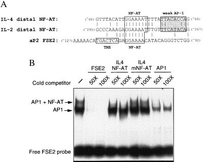
An examination of the region of the aP2 promoter footprinted by adipocyte extracts revealed a perfect consensus NFAT binding site immediately upstream of the AP-1 site that has been shown to bind c-fos. The similarity of these sequences to NFAT/AP-1 sites in cytokine promoters (Fig. 1a) prompted us to test whether NFAT proteins could bind to the FSE2. EMSAs with nuclear extracts, prepared from activated T lymphocyte clones, and the FSE2 site (positions −127 to −101), as the probe (Fig. 1b), revealed two shifted complexes whose binding was competed with an excess of unlabeled FSE2 probe. The upper complex was competed with an oligonucleotide containing the NFAT site in the IL-4 promoter, but not by an oligonucleotide containing a mutant NFAT site, and both complexes were competed with an oligonucleotide containing the AP-1 site in the IL-4 promoter. We conclude that the upper complex is composed of NFAT plus AP-1 proteins and the lower complex is composed of only AP-1 proteins. Thus these experiments demonstrate that the FSE2 can bind NFAT/AP-1 proteins present in T lymphocytes.
Figure 1.
FSE2 element binds NFAT proteins. (A) The 5′ upstream promoter region of the aP2 gene with NFAT and AP-1 binding sites is highlighted and nucleotide sequence comparisons of NFAT/AP-1 binding sites in the aP2, IL-4, and IL-2 promoters are shown. (B) EMSA using T lymphocyte nuclear extracts and radiolabeled probe containing the FSE2 element (positions −127 to −101).
NFAT/AP-1 Proteins Are Present in Differentiated Adipocytes and Bind the FSE2.
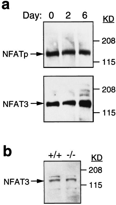
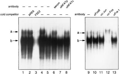
To determine whether adipocytes contain NFAT proteins, the 3T3-L1 preadipocyte cell line was differentiated in vitro as described (41) and cytoplasmic extracts were prepared from undifferentiated (day 0) and differentiated (days 2 and 6) cells. Western blot analysis using a mAb specific for NFATp and a polyclonal antiserum against NFAT3 demonstrated the presence of both of these NFAT proteins in the cytoplasm of both preadipocytes and differentiated adipocytes (Fig. 2a). As a control for the specificity of the polyclonal NFAT3 antiserum that could potentially also detect NFATp proteins, Western blot analysis was performed on cytoplasmic extracts prepared from mesenteric fat tissues of NFATp-deficient (−/−) (39) and control (+/−) mice (Fig. 2b). A protein of the correct molecular weight for NFAT3 was detected in NFATp-deficient mice, confirming the presence of both NFATp and NFAT3 proteins in adipocytes. To determine whether the NFAT proteins in adipocytes bind to FSE2 DNA, EMSA was performed with nuclear extracts prepared from day 4 differentiated 3T3-L1 cells. Similar to what was observed in T cell extracts, adipocyte nuclear extracts gave two complexes on FSE2 DNA that are competed with an excess of wild-type unlabeled FSE2 probe but not with a nonspecific oligonucleotide containing PPARγ2 (ARE6) sites (Fig. 3, lanes 1–3). Supershift analysis using the mAb against NFATp and anti-c-fos and c-jun antibodies revealed that the upper complex (complex a) contained NFATp and the lower complex (complex b) contained c-fos (Fig. 3, lanes 6–13). Interestingly, in contrast to what was observed in T cell extracts, the anti-cjun antibody did not supershift these complexes, consistent with the previously demonstrated binding of c-fos alone and not c-jun to the FSE2. We could not determine whether NFAT3 was present in the upper complex because the available antiserum does not recognize NFAT3 in EMSA.
Figure 2.
NFAT/AP-1 proteins are present in differentiated adipocytes and bind the FSE2. (a). Western blot analysis of cell extracts from differentiated 3T3L1 cells with antibodies to NFATp and NFAT3 is shown. (b) Western blot analysis of extracts prepared from the mesenteric fat of NFATp −/− and control NFAT +/+ mice with antibodies to NFAT3 is shown.
Figure 3.
NFATp binds to FSE2 DNA only in differentiated adipocytes. EMSA and supershift experiments were performed with nuclear extracts from either undifferentiated (lane 5) or day 4 differentiated (the remaining lanes) 3T3-L1 cells, an FSE2 radiolabeled probe, and antibodies to NFATp and AP-1 proteins.
NFATp Binds to FSE2 DNA Only in Differentiated Adipocytes.
NFATp and NFAT3 proteins are present in the cytoplasm of both preadipocytes and differentiated adipocytes (Fig. 2a). However, in T cells, NFAT proteins are present in the cytoplasm in resting cells and translocate to the nucleus upon T cell activation where they then bind DNA. We wondered whether a similar situation existed in adipocyte differentiation so that NFAT proteins in preadipocytes would not be present in the nucleus. To test this, nuclear extracts were prepared from undifferentiated and day 4 differentiated 3T3-L1 cells and EMSA was performed with a FSE2 labeled probe. Fig. 3, lanes 4 and 5, demonstrated that only differentiated adipocytes contained NFAT binding activity. In contrast, AP-1 binding activity was observed in both preadipocytes and differentiated adipocytes as demonstrated by the presence of the lower complex in all lanes. These data confirm that the upper complex contains NFAT proteins and the lower complex consists of AP-1 proteins. We conclude that NFAT proteins are present in both preadipocytes and adipocytes but only translocate into the nucleus upon fat cell differentiation.
NFATp Transactivates the aP2 Promoter in Fat Cells.
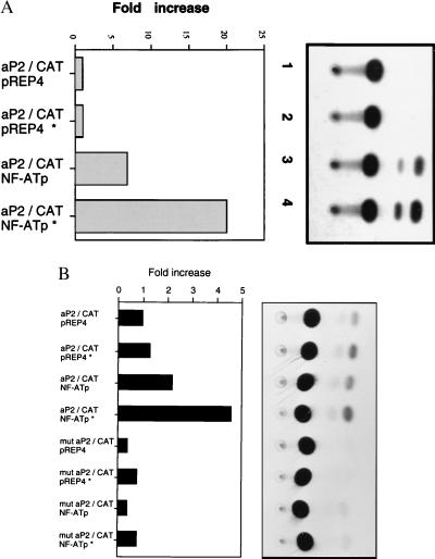
To directly test the functional contribution of NFAT proteins to aP2 regulation, an aP2 promoter reporter construct containing bases −168 to +20 of the aP2 promoter fused to the CAT reporter gene was cotransfected along with an NFATp expression construct into differentiating 3T3-L1 adipocytes. Very low basal activity of the aP2 reporter construct was detectable, and in the presence of NFATp, a substantial increase (approximately 7-fold) in CAT activity was observed (Fig. 4A). Treatment of the cells with phorbol 12-myristate 13-acetate and ionomycin, which has been shown to increase nuclear translocation of NFAT proteins, resulted in a further increase in promoter transactivation (approximately 20-fold). Further evidence that NFATp transactivates the aP2 reporter was obtained from experiments in which an aP2 reporter with a mutation in the NFAT site was used. The mutant reporter was much less active (3-fold) than the wild-type reporter and could not be transactivated by introduction of NFATp (Fig. 4B). These data demonstrate a functional role for NFATp in regulating the aP2 gene in vivo.
Figure 4.
NFATp transactivates the aP2 promoter in adipocytes. (A) 3T3L1 cells were cotransfected with 20 μg of an aP2-CAT reporter (positions −168 to +20) and 20 μg of an NFATp expression plasmid (pRep4 NFATp) or control pRep4 plasmid in the presence or absence of phorbol 12-myristate 13-acetate (2.5 nM) and ionomycin (2 μM). CAT activity determined 48 hr later. (B) The experiment in A was repeated with either the wild-type aP2-CAT reporter or an aP2 reporter with a mutation in the NFAT site. The data were normalized to the activity of the pRep control vector (=1). One representative experiment of three is shown.
Cyclosporin A Inhibits Adipocyte Differentiation.
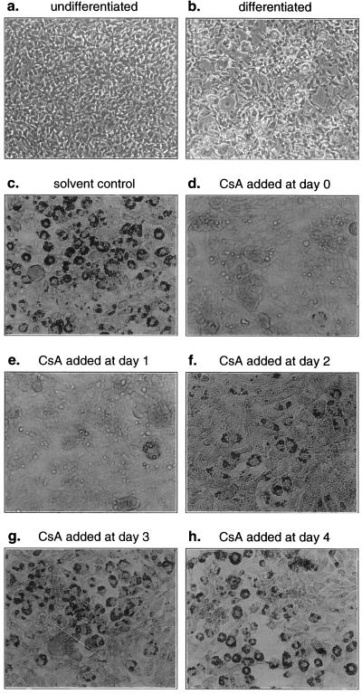
To further explore the role of NFAT proteins in adipogenesis, we took advantage of the known effect of the immunosuppressive drug CsA in inhibiting NFAT function in lymphocytes. Given the results shown above, we reasoned that if NFAT proteins were important in adipogenesis, then fat cell differentiation might be inhibited in the presence of CsA by virtue of its interference with NFAT nuclear translocation. The 3T3-L1 cells were therefore differentiated in vivo in the presence or absence of various concentrations of CsA or control solvent alone, and differentiation was assessed by staining with red oil O to detect triglyceride deposit accumulation. A striking effect of CsA in inhibiting fat cell differentiation was observed at a dose of 10 μg/ml and a significant effect was observed at 1 μg/ml (Fig. 5 a–d). Levels of C/EBPα and aP2 were greatly decreased in CsA-treated adipocytes (data not shown) consistent with the visualized inhibition of fat cell differentiation. Differentiation of 3T3-L1 fibroblasts was inhibited when CsA was introduced early (days 0 or 1), was less dramatic when introduced at day 2, and was not observed when CsA was added at later time points (days 3 or 4) (Fig. 5 d–h). Attempts to directly visualize the subcellular location of NFATp in 3T3L1 adipocytes by immunostaining were unfortunately unsuccessful.
Figure 5.
CsA inhibits adipocyte differentiation. (a) Undifferentiated 3T3-L1 cells. (b_–_d) T3L1 cells were differentiated for 6 days in the absence of control solvent (b), the presence of control solvent (c), or in CsA (10 μg/ml) (d) and stained with red oil O. The 3T3-L1 cells were differentiated in the presence of CsA (10 μg/ml) added at day 0 (d), day 1 (e), day 2 (f), day 3 (g), or day 4 (h).
DISCUSSION
Analysis of regulatory control regions of the fat-cell-specific gene aP2 has led to the identification of transcription factors critical for its basal and tissue-specific expression. The best characterized of these are members of the C/EBP and AP-1 families and the PPARγ2 (32, 35, 36, 41). Herein we present strong evidence that members of the NFAT transcription factor family are also important in aP2 gene regulation. NFATp and NFAT3 proteins are present in adipocytes and NFATp can bind to the FSE2 element and transactivate an aP2 reporter in vivo in fat cells. Further, the action of NFAT in regulating aP2 is controlled by differentiation because NFAT binding activity to the FSE2 was present in differentiated but not undifferentiated adipocytes. Finally, we demonstrate that CsA, an agent known to inhibit the nuclear localization of NFAT in lymphocytes inhibits fat cell differentiation. Given the presence of NFATp and NFAT3 proteins in undifferentiated adipocytes, these latter findings are most likely explained by the failure of NFAT to translocate into the nucleus although we were unable to directly visualize NFATp in adipocytes by immunostaining. Thus, our data provide evidence for a role of NFAT in regulating the expression of the fat-cell-specific gene aP2 and are consistent with a role for NFAT family members in adipogenesis.
The basic-region–leucine-zipper factor C/EBPα binds to the promoter region of several fat-cell-specific genes and appears to be key in terminal adipocyte differentiation. Overexpression of C/EBPα promotes adipogenesis in 3T3-L1 fibroblast cell lines (32, 33, 42–46). Recently, it has been demonstrated that forced expression of two other members of the C/EBP family, C/EBPδ and C/EBP/β, which are expressed at the onset of differentiation, accelerates adipogenesis in the presence of hormonal stimuli and leads to the activation of C/EBPα, thus placing them proximal to C/EBPα in the cascade of events leading to a fully differentiated fat cell (41, 47). PPARγ2 is an adipocyte-specific nuclear hormone receptor critical for the regulation of two fat cell enhancers (35). Retroviral expression of PPARγ2 promotes adipogenesis in NIH 3T3 fibroblasts and, when coexpressed with C/EBPα, acts synergistically to promote adipocyte differentiation (36). Ligand activation of PPARγ also promotes adipogenesis in vivo. Indeed, ectopic expression of C/EBPβ and C/EBPδ in the presence of glucocorticoids activates PPARγ2 gene expression (47). Nevertheless, mice lacking C/EBPβ and/or C/EBPδ do express PPARγ and C/EBPα despite impaired adipocyte differentiation (48). Thus, complete adipocyte differentiation likely requires all four of these factors. The C/EBPα and PPARγ2 proteins thus act in concert to achieve adipocyte conversion from mesodermal precursor cells. Exactly where NFAT fits in the constellation of transcription factors already known to be involved in adipogenesis is unclear, but possibilities may be inferred from the inhibition of adipogenesis by the immunosuppressive agent CsA.
The mechanism by which CsA impedes adipogenesis is likely multifactorial, but the stage at which CsA must be added to accomplish this may provide clues as to the role of NFAT in regulating adipogenesis. Triglyceride formation could be inhibited if CsA was added at day 0 and day 1 but was only inhibited when it was added before day 2 (Fig. 5). This result demonstrates both that CsA needs to be introduced early in the differentiation process to inhibit adipogenesis and that CsA cannot reverse this process once it has passed a critical point. Further, we observed that levels of PPARγ2 were unchanged in the presence of CsA, although levels of C/EBPα were diminished (unpublished observations). Thus these data suggest that NFAT acts at a time subsequent to PPARγ2 induction and before C/EBPα induction. This raises the possibility that the induction of the C/EBP β, δ, or α genes themselves are controlled in part by NFAT family members. Sequence examination of the promoter regions of the C/EBPα and β genes did not reveal obvious NFAT target elements, but only limited sequence information is available (49–52).
It has been reported that fat cell differentiation could be inhibited by rapamycin but not by either CsA or FK506 (53). Those experiments used the preparation of CsA given to patients that is dissolved in a cremophore carrier, whereas our experiments were performed with two separate batches of the pure crystalline powder. In an attempt to reconcile our findings with the published report, we repeated our experiments with the cremophore-based CsA preparation. We found that this preparation had only a modest, although clearly detectable, inhibitory effect on fat cell differentiation compared with the pure preparations used (I.-C.H. and H.-J.K., unpublished data). The discrepant results obtained are then likely due to differences in the purity of the CsA. It is possible that the cremophore carrier diminishes the efficiency of uptake of CsA by the 3T3-L1 cells compared with the purified powder form that has most frequently been used in in vitro assays.
Given the tightly restricted expression of NFATc and NFAT4 to the lymphoid system in the adult, it is likely that NFATp and/or NFAT3 will be the NFAT family members involved in adipogenesis. Indeed, the majority of NFAT binding activity in fat cell extracts could be supershifted with the anti-NFATp antibody. However, NFATp-deficient mice manifest no defects in fat tissue either by gross visual inspection or by histological analysis (I.-C.H. and L.H.G., unpublished data). This raises the possibility that NFAT3 may be important in fat cell development or, alternatively, that the two proteins can compensate for each other. The production and analysis of NFAT3-deficient mice and their intercross with the NFATp-deficient strain already available should be valuable in addressing this issue.
Acknowledgments
This work was supported by National Institutes of Health Grant AI/AG37833 (L.H.G.), a grant from The G. Harold and Leila Y. Mathers Charitable Foundation (L.G.H.), a Howard Hughes Medical Institute Fellowship (I.-C.H.). and The Arthritis Foundation (I.-C.H.).
ABBREVIATIONS
NFAT
nuclear factor of activated T cells
IL
interleukin
CsA
cyclosporin A
FSE2
fat-specific element 2
PPARγ2
peroxisome proliferator-activated receptor γ2
Footnotes
This paper was submitted directly (Track II) to the Proceedings Office.
References
- 1.Rao A, Luo C, Hogan P G. Annu Rev Immunol. 1997;15:707–747. doi: 10.1146/annurev.immunol.15.1.707. [DOI] [PubMed] [Google Scholar]
- 2.Durand D, Shaw J, Bush M, Replogle R, Belagaje R, Crabtree G. Mol Cell Biol. 1988;8:1715–1724. doi: 10.1128/mcb.8.4.1715. [DOI] [PMC free article] [PubMed] [Google Scholar]
- 3.Shaw J, Utz P, Durand D, Toole J, Emmel E, Crabtree G. Science. 1988;241:202–205. [PubMed] [Google Scholar]
- 4.Crabtree G. Science. 1989;249:355–360. doi: 10.1126/science.2783497. [DOI] [PubMed] [Google Scholar]
- 5.Northrop J P, Ho S N, Chen L, Thomas D J, Timmerman L A, Nolan G P, Admon A, Crabtree G R. Nature (London) 1994;369:497–502. doi: 10.1038/369497a0. [DOI] [PubMed] [Google Scholar]
- 6.McCaffrey P G, Luo C, Kerppola T K, Jain J, Badalian T M, Ho A M, Burgeon E, Lane W S, Lambert J N, Curran T, et al. Science. 1993;262:750–754. doi: 10.1126/science.8235597. [DOI] [PubMed] [Google Scholar]
- 7.Hoey T, Sun Y-L, Williamson K, Xu X. Immunity. 1995;2:461–472. doi: 10.1016/1074-7613(95)90027-6. [DOI] [PubMed] [Google Scholar]
- 8.Masuda E S, Naito Y, Tokumitsu H, Campbell D, Saito F, Hannum C, Arai K-i, Arai N. Mol Cell Biol. 1995;15:2697–2706. doi: 10.1128/mcb.15.5.2697. [DOI] [PMC free article] [PubMed] [Google Scholar]
- 9.Rooney J W, Hoey T, Glimcher L H. Immunity. 1995;2:545–553. [Google Scholar]
- 10.Jain J, McCaffrey P G, Valge-Archer V E, Rao A. Nature (London) 1992;356:801–804. doi: 10.1038/356801a0. [DOI] [PubMed] [Google Scholar]
- 11.Jain J, McCaffrey P G, Miner Z, Kerppola T K, Lambert J N, Verdine G L, Curran T, Rao A. Nature (London) 1993;365:352–355. doi: 10.1038/365352a0. [DOI] [PubMed] [Google Scholar]
- 12.Rooney J, Sun Y-L, Glimcher L H, Hoey T. Mol Cell Biol. 1995;15:6299–6310. doi: 10.1128/mcb.15.11.6299. [DOI] [PMC free article] [PubMed] [Google Scholar]
- 13.Boise L H, Petryniak B, Mao X, June C H, Wang C -Y, Lindsten T, Bravo R, Kovary K, Leiden J M, Thompson C B. Mol Cell Biol. 1993;13:1911–1919. doi: 10.1128/mcb.13.3.1911. [DOI] [PMC free article] [PubMed] [Google Scholar]
- 14.Beals C R, Clipstone N A, Ho S N, Crabtree G R. Genes Dev. 1997;11:824–834. doi: 10.1101/gad.11.7.824. [DOI] [PubMed] [Google Scholar]
- 15.Clipstone N A, Crabtree G R. Nature (London) 1992;357:695–697. doi: 10.1038/357695a0. [DOI] [PubMed] [Google Scholar]
- 16.Flanagan W M, Corthesy B, Bram R J, Crabtree G R. Nature (London) 1991;352:803–807. doi: 10.1038/352803a0. [DOI] [PubMed] [Google Scholar]
- 17.Emmel E A, Verweij C L, Durand D B, Higgins K M, Lacy E, Crabtree G R. Science. 1989;246:1617–1620. doi: 10.1126/science.2595372. [DOI] [PubMed] [Google Scholar]
- 18.Beals C R, Sheridan C M, Turck C W, Gardner P, Crabtree G R. Science. 1997;275:1930–1933. doi: 10.1126/science.275.5308.1930. [DOI] [PubMed] [Google Scholar]
- 19.Ranger A M, Grusby M J, Hodge M R, Gravallese E M, Charles de la Brousse F, Hoey T, Mickanin C, Baldwin H S, Glimcher L H. Nature (London) 1998;392:186–190. doi: 10.1038/32426. [DOI] [PubMed] [Google Scholar]
- 20.He X, Saint-Jeannet J P, Woodgett J R, Varmus H E, Dawid I B. Nature (London) 1995;374:617–622. doi: 10.1038/374617a0. [DOI] [PubMed] [Google Scholar]
- 21.Bourouis M, Moore P, Ruel L, Grau Y, Heitzler P, Simpson P. EMBO J. 1990;9:2877–2884. doi: 10.1002/j.1460-2075.1990.tb07477.x. [DOI] [PMC free article] [PubMed] [Google Scholar]
- 22.Cook D, Fry M J, Hughes K, Sumathipala R, Woodgett J R, Dale T C. EMBO J. 1996;15:4526–4536. [PMC free article] [PubMed] [Google Scholar]
- 23.Green H, Kehinde O. Cell. 1974;1:113–116. [Google Scholar]
- 24.Green H, Kehinde O. Cell. 1975;5:19–27. doi: 10.1016/0092-8674(75)90087-2. [DOI] [PubMed] [Google Scholar]
- 25.Student A K, Hsu R Y, Lane M D. J Biol Chem. 1980;255:4745–4750. [PubMed] [Google Scholar]
- 26.Bernlohr D A, Angus C W, Lane M D, Bolanowski M A, Kelly T J. Proc Natl Acad Sci USA. 1994;81:5468–5472. doi: 10.1073/pnas.81.17.5468. [DOI] [PMC free article] [PubMed] [Google Scholar]
- 27.Cook K S, Hunt C R, Spiegelman B M. J Cell Biol. 1985;100:514–520. doi: 10.1083/jcb.100.2.514. [DOI] [PMC free article] [PubMed] [Google Scholar]
- 28.Spiegelman B M, Frank M, Green H. J Biol Chem. 1983;258:10083–10089. [PubMed] [Google Scholar]
- 29.Distel R J, Ro H-S, Rosen B S, Groves D L, Spiegelman B M. Cell. 1987;49:835–844. doi: 10.1016/0092-8674(87)90621-0. [DOI] [PubMed] [Google Scholar]
- 30.Hunt C R, Ro J H-S, Dobson D, Min H Y, Spiegelman B M. Proc Natl Acad Sci USA. 1986;83:3786–3790. doi: 10.1073/pnas.83.11.3786. [DOI] [PMC free article] [PubMed] [Google Scholar]
- 31.Phillips M, Djian P, Green H. J Biol Chem. 1986;261:10821–10827. [PubMed] [Google Scholar]
- 32.Lin F T, Lane M D. Proc Natl Acad Sci USA. 1994;91:8757–8761. doi: 10.1073/pnas.91.19.8757. [DOI] [PMC free article] [PubMed] [Google Scholar]
- 33.Christy R J, Yang V W, Ntambi J M, Geiman D E, Landschulz W H, Friedman A D, Nakabeppu Y, Kelly T J, Lane M D. Genes Dev. 1989;3:1323–1335. doi: 10.1101/gad.3.9.1323. [DOI] [PubMed] [Google Scholar]
- 34.Ross S R, Graves R A, Greenstein A, Platt K A, Shyu H-L, Mellovitz B, Spiegelman B M. Proc Natl Acad Sci USA. 1990;87:9590–9594. doi: 10.1073/pnas.87.24.9590. [DOI] [PMC free article] [PubMed] [Google Scholar]
- 35.Tontonoz P, Hu E, Spiegelman B M. Cell. 1994;79:1147–2256. doi: 10.1016/0092-8674(94)90006-x. [DOI] [PubMed] [Google Scholar]
- 36.Tontonoz P, Hu E, Graves R A, Budavari A I, Spiegelman B M. Genes Dev. 1994;8:1224–1234. doi: 10.1101/gad.8.10.1224. [DOI] [PubMed] [Google Scholar]
- 37.Hecht T T, Longo D L, Matis L A. J Immunol. 1983;131:1049–1055. [PubMed] [Google Scholar]
- 38.Kaye J, Porcelli S, Tite J, Jones B, Janeway C A. J Exp Med. 1983;158:836–856. doi: 10.1084/jem.158.3.836. [DOI] [PMC free article] [PubMed] [Google Scholar]
- 39.Hodge M R, Ranger A M, Charles de la Brousse F, Hoey T, Grusby M J, Glimcher L H. Immunity. 1996;4:1–20. doi: 10.1016/s1074-7613(00)80253-8. [DOI] [PubMed] [Google Scholar]
- 40.Hodge M R, Chun H J, Rengarajan J, Alt A, Lieberson R, Glimcher L H. Science. 1996;274:1903–1905. doi: 10.1126/science.274.5294.1903. [DOI] [PubMed] [Google Scholar]
- 41.Yeh W-C, Cao Z, Classon M, McKnight S L. Genes Dev. 1995;9:168–181. doi: 10.1101/gad.9.2.168. [DOI] [PubMed] [Google Scholar]
- 42.Lin F-T, Lane M D. Genes Dev. 1992;6:533–544. doi: 10.1101/gad.6.4.533. [DOI] [PubMed] [Google Scholar]
- 43.Samuelsson L, Stromberg K, Vikman K, Bjursell G, Enerback S. EMBO J. 1991;10:3787–3793. doi: 10.1002/j.1460-2075.1991.tb04948.x. [DOI] [PMC free article] [PubMed] [Google Scholar]
- 44.Umek R, Friedman A, McKnight S L. Science. 1991;251:288–292. doi: 10.1126/science.1987644. [DOI] [PubMed] [Google Scholar]
- 45.Freytag S, Geddes T. Science. 1992;256:379–382. doi: 10.1126/science.256.5055.379. [DOI] [PubMed] [Google Scholar]
- 46.Kaestner K, Christy R, Lane M D. Proc Natl Acad Sci USA. 1990;87:251–255. doi: 10.1073/pnas.87.1.251. [DOI] [PMC free article] [PubMed] [Google Scholar]
- 47.Wu Z, Bucher N L R, Farmer S R. Mol Cell Biol. 1996;16:4128–4136. doi: 10.1128/mcb.16.8.4128. [DOI] [PMC free article] [PubMed] [Google Scholar]
- 48.Tanaka T, Yoshida N, Kishimoto T, Akira S. EMBO J. 1997;16:7432–7443. doi: 10.1093/emboj/16.24.7432. [DOI] [PMC free article] [PubMed] [Google Scholar]
- 49.Talbot D, Descombes P, Schibler U. Nuc Acid Res. 1994;22:756–766. doi: 10.1093/nar/22.5.756. [DOI] [PMC free article] [PubMed] [Google Scholar]
- 50.Timchenko N, Wilson D R, Taylor L R, Abdelsayed S, Wilde M, Sawadogo M, Darlington G J. Mol Cell Biol. 1995;15:1192–1202. doi: 10.1128/mcb.15.3.1192. [DOI] [PMC free article] [PubMed] [Google Scholar]
- 51.Chang C-J, Shen B-J, Lee S-C. DNA Cell Biol. 1995;14:529–537. doi: 10.1089/dna.1995.14.529. [DOI] [PubMed] [Google Scholar]
- 52.Niehof M, Manns M P, Trautwein C. Mol Cell Biol. 1997;17:3600–3613. doi: 10.1128/mcb.17.7.3600. [DOI] [PMC free article] [PubMed] [Google Scholar]
- 53.Yeh W-C, Bierer B E, McKnight S L. Proc Natl Acad Sci USA. 1995;92:11086–11090. doi: 10.1073/pnas.92.24.11086. [DOI] [PMC free article] [PubMed] [Google Scholar]
