Cerebral cortical blood flow maps are reorganized in MAOB-deficient mice - PubMed (original) (raw)
Cerebral cortical blood flow maps are reorganized in MAOB-deficient mice
O U Scremin et al. Brain Res. 1999.
Abstract
Cerebral cortical blood flow (CBF) was measured autoradiographically in conscious mice without the monoamine oxidase B (MAOB) gene (KO, n=11) and the corresponding wild-type animals (WILD, n=11). Subgroups of animals of each genotype received a continuous intravenous infusion over 30 min of phenylethylamine (PEA), an endogenous substrate of MAOB, (8 nmol g-1 min-1 in normal saline at a volume rate of 0.11 microl g-1 min-1) or saline at the same volume rate. Maps of relative CBF distribution showed predominance of midline motor and sensory area CBF in KO mice over WILD mice that received saline. PEA enhanced CBF in lateral frontal and piriform cortex in both KO and WILD mice. These changes may reflect a differential activation due to chronic and acute PEA elevations on motor and olfactory function, as well as on the anxiogenic effects of this amine. In addition to its effects on regional CBF distribution, PEA decreased CBF globally in KO mice (range -31% to -41% decrease from control levels) with a lesser effect in WILD mice. It is concluded that MAOB may normally regulate CBF distribution and its response to blood PEA.
Copyright 1999 Elsevier Science B.V.
Figures
Fig. 1
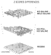
Bars represent _Z_-score differences between KO mice and WILD mice receiving saline (top figure), and KO mice receiving saline or PEA (middle figure). Negative values are shown with bars projecting below the zero plane (black top). Coronal slices are numbered from rostral to caudal, with distance to bregma as follows (positive values being rostral to this landmark): (1) 1.54 mm, (2) 1.18 mm, (3) 0.74 mm, (4) 0.26 mm, (5) −0.34 mm, (6) −0.82 mm, (7) −1.34 mm, (8) −1.82 mm, (9) −2.46 mm, (10) −2.92 mm, (11) −3.64 mm. The cerebral cortical locations corresponding to each measurement are numbered from dorsal midline to lateral and ventral. These are shown in the bottom panel. Abbreviations: AI = agranular insular, AU = auditory, BF = barrel field, C1 = cingulate, CA = amygdaloid, EC = ectorhinal, EN = entorhinal, GI = granular insular, M1 = primary motor, M2 = secondary motor, PI = piriform, PR = perirhinal, RS = retrosplenial, S1 = primary somatosensory, S2 = secondary somatosensory, TA = transition between barrel field and secondary somatosensory, TB = transition between primary somatosensory and primary motor, TC = transition between secondary somatosensory and perirhinal, TD = transition between cingulate and motor, V1 = primary visual, V2 = secondary visual. Outlined areas are those listed in Table 1.
Fig. 2
Canonical plot for the two first canonical variables providing discrimination between the relative CBF (_Z_-scores) of KO and WILD mice receiving saline, and by infusion type (PEA or saline) for both KO and WILD mice. For details see text.
Fig. 3
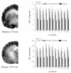
Two representative iodo-14C-antipyrine autoradiographs of slice 4 and slice 6 are shown in the left panels. Regions sampled are indicated by numerals. The corpus callosum and anterior commissure (slice 4) and the corpus callosum and hippocampal commissure (slice 6) are outlined as reference. The bar graphs (right panels) indicate group means and standard errors of CBF of the regions (numerals), as well as the slice means (ALL) in units of ml g−1 min−1. Abbreviations: WILD + S = WILD mice receiving saline; WILD + PEA = WILD mice receiving PEA; KO + S = KO mice receiving saline; KO + PEA = KO mice receiving PEA. * Statistically significant when compared to WILD + S, P < 0.05, Bonferroni correction for three contrasts.
Similar articles
- Regional cerebral cortical activation in monoamine oxidase A-deficient mice: differential effects of chronic versus acute elevations in serotonin and norepinephrine.
Holschneider DP, Scremin OU, Huynh L, Chen K, Seif I, Shih JC. Holschneider DP, et al. Neuroscience. 2000;101(4):869-77. doi: 10.1016/s0306-4522(00)00436-x. Neuroscience. 2000. PMID: 11113335 Free PMC article. - Phenethylamine is a substrate of monoamine oxidase B in the paraventricular thalamic nucleus.
Obata Y, Kubota-Sakashita M, Kasahara T, Mizuno M, Nemoto T, Kato T. Obata Y, et al. Sci Rep. 2022 Jan 7;12(1):17. doi: 10.1038/s41598-021-03885-6. Sci Rep. 2022. PMID: 34996979 Free PMC article. - Increased stress response and beta-phenylethylamine in MAOB-deficient mice.
Grimsby J, Toth M, Chen K, Kumazawa T, Klaidman L, Adams JD, Karoum F, Gal J, Shih JC. Grimsby J, et al. Nat Genet. 1997 Oct;17(2):206-10. doi: 10.1038/ng1097-206. Nat Genet. 1997. PMID: 9326944 - [MAOB: a modifier gene in phenylketonuria?].
Ghozlan A, Munnich A. Ghozlan A, et al. Med Sci (Paris). 2004 Oct;20(10):929-32. doi: 10.1051/medsci/20042010929. Med Sci (Paris). 2004. PMID: 15461973 Review. French. - Cloning, after cloning, knock-out mice, and physiological functions of MAO A and B.
Shih JC. Shih JC. Neurotoxicology. 2004 Jan;25(1-2):21-30. doi: 10.1016/S0161-813X(03)00112-8. Neurotoxicology. 2004. PMID: 14697877 Review.
Cited by
- Biochemical, behavioral, physiologic, and neurodevelopmental changes in mice deficient in monoamine oxidase A or B.
Holschneider DP, Chen K, Seif I, Shih JC. Holschneider DP, et al. Brain Res Bull. 2001 Nov 15;56(5):453-62. doi: 10.1016/s0361-9230(01)00613-x. Brain Res Bull. 2001. PMID: 11750790 Free PMC article. Review. - Low monoamine oxidase B in peripheral organs in smokers.
Fowler JS, Logan J, Wang GJ, Volkow ND, Telang F, Zhu W, Franceschi D, Pappas N, Ferrieri R, Shea C, Garza V, Xu Y, Schlyer D, Gatley SJ, Ding YS, Alexoff D, Warner D, Netusil N, Carter P, Jayne M, King P, Vaska P. Fowler JS, et al. Proc Natl Acad Sci U S A. 2003 Sep 30;100(20):11600-5. doi: 10.1073/pnas.1833106100. Epub 2003 Sep 12. Proc Natl Acad Sci U S A. 2003. PMID: 12972641 Free PMC article. - Monoamine oxidases in development.
Wang CC, Billett E, Borchert A, Kuhn H, Ufer C. Wang CC, et al. Cell Mol Life Sci. 2013 Feb;70(4):599-630. doi: 10.1007/s00018-012-1065-7. Epub 2012 Jul 11. Cell Mol Life Sci. 2013. PMID: 22782111 Free PMC article. Review. - Monoamine oxidase inactivation: from pathophysiology to therapeutics.
Bortolato M, Chen K, Shih JC. Bortolato M, et al. Adv Drug Deliv Rev. 2008 Oct-Nov;60(13-14):1527-33. doi: 10.1016/j.addr.2008.06.002. Epub 2008 Jul 4. Adv Drug Deliv Rev. 2008. PMID: 18652859 Free PMC article. Review. - Regional cerebral cortical activation in monoamine oxidase A-deficient mice: differential effects of chronic versus acute elevations in serotonin and norepinephrine.
Holschneider DP, Scremin OU, Huynh L, Chen K, Seif I, Shih JC. Holschneider DP, et al. Neuroscience. 2000;101(4):869-77. doi: 10.1016/s0306-4522(00)00436-x. Neuroscience. 2000. PMID: 11113335 Free PMC article.
References
- B A. Bailey, S R. Philips, A A. Boulton. In vivo release of endogenous dopamine, 5-hydroxytryptamine and some of their metabolites from rat caudate nucleus by phenylethylamine. Neurochem. Res. 1987;12:173–178. - PubMed
- Boulton AA. Some aspects of basic psychopharmacology: the trace amines, Prog. Neuro-Psychopharmacol. Biol. Psychiatry. 1982;6:563–570. - PubMed
- Bussone G, Giovannini P, Boiardi A, Boeri R. A study of the activity of platelet monoamine oxidase in patients with migraine headaches or with ‘cluster headaches’. Eur. Neurol. 1977;15:157–162. - PubMed
- Caramona MM, Cotrim MD, Ribeiro CF, Macedo T. Monoamine oxidase activity in blood platelets of migraine patients. J. Neural Transm. Suppl. 1990;32:161–164. - PubMed
- Clark C, Carson R, Kessler R, Margolin R, Buchsbaum M, DeLisi L, King C, Cohen R. Alternative statistical models for the examination of clinical positron emission tomography/fluorodeoxyglucose data. J. Cereb. Blood Flow Metab. 1985;5:142–150. - PubMed
Publication types
MeSH terms
Substances
Grants and funding
- 5-K12-AG-00521/AG/NIA NIH HHS/United States
- K05 MH 00796/MH/NIMH NIH HHS/United States
- K12 AG000521/AG/NIA NIH HHS/United States
- R37 MH39085/MH/NIMH NIH HHS/United States
- R37 MH039085/MH/NIMH NIH HHS/United States
LinkOut - more resources
Full Text Sources
Research Materials
Miscellaneous


