P-Selectin or intercellular adhesion molecule (ICAM)-1 deficiency substantially protects against atherosclerosis in apolipoprotein E-deficient mice - PubMed (original) (raw)
P-Selectin or intercellular adhesion molecule (ICAM)-1 deficiency substantially protects against atherosclerosis in apolipoprotein E-deficient mice
R G Collins et al. J Exp Med. 2000.
Abstract
The expression of leukocyte and endothelial cell adhesion molecules (CAMs) is essential for the emigration of leukocytes during an inflammatory response. The importance of the inflammatory response in the development of atherosclerosis is indicated by the increased expression of adhesion molecules, proinflammatory cytokines, and growth factors in lesions and lesion-prone areas and by protection in mice deficient in various aspects of the inflammatory response. We have quantitated the effect of deficiency for intercellular adhesion molecule (ICAM)-1, P-selectin, or E-selectin on atherosclerotic lesion formation at 20 wk of age in apolipoprotein (apo) E(-/-) (deficient) mice fed a normal chow diet. All mice were apo E(-/-) and CAM(+/+) or CAM(-/-) littermates, and no differences were found in body weight or cholesterol levels among the various genotypes during the study. ICAM-1(-/-) mice had significantly less lesion area than their ICAM-1(+/+) littermates: 4.08 +/- 0.70 mm(2) for -/- males vs. 5.87 +/- 0.66 mm(2) for +/+ males, and 3.95 +/- 0. 65 mm(2) for -/- females vs. 5.59 +/- 1.131 mm(2) for +/+ females, combined P < 0.0001. An even greater reduction in lesion area was observed in P-selectin(-/-) mice: 3.06 +/- 1.04 mm(2) for -/- males vs. 5.09 +/- 1.22 mm(2) for +/+ males, and 2.85 +/- 1.26 mm(2) for -/- females compared with 5.60 +/- 1.19 mm(2) for +/+ females, combined P < 0.001. The reduction in lesion area for the E-selectin null mice, although less than that seen for ICAM-1 or P-selectin, was still significant (4.54 +/- 2.14 mm(2) for -/- males vs. 5.92 +/- 0.63 mm(2) for +/+ males, and 4.38 +/- 0.85 mm(2) for -/- females compared with 5.94 +/- 1.44 mm(2) for +/+ females, combined P < 0.01). These results, coupled with the closely controlled genetics of this study, indicate that reductions in the expression of P-selectin, ICAM-1, or E-selectin provide direct protection from atherosclerotic lesion formation in this model.
Figures
Figure 1
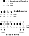
Breeding scheme for generating study animals. All animals in the study were apo E−/−. The animals for each CAM mutation are all descendants of a single founder pair of mice.
Figure 3
Histopathologic comparison of aortas from study animals. Sections shown are typical of the most advanced lesions found in animals of each genotype. (A) C57BL/6 wild-type murine aorta with intact intimal (top), unremarkable media (middle), and adventitial (bottom) layers with no evidence of atheromatous lesion. (B) Aorta from apo E−/− mouse with no CAM deficiency showing well formed atheromatous plaque characterized by foam cells, cell debris, cholesterol clefts, and calcification (arrow) within the expanded intima. Lesions exhibiting the calcification seen only in very advanced lesions were only found in apo E−/− mice with no CAM deficiency. (C) Aorta from an apo E−/−ICAM-1−/− animal with lesion composed of foam cells, cholesterol clefts, and extracellular lipid in expanded intima (arrow). (D) Aorta from apo E−/− P-selectin−/− animal with less developed atherosclerotic lesions (arrows) composed of frequent foam cells within expanded intima. Note relatively normal aortic segment between atheromatous lesions. (All hematoxylin and eosin stain with original magnification of 200).
Figure 2
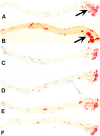
Oil Red O–stained lesions on aortas from apo E−/− mice. (A and B) Representative aortas from P-selectin+/+ and ICAM-1+/+ males, respectively. Total lesion area on these aortas was 5.81 mm2 for A and 6.26 mm2 for B. Arrows point to more advanced lesions in the aortic arch. (C and D) Aortas from two P-selectin−/− males with total lesion areas of 3.08 and 2.91 mm2, respectively. Note the relative absence of lesions throughout the aorta compared with CAM+/+ mice in A and B. (E and F) Aortas from ICAM-1−/− mice with lesion areas of 4.38 and 3.90 mm2, respectively. Early lesions are present in the aortic arch.
Figure 4
Effect of CAM deficiency on lesion area in apo E−/− mice. Each data point represents total lesion area (mm2) in an animal's aorta between the aortic valve and the iliac branch for the sex and genotype shown. Error bars represent mean ± SD. Significant reduction in lesion area was seen in the animals of each experimental group (CAM mutation) with the P values shown. Along with the rigid control of genetics and husbandry, two-way ANOVA indicated that the protection seen was due to genotype, with no significant contribution from gender.
Similar articles
- Deficiency of inflammatory cell adhesion molecules protects against atherosclerosis in mice.
Nageh MF, Sandberg ET, Marotti KR, Lin AH, Melchior EP, Bullard DC, Beaudet AL. Nageh MF, et al. Arterioscler Thromb Vasc Biol. 1997 Aug;17(8):1517-20. doi: 10.1161/01.atv.17.8.1517. Arterioscler Thromb Vasc Biol. 1997. PMID: 9301629 - The atheroprotective effect of 17 beta-estradiol is not altered in P-selectin- or ICAM-1-deficient hypercholesterolemic mice.
Gourdy P, Mallat Z, Castano C, Garmy-Susini B, Mac Gregor JL, Tedgui A, Arnal JF, Bayard F. Gourdy P, et al. Atherosclerosis. 2003 Jan;166(1):41-8. doi: 10.1016/s0021-9150(02)00322-2. Atherosclerosis. 2003. PMID: 12482549 - E/P-selectin-deficient mice: an optimal mutation for abrogating antigen but not tumor necrosis factor-alpha-induced immune responses.
McCafferty DM, Kanwar S, Granger DN, Kubes P. McCafferty DM, et al. Eur J Immunol. 2000 Aug;30(8):2362-71. doi: 10.1002/1521-4141(2000)30:8<2362::AID-IMMU2362>3.0.CO;2-F. Eur J Immunol. 2000. PMID: 10940927 - Endothelial-leukocyte adhesive interactions in inflammatory diseases.
Munro JM. Munro JM. Eur Heart J. 1993 Dec;14 Suppl K:72-7. Eur Heart J. 1993. PMID: 7510638 Review.
Cited by
- Impact of Probiotic Lactiplantibacillus plantarum ATCC 14917 on atherosclerotic plaque and its mechanism.
Hassan A, Luqman A, Zhang K, Ullah M, Din AU, Xiaoling L, Wang G. Hassan A, et al. World J Microbiol Biotechnol. 2024 May 10;40(7):198. doi: 10.1007/s11274-024-04010-1. World J Microbiol Biotechnol. 2024. PMID: 38727952 - Anti-atherosclerotic effects and molecular targets of ginkgolide B from Ginkgo biloba.
Ye W, Wang J, Little PJ, Zou J, Zheng Z, Lu J, Yin Y, Liu H, Zhang D, Liu P, Xu S, Ye W, Liu Z. Ye W, et al. Acta Pharm Sin B. 2024 Jan;14(1):1-19. doi: 10.1016/j.apsb.2023.09.014. Epub 2023 Sep 23. Acta Pharm Sin B. 2024. PMID: 38239238 Free PMC article. Review. - Role of Flow-Sensitive Endothelial Genes in Atherosclerosis and Antiatherogenic Therapeutics Development.
Baek KI, Ryu K. Baek KI, et al. J Cardiovasc Transl Res. 2024 Jun;17(3):609-623. doi: 10.1007/s12265-023-10463-w. Epub 2023 Nov 27. J Cardiovasc Transl Res. 2024. PMID: 38010480 Review. - Atherosclerosis from Newborn to Adult-Epidemiology, Pathological Aspects, and Risk Factors.
Luca AC, David SG, David AG, Țarcă V, Pădureț IA, Mîndru DE, Roșu ST, Roșu EV, Adumitrăchioaiei H, Bernic J, Cojocaru E, Țarcă E. Luca AC, et al. Life (Basel). 2023 Oct 14;13(10):2056. doi: 10.3390/life13102056. Life (Basel). 2023. PMID: 37895437 Free PMC article. Review.
References
- McGill H.C., Jr. Overview. In: Fuster V., Ross R., Topol E.J., editors. Atherosclerosis and Coronary Artery Disease. Lippincott-Raven; Philadelphia : 1996. pp. 25–41.
- Ross R. The pathogenesis of atherosclerosisa perspective for the 1990s. Nature. 1993;362:801–809 . - PubMed
- Ross R. Atherosclerosis—an inflammatory disease. N. Engl. J. Med. 1999;340:115–126 . - PubMed
- Springer T.A. Traffic signals for lymphocyte recirculation and leukocyte emigrationthe multistep paradigm. Cell. 1994;76:301–314 . - PubMed
- Richardson M., Hadcock S.J., DeReske M., Cybulsky M.I. Increased expression in vivo of VCAM-1 and E-selectin by the aortic endothelium of normolipemic and hyperlipemic diabetic rabbits. Arterioscler. Thromb. 1994;14:760–769 . - PubMed
Publication types
MeSH terms
Substances
LinkOut - more resources
Full Text Sources
Other Literature Sources
Molecular Biology Databases
Miscellaneous


