A nuclear export signal in the N-terminal regulatory domain of IkappaBalpha controls cytoplasmic localization of inactive NF-kappaB/IkappaBalpha complexes - PubMed (original) (raw)
A nuclear export signal in the N-terminal regulatory domain of IkappaBalpha controls cytoplasmic localization of inactive NF-kappaB/IkappaBalpha complexes
T T Huang et al. Proc Natl Acad Sci U S A. 2000.
Abstract
Appropriate subcellular localization is crucial for regulation of NF-kappaB function. Herein, we show that latent NF-kappaB complexes can enter and exit the nucleus in preinduction states. The nuclear export inhibitor leptomycin B (LMB) sequestered NF-kappaB/IkappaBalpha complexes in the nucleus. Using deletion and site-directed mutagenesis, we identified a previously uncharacterized nuclear export sequence in residues 45-54 of IkappaBalpha that was required for cytoplasmic localization of inactive complexes. This nuclear export sequence also caused nuclear exclusion of heterologous proteins in a LMB-sensitive manner. Importantly, a LMB-insensitive CRM1 mutant (Crm1-K1) abolished LMB-induced nuclear accumulation of the inactive complexes. Moreover, a cell-permeable p50 NF-kappaB nuclear localization signal peptide also blocked these LMB effects. These results suggest that NF-kappaB/IkappaBalpha complexes shuttle between the cytoplasm and nucleus by a nuclear localization signal-dependent nuclear import and a CRM1-dependent nuclear export. The LMB-induced nuclear complexes could not bind DNA and were inaccessible to signaling events, because LMB inhibited NF-kappaB activation without affecting the subcellular localization of upstream kinases IKKbeta and NIK. Our findings indicate that the dominant nuclear export over nuclear import contributes to the largely cytoplasmic localization of the inactive complexes to achieve efficient NF-kappaB activation by extracellular signals.
Figures
Figure 1
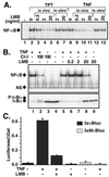
LMB inhibits signal-inducible IκBα degradation and NF-κB activation. (A) PC-3 cells were treated with topotecan (TPT; 30 μM for 60 min; lanes 2–5) or TNFα (10 ng/ml for 15 min; lanes 8–11) in the presence or absence of various doses of LMB pretreatment for 30 min. Nuclear extracts were analyzed by EMSA by using an Igκ-κB probe. LMB was also added directly to nuclear extracts (as in lanes 2 and 8) and processed as described above (lanes 6, 7, 12, and 13). A section of the autoradiogram containing NF-κB complexes is shown. (B) HeLa cells were pretreated with CI-I (100 μM for 30 min; lanes 2 and 3) or LMB (ng/ml for 30 min; lanes 6–9) and then treated with TNFα (10 ng/ml for 15 min; lanes 1, 2, and 5–8). Nuclear extracts were analyzed by EMSA as described above (Upper). NS refers to a nonspecific band. Total cell extracts from parallel cell samples described in B were analyzed by Western blotting with IκBα antibody. P-IκBα refers to the position of the phosphorylated IκBα (Lower). (C) HEK293 cells were transiently transfected with a NF-κB-dependent reporter plasmid (3xκB-Luc or 3xMκB-Luc for control) and an internal control for transfection efficiency (CMV-β-Gal). At 36 h after transfection, cells were treated with or without TNFα in the presence or absence of LMB treatment. Cell extracts were analyzed for luciferase and β-galactosidase activities. Error bars are SD.
Figure 2
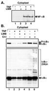
LMB fails to inhibit TNFα-induced IκBα degradation and NF-κB activation in enucleated cells. (A) HeLa cells were enucleated as described in Materials and Methods, and the resulting cytoplasts were pretreated with LMB (20 ng/ml for 30 min; lanes 2 and 3) and then treated with TNFα (10 ng/ml for 15 min; lanes 3 and 4). Total cell extracts were prepared and analyzed by EMSA. (B Upper) Cytoplasts as described in A were pretreated for 30 min with either LMB (20 ng/ml, lanes 3 and 4) or CI-I (100 μM, lanes 5 and 6) and then treated with TNFα (lanes 2, 3, and 6). Samples were analyzed by Western blotting with IκBα antibody. (B Lower) A longer exposure of the Western blot used for B Upper is shown.
Figure 3
LMB causes nuclear accumulation of IκBα without de novo protein synthesis or basal IκBα degradation. (A) A pool of HEK293 cells stably expressing GFP-IκBα was treated with TNFα (20 ng/ml) for the indicated times (lanes 2–6) and analyzed by Western blotting with IκBα antibody. Equal cell extracts prepared from untreated (lane 7) or LMB-treated (20 ng/ml for 60 min; lane 8) cells were processed for immunoprecipitation with p65 A antibody and analyzed by Western blotting with IκBα antibody. Horseradish peroxidase-conjugated protein A was used to detect bands. (B) HEK293 cells stably expressing the GFP-IκBα protein were untreated or treated with LMB (10 ng/ml for 30 min) and visualized under fluorescein-aided fluorescent microscopy. (C) PC-3 cells were untreated or treated with LMB as described for B, fixed, and stained with either IκBα (Upper) or p65 C20 (Lower) antibodies. (D) PC-3 cells were treated as described for C or with TNFα (10 ng/ml for 15 min), and the cell extracts were immunoprecipitated (IP) with p65 A antibody and analyzed by IκBα Western blotting with horseradish peroxidase-conjugated goat anti-rabbit antibody (lanes 1–3). (E) HEK293 cells stably expressing GFP-IκBα were pretreated with cycloheximide (CX; 20 μg/ml for 60 min), untreated or treated with LMB (20 ng/ml for 15 min), and then visualized as described above. (F) HEK293 cells stably expressing GFP-IκBα were pretreated with CI-I (100 μM for 60 min), untreated or treated with LMB, and then visualized.
Figure 4
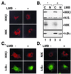
LMB does not cause nuclear accumulation of IKKβ and NIK. (A) Cos-7 cells were transfected with either Flag-tagged IKKβ (2 μg) or NIK (2 μg) expression vector and treated or left untreated with LMB (20 ng/ml for 60 min) 24 h after transfection. Cells were visualized with IKKβ and NIK antibodies, respectively. (B) PC-3 cells were untreated (lanes 1 and 2) or treated with LMB (20 ng/ml for 60 min; lanes 3 and 4), and crude cytoplasmic and nuclear extracts prepared as described (25) were analyzed by Western blotting with IKKβ-, NIK-, and IκBα-specific antibodies. N.S. refers to a nonspecific band. (C) Cos-7 cells were cotransfected with IκBα (1.0 μg), p50 (1.0 μg), and p65 (1.0 μg) constructs along with a Flag-tagged IKKβ (2 μg) construct, untreated or treated with LMB as described for A, and stained with IκBα and Flag antibodies. (D) Cos-7 cells were transfected and visualized as described for B, except that Flag-tagged NIK (2 μg) was used instead of Flag-tagged IKKβ.
Figure 5
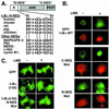
Identification of N-NES of IκBα. (A) N-NESs in different species are aligned with those of previously characterized shuttling proteins. The critical hydrophobic residues are boxed. ANK, ankyrin repeats; PEST, region rich in proline (P), glutamate (E), serine (S), and threonine (T). (B) GFP-IκBα wild type (WT) (1.0 μg) and GFP-IκBα mutants (1.0 μg) were transfected with p50 and p65 (1.0 μg each) expression vectors into Cos-7 cells and left untreated or treated with LMB (20 ng/ml for 60 min). GFP-IκBα and p65 were covisualized with GFP fluorescence (green) and by staining with p65 antibody (red). In the GFP-IκBα mutants, critical hydrophobic residues (bold) in C-NES (IQQQLGQLTLENLQML) and N-NES (MVKELQEIRLEP) were substituted with Ala. (C) GFP alone, IκBα N-terminal 55, and 55 residues with NES mutations as described above were fused C-terminally to GFP and transfected into Cos-7 cells. GFP and GFP fusion proteins were visualized directly in living cells with GFP fluorescence with or without LMB treatment.
Figure 6
Inhibition of NF-κB/IκBα nucleocytoplasmic shuttling by a p50 NLS peptide and a LMB-insensitive CRM1 mutant. (A) Empty vector, CRM1 wild type (WT), or LMB-insensitive Crm1-K1 mutant was transfected into HEK293 cells stably expressing GFP-IκBα. Cells were then treated or untreated with LMB (20 ng/ml for 15 min) and visualized with GFP fluorescence. (B) HeLa cells were pretreated with either LMB (20 ng/ml for 30 min; lane 4) or cell-permeable p50 NLS peptide (NF-κB SN50) for 1 h (100 μg/ml; lane 3) and stimulated with TNFα (10 ng/ml for 15 min; lanes 2 and 3). Nuclear extracts were analyzed for NF-κB activity with EMSA (Upper). Cytoplasmic extracts from the same samples were analyzed by Western blotting for IκBα and α-tubulin (loading control). (C) HeLa cells were untreated or treated with SN50 (100 μg/ml for 1 h) and then either untreated (Top) or treated with LMB (20 ng/ml for 30 min; Middle) and TNFα (10 ng/ml for 30 min; Lower). Cells were then stained with p65 antibody as described above. (D) Model of nucleocytoplasmic shuttling of the inactive NF-κB/IκBα complexes. See the text for details.
Similar articles
- IkappaBalpha and IkappaBalpha /NF-kappa B complexes are retained in the cytoplasm through interaction with a novel partner, RasGAP SH3-binding protein 2.
Prigent M, Barlat I, Langen H, Dargemont C. Prigent M, et al. J Biol Chem. 2000 Nov 17;275(46):36441-9. doi: 10.1074/jbc.M004751200. J Biol Chem. 2000. PMID: 10969074 - Regulation of RelA subcellular localization by a putative nuclear export signal and p50.
Harhaj EW, Sun SC. Harhaj EW, et al. Mol Cell Biol. 1999 Oct;19(10):7088-95. doi: 10.1128/MCB.19.10.7088. Mol Cell Biol. 1999. PMID: 10490645 Free PMC article. - Evidence for a role of CRM1 in signal-mediated nuclear protein export.
Ossareh-Nazari B, Bachelerie F, Dargemont C. Ossareh-Nazari B, et al. Science. 1997 Oct 3;278(5335):141-4. doi: 10.1126/science.278.5335.141. Science. 1997. PMID: 9311922 - Nucleo-cytoplasmic transport of proteins as a target for therapeutic drugs.
Yashiroda Y, Yoshida M. Yashiroda Y, et al. Curr Med Chem. 2003 May;10(9):741-8. doi: 10.2174/0929867033457791. Curr Med Chem. 2003. PMID: 12678777 Review. - Trichostatin and leptomycin. Inhibition of histone deacetylation and signal-dependent nuclear export.
Yoshida M, Horinouchi S. Yoshida M, et al. Ann N Y Acad Sci. 1999;886:23-36. doi: 10.1111/j.1749-6632.1999.tb09397.x. Ann N Y Acad Sci. 1999. PMID: 10667200 Review.
Cited by
- URG4/URGCP enhances the angiogenic capacity of human hepatocellular carcinoma cells in vitro via activation of the NF-κB signaling pathway.
Xing S, Zhang B, Hua R, Tai WC, Zeng Z, Xie B, Huang C, Xue J, Xiong S, Yang J, Liu S, Li H. Xing S, et al. BMC Cancer. 2015 May 7;15:368. doi: 10.1186/s12885-015-1378-7. BMC Cancer. 2015. PMID: 25947641 Free PMC article. - NFAT and NFkappaB activation in T lymphocytes: a model of differential activation of gene expression.
Fisher WG, Yang PC, Medikonduri RK, Jafri MS. Fisher WG, et al. Ann Biomed Eng. 2006 Nov;34(11):1712-28. doi: 10.1007/s10439-006-9179-4. Epub 2006 Oct 10. Ann Biomed Eng. 2006. PMID: 17031595 Free PMC article. - NF-κB in monocytes and macrophages - an inflammatory master regulator in multitalented immune cells.
Mussbacher M, Derler M, Basílio J, Schmid JA. Mussbacher M, et al. Front Immunol. 2023 Feb 23;14:1134661. doi: 10.3389/fimmu.2023.1134661. eCollection 2023. Front Immunol. 2023. PMID: 36911661 Free PMC article. Review. - Arid5a Regulation and the Roles of Arid5a in the Inflammatory Response and Disease.
Nyati KK, Agarwal RG, Sharma P, Kishimoto T. Nyati KK, et al. Front Immunol. 2019 Dec 5;10:2790. doi: 10.3389/fimmu.2019.02790. eCollection 2019. Front Immunol. 2019. PMID: 31867000 Free PMC article. Review. - Exclusive ubiquitination and sumoylation on overlapping lysine residues mediate NF-kappaB activation by the human T-cell leukemia virus tax oncoprotein.
Lamsoul I, Lodewick J, Lebrun S, Brasseur R, Burny A, Gaynor RB, Bex F. Lamsoul I, et al. Mol Cell Biol. 2005 Dec;25(23):10391-406. doi: 10.1128/MCB.25.23.10391-10406.2005. Mol Cell Biol. 2005. PMID: 16287853 Free PMC article.
References
- Baeuerle P A, Baltimore D. Cell. 1996;87:13–20. - PubMed
- Verma I M, Stevenson J K, Schwarz E M, Van Antwerp D, Miyamoto S. Genes Dev. 1995;9:2723–2735. - PubMed
- Baeuerle P A, Baltimore D. Cell. 1988;53:211–217. - PubMed
- Arenzana-Seisdedos F, Turpin T, Rodriguez M, Thomas D, Hay R T, Virelizier J-L, Dargemont C. J Cell Sci. 1997;110:369–378. - PubMed
- Cheng Q, Cant C A, Moll T, Hofer-Warbinek R, Wagner E, Birnstiel M L, Bach F H, de Martin R. J Biol Chem. 1994;269:13551–13557. - PubMed
Publication types
MeSH terms
Substances
LinkOut - more resources
Full Text Sources
Other Literature Sources
Molecular Biology Databases
Research Materials
Miscellaneous


