Targeted deletion of keratins 18 and 19 leads to trophoblast fragility and early embryonic lethality - PubMed (original) (raw)
Targeted deletion of keratins 18 and 19 leads to trophoblast fragility and early embryonic lethality
M Hesse et al. EMBO J. 2000.
Abstract
It has been reported previously that keratin 8 (K8)-deficient mice of one strain die from a liver defect at around E12.5, while those of another strain suffer from colorectal hyperplasia. These findings have generated considerable confusion about the function of K8, K18 and K19 that are co-expressed in the mouse blastocyst and internal epithelia. To resolve this issue, we produced mice doubly deficient for K18 and K19 leading to complete loss of keratin filaments in early mouse development. These embryos died at around day E9.5 with 100% penetrance. The absence of keratins caused cytolysis restricted to trophoblast giant cells, followed by haematomas in the trophoblast layer. Up to that stage, embryonic development proceeded unaffected in the absence of keratin filaments. K18/19-deficient mouse embryos die earlier than any other intermediate filament knockouts reported so far, suggesting that keratins, in analogy to their well established role in epidermis, are essential for the integrity of a specialized embryonic epithelium. Our data also offer a rationale to explore the involvement of keratin mutations in early abortions during human pregnancies.
Figures
Fig. 1. Appearance of K18–/–K19–/– extra-embryonic tissues and embryos. (A) An E10.5 doubly deficient embryo (middle) with its littermates adjacent in its implantation site in utero. Note the decreased size and the haematoma (H) located on the anti-mesometrial side (arrow). (B) Dissected E10.5 doubly deficient embryo in its implantation site. The uterine wall and parts of the decidua were dissected. Bleeding occurred between maternal decidual tissue (Dc) and the yolk sac (YS). (C and D) Comparison between yolk sacs of wild-type (wt) and doubly deficient mice. (C) At E9.5, yolk sacs of K18–/–K19–/– mice were slightly smaller and of pale appearance. (D) Yolk sacs of E10.5 K18–/–K19–/–-mice were significantly smaller, pale and deformed. (E and F) Comparison of wild-type and doubly deficient embryos. (E) Doubly deficient embryos on E9.5 were slightly growth retarded but otherwise appeared normal. (F) One day later, double mutant embryos were significantly growth retarded, necrotic and in the process of absorption.
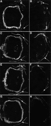
Fig. 2. Lack of K7, K18 and K19 in K18–/–K19–/– mice. (A and B) Immunofluorescence analysis of K7. (A) Strong staining of trophoblast giant cell layer in wild-type implantation sites. Maternal uterine glands and epithelia were stained weakly (arrows). (B) Positive staining of uterine epithelia and glands (arrows) but absence in doubly deficient extra-embyronic and embryonic compartments. (C and D) Staining for K8. (C) In wild-type animals, trophoblast giant cells, early placenta and maternal uterine glands and epithelia (arrows) were strongly positive. Parietal and visceral yolk sac, amnion, embryonic surface ectoderm, notochord and gut were also positive for K8. (D) Implantation site containing a doubly deficient embryo displayed strong staining in maternal uterine epithelia and glands (arrows). Weak staining was noted in the trophoblast giant cell layer and embryonic gut corresponding to K8 aggregates. (E and F) Staining for K18. (E) In wild type, the trophoblast giant cell layer and visceral yolk sac were strongly positive for K18. The amnion, embryonic ectoderm, gut and notochord were also positive. Maternal uterine glands and epithelia were stained. (F) Only maternal epithelia and glands were positive in doubly deficient implantation sites (arrows). (G and H) Staining for K19. (G) The trophoblast giant cell layer and maternal uterine epithelia were strongly positive. Weaker staining was observed in amnion, surface ectoderm, gut and notochord. (H) No staining was noted in doubly deficient embryos. The lack of staining in maternal tissues was due to the K19–/– genotype of parental mice. Bar, 1 mm.
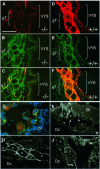
Fig. 3. K8 aggregates in extra-embryonic compartments of K18–/–K19–/– (–/–) mice. (A–F) Indirect immunofluorescence staining of E9.5 cryosections of wild-type and K18–/–K19–/– extra-embryonic compartments. (A) Staining for K8 in the visceral yolk sac (VYS) and giant trophoblast (gT) cell layer. Note the apically located aggregates in endodermal cells of the visceral yolk sac and in the trophoblast giant cells. (B) Staining for desmoplakin of the same section as in (A). Note the large number of desmosomes in the trophoblast giant cells not altered compared with wild-type. (C) Double staining for K8 and desmoplakin. (D) Staining for K8 in wild-type (+/+) trophoblast giant cells and visceral yolk sac. Both compartments were strongly positive for K8. (E) Staining of the same section as in (D) for desmoplakin. (F) Double staining for K8 and desmoplakin. Desmosomes co-localized with the keratin filaments. (G–J) Immunofluorescence analysis of fragile trophoblast giant cells at the edges of tissue separation and haematoma 9.5 days post-coitum. The area displayed here was similiar to that shown in Figure 4C. (G) Immunofluorescence staining of giant trophoblast cells at the border of a split between decidual tissue (Dc) and parietal yolk sac, partly in the process of cytolysis. Triple immunofluorescence (K8, desmoplakin, DAPI) revealing aggregates in the cytoplasm of trophoblast giant cells (K8, red fluorescence) and showing colocalization between some but not all K8 aggregates and desmosomes (yellow fluorescence). Cytolysing trophoblast giant cells lacked desmoplakin staining (green fluorescence) at the edge of the split. Note the enormous size of giant trophoblast nuclei (blue fluorescence). (H–J) Immunofluorescence analysis of desmosomes in trophoblast giant cells (n = nucleus) of doubly deficient embryos. (H) Note the large amount of apically located desmosomes in intact trophoblast giant cells. (I and J) Trophoblast giant cells at the edge of maternal blood with cytolysis and loss of contact. Desmoplakin staining revealed a discontinous distribution or missing of desmosomes (arrows in I). Bar, 100 µm.
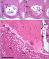
Fig. 4. Bleeding and giant trophoblast fragility in K18–/–K19–/– mice (A and B) Sagittally sectioned wild-type and doubly deficient E9.5 embryos. (A) In the wild-type embryos the barrier between embryonic and maternal compartments is formed by giant trophoblast (gT) cells (arrows). The visceral yolk sac (VYS), placenta (P) and amnion (A) are clearly visible. (B) Note the haematoma (H) in the mutant embryo, which is deforming the parietal and visceral yolk sacs. In the region with the haematoma, trophoblast giant cells surrounding the conceptus (arrows) were destroyed by cytolysis. Bar, 500 µm. (C) Higher magnification of (B). The layer of trophoblast giant cells is disrupted, with trophoblast giant cells in the process of cytolysis (arrows). Cytolysed trophoblast giant cells were surrounded by granulocytes. The origin of blood was a maternal vessel in the decidual tissue (Dcv). After breakdown of the trophoblast giant cell layer, maternal blood entered between the parietal yolk sac (PYS) and decidual tissue (Dc) unimpaired. The haematoma consisted of maternally derived erythrocytes, fibrin (Fi) and infiltrated granulocytes. Note that Reichert’s membrane (basal lamina in PYS) was not penetrated by blood. Em, embryo; ∗, visceral yolk sac. Bar, 200 µm.
Fig. 5. Histological analysis of E10.5 double-null and wild-type embryos. (A) Wild-type embryo with intact extra-embryonic membranes, like visceral yolk sac (VYS), placenta (P) and decidual tissue (Dc). The embryo was sectioned through the head, showing rhombencephalon and otic vesicles. (B) Same magnification as (A). The K18–/–K19–/– embryo showed intact parietal and visceral yolk sac, but severe necrosis of giant trophoblast and decidual cells accompanied by bleeding (H) into the uterine lumen. The placenta, which is not visible in this section, was smaller than that of wild-type embryos. Bar, 1 mm. (C) Higher magnification of (B). The embryo showed necroses and died on ∼E10. Bar, 300 µm. (D) Higher magnification of (B). Necrosis of giant trophoblast (arrows) and decidual cells (Dc). The small triangle marks the visceral yolk sac and the large triangle marks the uterine epithelium. Fibrin and infiltrated granulocytes were noted between the dead cells. Bar, 100 µm.
Fig. 6. Placentas of K18–/–K19–/– (–/–) and wild type (wt) mice 9.5 days post-coitum. (A and C) Placentas of wild-type mice. Note the well developed labyrinthian layer and the deeply invaded giant trophoblast cells (arrows). (B and D) Placentas of K18–/–K19–/– mice. Note the cleft between the trophoblast giant cells and the spongiotrophoblast (arrows in B) and the poorly developed labyrinth (arrow in D). Bar, 200 µm.
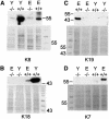
Fig. 7. Western blot analysis of total proteins from K18–/–K19–/– (–/–) and wild-type (+/+) embryos (E) and yolk sacs (Y) separated by SDS–PAGE. Detection of (A) K8, (B) K18, (C) K19 and (D) K7. (A) Note the large amount of K8 in wild-type yolk sacs and embryos compared with the small amount in K18–/–K19–/– embryos and yolk sacs. K8 detectable by western blotting probably corresponded to K8 aggregates detected by immunofluorescence. (B) K18 was absent in doubly deficient yolk sacs and embryos but present in normal amounts in wild-type embryos. There was much more K8 and K18 in wild-type yolk sacs than in wild-type embryos (A and B). K7 and K19 were only detectable in wild-type embryos and not in doubly deficient embryos (C and D); they were absent in wild-type and doubly deficient yolk sacs.
Similar articles
- A mutation of keratin 18 within the coil 1A consensus motif causes widespread keratin aggregation but cell type-restricted lethality in mice.
Hesse M, Grund C, Herrmann H, Bröhl D, Franz T, Omary MB, Magin TM. Hesse M, et al. Exp Cell Res. 2007 Aug 15;313(14):3127-40. doi: 10.1016/j.yexcr.2007.05.019. Epub 2007 May 25. Exp Cell Res. 2007. PMID: 17617404 - Rescue of keratin 18/19 doubly deficient mice using aggregation with tetraploid embryos.
Hesse M, Watson ED, Schwaluk T, Magin TM. Hesse M, et al. Eur J Cell Biol. 2005 Mar;84(2-3):355-61. doi: 10.1016/j.ejcb.2004.12.014. Eur J Cell Biol. 2005. PMID: 15819413 - Type II keratins precede type I keratins during early embryonic development.
Lu H, Hesse M, Peters B, Magin TM. Lu H, et al. Eur J Cell Biol. 2005 Aug;84(8):709-18. doi: 10.1016/j.ejcb.2005.04.001. Eur J Cell Biol. 2005. PMID: 16180309 - Keratin transgenic and knockout mice: functional analysis and validation of disease-causing mutations.
Vijayaraj P, Söhl G, Magin TM. Vijayaraj P, et al. Methods Mol Biol. 2007;360:203-51. doi: 10.1385/1-59745-165-7:203. Methods Mol Biol. 2007. PMID: 17172732 Review. - Keratins in the human trophoblast.
Gauster M, Blaschitz A, Siwetz M, Huppertz B. Gauster M, et al. Histol Histopathol. 2013 Jul;28(7):817-25. doi: 10.14670/HH-28.817. Epub 2013 Mar 1. Histol Histopathol. 2013. PMID: 23450430 Review.
Cited by
- Unique amino acid signatures that are evolutionarily conserved distinguish simple-type, epidermal and hair keratins.
Strnad P, Usachov V, Debes C, Gräter F, Parry DA, Omary MB. Strnad P, et al. J Cell Sci. 2011 Dec 15;124(Pt 24):4221-32. doi: 10.1242/jcs.089516. Epub 2012 Jan 3. J Cell Sci. 2011. PMID: 22215855 Free PMC article. - Cytoskeletal regulation of calcium-permeable cation channels in the human syncytiotrophoblast: role of gelsolin.
Montalbetti N, Li Q, Timpanaro GA, González-Perrett S, Dai XQ, Chen XZ, Cantiello HF. Montalbetti N, et al. J Physiol. 2005 Jul 15;566(Pt 2):309-25. doi: 10.1113/jphysiol.2005.087072. Epub 2005 Apr 21. J Physiol. 2005. PMID: 15845576 Free PMC article. - Mitofusins Mfn1 and Mfn2 coordinately regulate mitochondrial fusion and are essential for embryonic development.
Chen H, Detmer SA, Ewald AJ, Griffin EE, Fraser SE, Chan DC. Chen H, et al. J Cell Biol. 2003 Jan 20;160(2):189-200. doi: 10.1083/jcb.200211046. Epub 2003 Jan 13. J Cell Biol. 2003. PMID: 12527753 Free PMC article. - Absence of keratin 8 confers a paradoxical microflora-dependent resistance to apoptosis in the colon.
Habtezion A, Toivola DM, Asghar MN, Kronmal GS, Brooks JD, Butcher EC, Omary MB. Habtezion A, et al. Proc Natl Acad Sci U S A. 2011 Jan 25;108(4):1445-50. doi: 10.1073/pnas.1010833108. Epub 2011 Jan 10. Proc Natl Acad Sci U S A. 2011. PMID: 21220329 Free PMC article. - The intermediate filament network in cultured human keratinocytes is remarkably extensible and resilient.
Fudge D, Russell D, Beriault D, Moore W, Lane EB, Vogl AW. Fudge D, et al. PLoS One. 2008 Jun 4;3(6):e2327. doi: 10.1371/journal.pone.0002327. PLoS One. 2008. PMID: 18523546 Free PMC article.
References
- Bancroft J.D. and Cook,H.C. (1994) Manual of Histological Techniques and their Diagnostic Application. Churchill Livingstone, Edinburgh.
- Baribault H., Price,J., Miyai,K. and Oshima,R.G. (1993) Mid-gestational lethality in mice lacking keratin 8. Genes Dev., 7, 1191–1202. - PubMed
- Baribault H., Penner,J., Iozzo,R.V. and Wilson,H.M. (1994) Colorectal hyperplasia and inflammation in keratin 8-deficient FVB/N mice. Genes Dev., 8, 2964–2973. - PubMed
- Bierkamp C., Mclaughlin,K.J., Schwarz,H., Huber,O. and Kemler,R. (1996) Embryonic heart and skin defects in mice lacking plakoglobin. Dev. Biol., 180, 780–785. - PubMed
Publication types
MeSH terms
Substances
LinkOut - more resources
Full Text Sources
Molecular Biology Databases
Research Materials


