Dysregulation of CD30+ T cells by leukemia impairs isotype switching in normal B cells - PubMed (original) (raw)
Dysregulation of CD30+ T cells by leukemia impairs isotype switching in normal B cells
A Cerutti et al. Nat Immunol. 2001 Feb.
Erratum in
- Nat Immunol 2001 Apr;2(4):368
Abstract
Chronic lymphocytic leukemia (CLL) is associated with impaired immunoglobulin (Ig) class-switching from IgM to IgG and IgA, a defect that leads to recurrent infections. When activated in the presence of leukemic CLL B cells, T cells rapidly up-regulate CD30 through an OX40 ligand and interleukin 4 (IL-4)-dependent mechanism. These leukemia-induced CD30+ T cells inhibit CD40 ligand (CD40L)-mediated S mu-->S gamma and S mu-->S alpha class-switch DNA recombination (CSR) by engaging CD30 ligand (CD30L), a molecule that interferes with the assembly of the CD40-tumor necrosis factor receptor-associated factor (TRAF) complex in nonmalignant IgD+ B cells. In addition, engagement of T cell CD30 by CD30L on neoplastic CLL B cells down-regulates the CD3-induced expression of CD40L. These findings indicate that, in CLL, abnormal CD30-CD30L interaction impairs IgG and IgA production by interfering with the CD40-mediated differentiation of nonmalignant B cells.
Figures
Figure 1. CLL TS2 cells inhibit htCD40L-induced CSR in a CD30-dependent fashion
(a,b) The proportion of TS1 and TS2 cells was assessed by triple-staining peripheral blood lymphocytes (PBLs) for CD3, CD8 and CD28. The expression of CD30, IL-4Rα and IL-4 was assessed on gated TS1 (shaded histograms) or TS2 (open histograms) cells upon CD28 staining of purified CD8+ T cells. These data are representative of ten experiments that yielded similar results. (c) Normal IgD+ B cells were incubated with or without htCD40L and IL-4 and in the presence or absence of immobilized agonistic mAbs to CD30L or OX40L. Similar IgD+ B cells were cultured with normal TS1, normal TS2 cells, CLL TS1 cells or CLL TS2 cells preincubated with or without blocking mAbs to CD56 (anti-CD56bl) or CD30 (anti-CD30bl). After 4 days, genomic DNA from T cell–depleted B cells were used to amplify Sγ-Sµ switch circles and polymerase chain reaction (PCR) products were hybridized with a radiolabeled Sµ probe. Apoptotic B cells were assessed by annexin V staining. These data are from one of three experiments that yielded similar results.
Figure 2. CLL TS2 cells inhibit TH cell-induced CSR in a CD30-dependent fashion
(a) Normal IgD+ B cells were incubated with fixed CD4+D40L+ TH cells, IL-4 and IL-10 in the presence or absence of decreasing amounts of fixed TS1 or TS2 cells. After 7 days, equal amounts of genomic DNA from T cell–depleted B cells were used to amplify switch circles. Supernatants were collected to measure IgG and IgA (arrowheads indicate an immunoglobulin concentration of <100 ng/ml and bars are s.d. of the mean of triplicate experiments). (b) Normal IgD+ B cells were cocultured with fixed CLL CD4+D40L+ TH cells in the presence or absence of CLL TS1 cells or CLL TS2 cells that were preincubated with or without blocking mAbs to CD56 or CD30. After 4 days, equal amounts of cDNA from T cell-depleted B cells were used to amplify Igβ, Iγ1-Cγ1 and Iα1-Cα1. (c) Expression of CD30 (filled histograms) on (unfixed) normal TS2 cells or CLL TS2 cells before and after a 4-day incubation with normal IgD+ B cells, htCD40L, IL-4 and IL-10. Shaded histograms show cell staining by an irrelevant mAb. These data are from one of two experiments that yielded similar results.
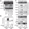
Figure 3. Engagement of CD30L inhibits CD40 signaling in normal IgD+ B cells
(a) Normal IgD+ B cells were incubated with htCD40L and IL-4 in the presence or absence of fixed CLL TS1 or CLL TS2 cells. Before culture, CLL T cells were fixed and preincubated with control MOPC-21 or blocking mAbs to CD56 or CD30. After 6 h, nuclear proteins were extracted from T cell-depleted B cells and the binding of NF-κB and STAT6 to specific radiolabeled oligonucleotides was assessed by EMSA. Arrowsheads (from top to bottom) correspond to p50-p65, p50-c-Rel and p50-p50 NF-κB–Rel complexes. After 4 days, B cells were purified and Igβ and Iγ3-Cγ3 transcripts were PCR-amplified from equal amounts of cDNA. (b) IgD+ CL-01 B cells transfected with a −238/−188 ECS-Iγ3-CD40 RE-SV40 minimal promoter or an Ig (κB2)-LUC vector were cultured for 24 h with or without IL-4 and/or htCD40L and in the presence of immobilized MOPC-21 or monoclonal anti-CD30L (data are from two similar experiments and bars indicate s.d. of mean). (c) Total proteins from 2-h stimulated normal IgD+ B cells were immunoprecipitated with a monoclonal anti-CD40 and then immunoblotted for CD40, TRAFs and TANK. Cytoplasmic proteins were immunoblotted for NIK, IKKα and IκBα or used to assess the IKKα activity. These data are from one of three experiments that yielded similar results.
Figure 4. Leukemic B cells induce reciprocal modulation of CD30 and CD40L on T cells
(a,b,e,f,i,j) Normal PBMCs, normal CD4+ T cells, CLL PBMCs or CLL CD4+ T cells were incubated with immobilized monoclonal anti-CD3. After 0, 6, 12, 72 or 120 h, cells were stained with monoclonal FITC–anti-CD4, PerCP–anti-CD3 and PE–anti-CD30 or PE–anti-CD40L or PE–anti-CD25. The CD30, CD40L and CD25 MFIs were evaluated on gated CD4+ T cells. (c,d,g,h,k,l) Normal CD4+ T cells or CLL CD4+ T cells were incubated for 12 or 120 h with immobilized monoclonal anti-CD3 and leukemic CLL B cells or irradiated MRC-5 fibroblasts (F) at different ratios. Data are mean of five experiments±s.d. (*_P_>0.05 and **_P_>0.005.)
Figure 5. Leukemic B cells up-regulate T cell CD30 in an OX40L and IL-4–dependent fashion
(a) Normal IgD+ B cells were incubated with htCD40L, IL-4 and IL-10 in the presence or absence of purified and fixed T1, T2, T3 or T4 cells that were preincubated with MOPC-21 (solid line) or blocking monoclonal anti-CD30 (broken line). IgG were measured after 8 days. The shaded area indicates IgG values obtained in the presence of htCD40L and cytokines only. The data are mean±s.d. of five experiments. (b) CLL CD4+T cells were CD3-activated in the presence of leukemic CLL B cells (1:4 T:B cell ratio) that had been preincubated with MOPC-21 or blocking mAbs to CD72, CD80 or OX40L. Neutralizing antibodies to IL-4 or IFN-γ and TAPI were also used. CD30 was measured on gated CD4+ T cells after 24 h (bars indicate s.d. of three experiments). (c) OX40 on normal CD4+T cells or CLL CD4+T cells incubated for 24 h with immobilized agonistic mAb to CD3. (d) OX40L on normal B cells or leukemic CLL B cells incubated for 24 h with htCD40L and IL-4 (filled histograms indicate cell stained with a control mAb). (e) CD30 on CLL CD4+ T cells cultured for 24 h with or without immobilized agonistic mAbs to CD3, CD28 and/or OX40 (shaded histogram indicates T cells stained with a control antibody). These data are from one of five experiments that yielded similar results.
Figure 6. Leukemic B cells induce CD30L-dependent down-regulation of CD40L on T cells
(a) CLL CD4+ T cells were CD3-activated for 120 h in the presence of leukemic CLL B cells (1:4 T:B cell ratio) and monoclonal control MOPC-21 (solid line) or blocking anti-CD30 (broken line). CD40L was measured on gated CD4+ T cells (data are mean±s.d. of five experiments). (b,c) CD30 and CD40L transcripts and protein in CD4+CD30+ Jurkat D1.1 T cells stimulated by an agonistic monoclonal anti-CD3 in the presence of immobilized MOPC-21 or agonistic monoclonal anti-CD30. (d) Normal CD4+ T cells or CLL CD4+ T cells were CD3-activated for 12 h with or without leukemic CLL B cells or MRC fibroblasts, and in the presence or absence of blocking mAbs to CD30 or CD5. After CLL B cell–depletion, T cells were fixed and cocultured with normal IgD+ B cells and cytokines in the presence or absence of a blocking monoclonal anti-CD40L. IgG were measured after 8 days. These data are from one of three experiments that yielded similar results (bars indicate s.d. of mean of triplicate experiments).
Figure 7. Engagement of CD30L enhances leukemic B cell accumulation
Leukemic CLL B cells were incubated with or without IL-4 and/or htCD40L in the presence of immobilized monoclonal MOPC-21 or agonistic anti-CD30L. Blocking mAbs to IL-6Rα, IL-6, TNFR2 and TNF-α were also used. TNF-α secretion, thymidine incorporation (SI) and viability were assessed after 4 days. Viable cells were assessed by exclusion dye test. These data are from one of three experiments that yielded similar results (bars indicate s.d. of mean of triplicate experiments).
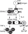
Figure 8. Dysregulated CD30-CD30L interaction impairs isotype switching in normal IgD+ B cells
In CLL, antigen-activated T cells transiently express CD40L that induces OX40L on CD40-expressing leukemic CLL B cells. By triggering OX40 on T cells, these malignant B cells elicit IL-4 secretion, which, in turn, rapidly induces CD30 on T cells. These CD30+ T cells inhibit switching to IgG and IgA by inducing CD30L-mediated CD40-inhibitory signals in nonmalignant IgD+ B cells. In addition, engagement of T cell CD30 by leukemic B cell CD30L down-modulates T cell CD40L and elicits TNF-α–TNFR2–dependent expansion of the neoplastic clone.
Similar articles
- Ongoing in vivo immunoglobulin class switch DNA recombination in chronic lymphocytic leukemia B cells.
Cerutti A, Zan H, Kim EC, Shah S, Schattner EJ, Schaffer A, Casali P. Cerutti A, et al. J Immunol. 2002 Dec 1;169(11):6594-603. doi: 10.4049/jimmunol.169.11.6594. J Immunol. 2002. PMID: 12444172 Free PMC article. - Engagement of CD153 (CD30 ligand) by CD30+ T cells inhibits class switch DNA recombination and antibody production in human IgD+ IgM+ B cells.
Cerutti A, Schaffer A, Goodwin RG, Shah S, Zan H, Ely S, Casali P. Cerutti A, et al. J Immunol. 2000 Jul 15;165(2):786-94. doi: 10.4049/jimmunol.165.2.786. J Immunol. 2000. PMID: 10878352 Free PMC article. - CD30 is a CD40-inducible molecule that negatively regulates CD40-mediated immunoglobulin class switching in non-antigen-selected human B cells.
Cerutti A, Schaffer A, Shah S, Zan H, Liou HC, Goodwin RG, Casali P. Cerutti A, et al. Immunity. 1998 Aug;9(2):247-56. doi: 10.1016/s1074-7613(00)80607-x. Immunity. 1998. PMID: 9729045 Free PMC article. - Immunoglobulin class switch recombination in chronic lymphocytic leukemia.
Lanasa MC, Weinberg JB. Lanasa MC, et al. Leuk Lymphoma. 2011 Jul;52(7):1398-400. doi: 10.3109/10428194.2011.568076. Leuk Lymphoma. 2011. PMID: 21699386 Review. - The role of CD40 ligand in costimulation and T-cell activation.
Grewal IS, Flavell RA. Grewal IS, et al. Immunol Rev. 1996 Oct;153:85-106. doi: 10.1111/j.1600-065x.1996.tb00921.x. Immunol Rev. 1996. PMID: 9010720 Review.
Cited by
- Chronic lymphocytic leukemia B cells can undergo somatic hypermutation and intraclonal immunoglobulin V(H)DJ(H) gene diversification.
Gurrieri C, McGuire P, Zan H, Yan XJ, Cerutti A, Albesiano E, Allen SL, Vinciguerra V, Rai KR, Ferrarini M, Casali P, Chiorazzi N. Gurrieri C, et al. J Exp Med. 2002 Sep 2;196(5):629-39. doi: 10.1084/jem.20011693. J Exp Med. 2002. PMID: 12208878 Free PMC article. - Secondary Immunodeficiency in Hematological Malignancies: Focus on Multiple Myeloma and Chronic Lymphocytic Leukemia.
Allegra A, Tonacci A, Musolino C, Pioggia G, Gangemi S. Allegra A, et al. Front Immunol. 2021 Oct 25;12:738915. doi: 10.3389/fimmu.2021.738915. eCollection 2021. Front Immunol. 2021. PMID: 34759921 Free PMC article. Review. - DCs induce CD40-independent immunoglobulin class switching through BLyS and APRIL.
Litinskiy MB, Nardelli B, Hilbert DM, He B, Schaffer A, Casali P, Cerutti A. Litinskiy MB, et al. Nat Immunol. 2002 Sep;3(9):822-9. doi: 10.1038/ni829. Epub 2002 Aug 5. Nat Immunol. 2002. PMID: 12154359 Free PMC article. - Chronic lymphocytic leukemia cells induce changes in gene expression of CD4 and CD8 T cells.
Görgün G, Holderried TA, Zahrieh D, Neuberg D, Gribben JG. Görgün G, et al. J Clin Invest. 2005 Jul;115(7):1797-805. doi: 10.1172/JCI24176. Epub 2005 Jun 16. J Clin Invest. 2005. PMID: 15965501 Free PMC article. - Ongoing in vivo immunoglobulin class switch DNA recombination in chronic lymphocytic leukemia B cells.
Cerutti A, Zan H, Kim EC, Shah S, Schattner EJ, Schaffer A, Casali P. Cerutti A, et al. J Immunol. 2002 Dec 1;169(11):6594-603. doi: 10.4049/jimmunol.169.11.6594. J Immunol. 2002. PMID: 12444172 Free PMC article.
References
- Stavnezer J. Antibody class switching. Adv. Immunol. 1996;61:79–146. - PubMed
- Van Kooten C, Banchereau J. CD40-CD40 ligand: a multifunctional receptor-ligand pair. Adv. Immunol. 1996;61:1–77. - PubMed
- Grewal IS, Flavell RA. CD40 and CD154 in cell-mediated immunity. Annu. Rev. Immunol. 1998;16:111–135. - PubMed
- Rothe M, Sarma V, Dixit VM, Geoddel DV. TRAF2-mediated activation of NF-κB by TNF receptor 2 and CD40. Science. 1995;269:1424–1427. - PubMed
Publication types
MeSH terms
Substances
Grants and funding
- AR40908/AR/NIAMS NIH HHS/United States
- R01 AR040908/AR/NIAMS NIH HHS/United States
- T32 AI 07621/AI/NIAID NIH HHS/United States
- T32 AI007621/AI/NIAID NIH HHS/United States
- R56 AI045011/AI/NIAID NIH HHS/United States
- AG 13910/AG/NIA NIH HHS/United States
- R01 AI045011/AI/NIAID NIH HHS/United States
- AI 45011/AI/NIAID NIH HHS/United States
LinkOut - more resources
Full Text Sources
Other Literature Sources
Research Materials
Miscellaneous


