Ligation of CD40 stimulates the induction of nitric-oxide synthase in microglial cells - PubMed (original) (raw)
Ligation of CD40 stimulates the induction of nitric-oxide synthase in microglial cells
M Jana et al. J Biol Chem. 2001.
Abstract
The present study was undertaken to investigate the role of CD40 ligation in the expression of inducible nitric-oxide synthase (iNOS) in mouse BV-2 microglial cells and primary microglia. Ligation of CD40 alone by either cross-linking antibodies against CD40 or a recombinant CD40 ligand (CD154) was unable to induce the production of NO in BV-2 microglial cells. The absence of induction of NO production by CD40 ligation alone even in CD40-overexpressed BV-2 microglial cells suggests that a signal transduced by the ligation of CD40 alone is not sufficient to induce NO production. However, CD40 ligation markedly stimulated interferon-gamma (IFN-gamma)-mediated NO production. Ligation of CD40 in CD40-overexpressed cells further stimulated IFN-gamma-induced production of NO. This stimulation of NO production was accompanied by stimulation of the iNOS protein and mRNA. In addition to BV-2 glial cells, CD40 ligation also stimulated IFN-gamma-mediated NO production in mouse primary microglia and peritoneal macrophages. To understand the mechanism of induction/stimulation of iNOS, we investigated the roles of nuclear factor kappaB (NF-kappaB) and CCAAT/enhancer-binding protein beta (C/EBPbeta), transcription factors responsible for the induction of iNOS. IFN-gamma alone was able to induce the activation of NF-kappaB as well as C/EBPbeta. However, CD40 ligation alone induced the activation of only NF-kappaB but not of C/EBPbeta, suggesting that the activation of NF-kappaB alone by CD40 ligation is not sufficient to induce the expression of iNOS and that the activation of C/EBPbeta is also necessary for the expression of iNOS. Consistently, dominant-negative mutants of p65 (Deltap65) and C/EBPbeta (DeltaC/EBPbeta) inhibited the expression of iNOS in BV-2 microglial cells that were stimulated with the combination of IFN-gamma and CD40 ligand. Stimulation of IFN-gamma-mediated activation of NF-kappaB but not of C/EBPbeta by CD40 ligation suggests that CD40 ligation stimulates the expression of iNOS in IFN-gamma-treated BV-2 microglial cells through the stimulation of NF-kappaB activation. This study illustrates a novel role for CD40 ligation in stimulating the expression of iNOS in microglial cells, which may participate in the pathogenesis of neuroinflammatory diseases.
Figures
Fig. 1. Stimulation of IFN-_γ_-mediated production of NO by CD40 ligation in BV-2 microglial cells
Cells were treated with 2 _μ_g/ml cross-linking antibodies against CD40 in the presence or absence of IFN-γ (25 units/ml) under serum-free conditions. At different hours of incubation supernatants were used for the nitrite assay using Griess reagent as described under “Materials and Methods.” Data are expressed as the mean of two separate experiments.
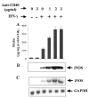
Fig. 2. CD40 ligation stimulates the expression of iNOS in IFN-_γ_-treated BV-2 microglial cells
Cells were treated with different concentrations of cross-linking antibodies against CD40 in the presence or absence of IFN-γ (25 units/ml) under serum-free conditions. A, after 24 h, supernatants were used for the nitrite assay as mentioned under “Materials and Methods.” Data are mean ± S.D. of three different experiments. B, cell homogenates were electrophoresed, transferred on nitrocellulose membrane, and immunoblotted with antibodies against mouse macrophage iNOS as described under “Materials and Methods.” C, after 6 h of incubation, cells were taken out directly by adding Ultraspec-II RNA reagent (Biotecx Laboratories Inc.) to the plates for isolation of total RNA, and Northern blot analysis for iNOS mRNA was carried out as described under “Materials and Methods.”
Fig. 3. Dose-dependent stimulation of iNOS expression by human recombinant CD40L in IFN-_γ_-treated BV-2 microglial cells
Cells were treated with different concentrations of human recombinant CD40L (CD154) in the presence or absence of IFN-γ (25 units/ml) under serum-free conditions. A, after 24 h, supernatants were used for the nitrite assay. Data are mean ± S.D. of three different experiments. B, after 6 h of incubation, total RNA was isolated from cells, and Northern blot analysis for iNOS mRNA was carried out.
Fig. 4. Effect of overexpression of CD40 on the induction of NO production in BV-2 microglial cells
Cells plated at 50–60% confluence in six-well plates were transfected with different amounts of either CD40 cDNA or an empty vector using LipofectAMINE Plus as described under “Materials and Methods.” After 24 h of transfection, cells were stimulated with anti-CD40 (1 _μ_g/ml) in the presence or absence of IFN-γ (10 units/ml) under serum-free conditions. After 24 h of stimulation, supernatants were used for the nitrite assay. Data are expressed as the mean of two separate experiments.
Fig. 5. Effect of overexpression of CD40 on the expression of iNOS mRNA in BV-2 microglial cells
Cells plated at 50–60% confluence in a 100-mm dish were transfected with 0.8 _μ_g of either CD40 cDNA or an empty vector. After 24 h of transfection, cells were stimulated with anti-CD40 (1 _μ_g/ml) in the presence or absence of IFN-γ (10 units/ml) under serum-free conditions. After 6 h of incubation, total RNA was isolated from cells, and Northern blot analysis for iNOS mRNA was carried out.
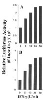
Fig. 6. IFN-γ induces the activation of both NF-κ_B (A) and C/EBP_β (B) in BV-2 microglial cells
Cells plated at 50–60% confluence in six-well plates were cotransfected with 1 _μ_g of either pBIIX-Luc(anNF-_κ_B-dependentreporterconstruct)orpC/EBP_β_-Luc(aC/EBP_β_-dependent reporter construct) and 50 ng of pRL-TK (a plasmid encoding Renilla luciferase, used as a transfection efficiency control). After 24 h of transfection, cells were stimulated with different concentrations of IFN-γ for 6 h under serum-free conditions. Firefly (ff-Luc) and Renilla luciferase (r-Luc) activities were obtained by analyzing the total cell extract as described under “Materials and Methods.” Data are expressed as the mean of two separate experiments. U, units.
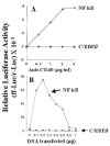
Fig. 7. Ligation of CD40 induces the activation of NF-κ_B but not of C/EBP_β in BV-2 microglial cells
A, cells plated at 50–60% confluence in six-well plates were cotransfected with 1 _μ_g of either pBIIX-Luc or pC/EBP_β_-Luc and 50 ng of pRL-TK. After 24 h of transfection, cells were stimulated with different concentrations of anti-CD40 for 6 h under serum-free conditions. Firefly (ff-Luc) and Renilla luciferase (r-Luc) activities were assayed as described above. Data are expressed as the mean of two separate experiments. B, cells plated at 50–60% confluence in six-well plates were cotransfected with different concentrations of CD40 cDNA and either 1 _μ_g of either pBIIX-Luc or pC/EBP_β_-Luc. All transfections also included 50 ng of pRL-TK. After 24 h of transfection, cells were stimulated with 1 _μ_g/ml anti-CD40 for 6 h under serum-free conditions. Firefly and Renilla luciferase activities were obtained by analyzing the total cell extract as described above. Data are expressed as the mean of two separate experiments.
Fig. 8. Dominant-negative mutants of p65 (Δp65) and C/EBP_β_ (ΔC/EBP_β_) inhibit the expression of iNOS in CD40-stimulated BV-2 microglial cells
A, cells plated at 50–60% confluence in six-well plates were transfected with 1 μ_g of either Δp65 or ΔC/EBP_β. After 24 h of transfection, cells were stimulated with the combination of anti-CD40 and IFN-γ for 24 h under serum-free conditions, and supernatants were used for the nitrite assay. Data are mean ± S.D. of three different experiments. B, similarly after 24 h of transfection, cells were stimulated with the combination of anti-CD40 and IFN-γ under serum-free conditions. After 6 h of incubation, total RNA was isolated, and Northern blot analysis for iNOS mRNA was carried out.
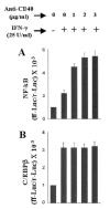
Fig. 9. Ligation of CD40 stimulates the activation of NF-κ_B but not of C/EBP_β in IFN-_γ_-stimulated BV-2 microglial cells
Cells plated at 50–60% confluence in six-well plates were cotransfected with 1_μ_g of either pBIIX-Luc (A) or pC/EBP_β_-Luc (B) and 50 ng of pRL-TK. After 24 h of transfection, cells were stimulated with 25 units/ml IFN-γ and different concentrations of anti-CD40 for 6 h under serum-free conditions. Firefly (ff-Luc) and Renilla luciferase (r-Luc) activities were obtained by analyzing the total cell extract. Data are mean ± S.D. of three different experiments.
Similar articles
- Regulation of tumor necrosis factor-alpha expression by CD40 ligation in BV-2 microglial cells.
Jana M, Dasgupta S, Liu X, Pahan K. Jana M, et al. J Neurochem. 2002 Jan;80(1):197-206. doi: 10.1046/j.0022-3042.2001.00691.x. J Neurochem. 2002. PMID: 11796758 - Human immunodeficiency virus type 1 (HIV-1) tat induces nitric-oxide synthase in human astroglia.
Liu X, Jana M, Dasgupta S, Koka S, He J, Wood C, Pahan K. Liu X, et al. J Biol Chem. 2002 Oct 18;277(42):39312-9. doi: 10.1074/jbc.M205107200. Epub 2002 Aug 7. J Biol Chem. 2002. PMID: 12167619 Free PMC article. - Induction of tumor necrosis factor-alpha (TNF-alpha) by interleukin-12 p40 monomer and homodimer in microglia and macrophages.
Jana M, Dasgupta S, Saha RN, Liu X, Pahan K. Jana M, et al. J Neurochem. 2003 Jul;86(2):519-28. doi: 10.1046/j.1471-4159.2003.01864.x. J Neurochem. 2003. PMID: 12871593 Free PMC article. - Induction of nitric-oxide synthase and activation of NF-kappaB by interleukin-12 p40 in microglial cells.
Pahan K, Sheikh FG, Liu X, Hilger S, McKinney M, Petro TM. Pahan K, et al. J Biol Chem. 2001 Mar 16;276(11):7899-905. doi: 10.1074/jbc.M008262200. Epub 2000 Dec 7. J Biol Chem. 2001. PMID: 11110796 Free PMC article. - Complementary roles of tumor necrosis factor alpha and interferon gamma in inducible microglial nitric oxide generation.
Mir M, Tolosa L, Asensio VJ, Lladó J, Olmos G. Mir M, et al. J Neuroimmunol. 2008 Nov 15;204(1-2):101-9. doi: 10.1016/j.jneuroim.2008.07.002. J Neuroimmunol. 2008. PMID: 18703234
Cited by
- Inhibition of IkappaB kinase-beta protects dopamine neurons against lipopolysaccharide-induced neurotoxicity.
Zhang F, Qian L, Flood PM, Shi JS, Hong JS, Gao HM. Zhang F, et al. J Pharmacol Exp Ther. 2010 Jun;333(3):822-33. doi: 10.1124/jpet.110.165829. Epub 2010 Feb 26. J Pharmacol Exp Ther. 2010. PMID: 20190013 Free PMC article. - Regulation of inducible nitric oxide synthase gene in glial cells.
Saha RN, Pahan K. Saha RN, et al. Antioxid Redox Signal. 2006 May-Jun;8(5-6):929-47. doi: 10.1089/ars.2006.8.929. Antioxid Redox Signal. 2006. PMID: 16771683 Free PMC article. Review. - Myelin basic protein-primed T cells induce nitric oxide synthase in microglial cells. Implications for multiple sclerosis.
Dasgupta S, Jana M, Liu X, Pahan K. Dasgupta S, et al. J Biol Chem. 2002 Oct 18;277(42):39327-33. doi: 10.1074/jbc.M111841200. Epub 2002 Aug 9. J Biol Chem. 2002. PMID: 12176974 Free PMC article. - Up-regulation of BDNF in astrocytes by TNF-alpha: a case for the neuroprotective role of cytokine.
Saha RN, Liu X, Pahan K. Saha RN, et al. J Neuroimmune Pharmacol. 2006 Sep;1(3):212-22. doi: 10.1007/s11481-006-9020-8. Epub 2006 May 16. J Neuroimmune Pharmacol. 2006. PMID: 18040799 Free PMC article. - Benfotiamine upregulates antioxidative system in activated BV-2 microglia cells.
Bozic I, Savic D, Stevanovic I, Pekovic S, Nedeljkovic N, Lavrnja I. Bozic I, et al. Front Cell Neurosci. 2015 Sep 4;9:351. doi: 10.3389/fncel.2015.00351. eCollection 2015. Front Cell Neurosci. 2015. PMID: 26388737 Free PMC article.
References
- Nathan C. FASEB J. 1992;6:3051–3064. - PubMed
- Jaffrey SR, Snyder SH. Annu Rev Cell Dev Biol. 1995;11:417–440. - PubMed
- Mitrovic B, Ignarro LJ, Montestruque S, Smoll A, Merril JE. Neuroscience. 1994;61:575–585. - PubMed
Publication types
MeSH terms
Substances
LinkOut - more resources
Full Text Sources
Other Literature Sources
Molecular Biology Databases
Research Materials


