Mutation of mouse p53 Ser23 and the response to DNA damage - PubMed (original) (raw)
Mutation of mouse p53 Ser23 and the response to DNA damage
Zhiqun Wu et al. Mol Cell Biol. 2002 Apr.
Abstract
Recent studies have suggested that phosphorylation of human p53 at Ser20 is important for stabilizing p53 in response to DNA damage through disruption of the interaction between MDM2 and p53. To examine the requirement for this DNA damage-induced phosphorylation event in a more physiological setting, we introduced a missense mutation into the endogenous p53 gene of mouse embryonic stem (ES) cells that changes serine 23 (S23), the murine equivalent of human serine 20, to alanine (A). Murine embryonic fibroblasts harboring the p53(S23A) mutation accumulate p53 as well as p21 and Mdm2 proteins to normal levels after DNA damage. Furthermore, ES cells and thymocytes harboring the p53(S23A) mutation also accumulate p53 protein to wild-type levels and undergo p53-dependent apoptosis similarly to wild-type cells after DNA damage. Therefore, phosphorylation of murine p53 at Ser23 is not required for p53 responses to DNA damage induced by UV and ionizing radiation treatment.
Figures
FIG. 1.
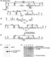
Generation of p53S23A ES cells. (A) The endogenous configuration of the p53 gene in AY ES cells. AY ES cells have one wild-type p53 allele and one mutant p53 allele (AY allele) with p53 exons 2 through 4 deleted. The initiation codon, ATG, of the p53 gene is located in exon 2, and the AY allele does not produce a truncated p53 protein. Open boxes represent the p53 exons, and the filled bars represent probes for Southern blot analysis. The wild-type 14-kbp _Eco_RI and 7-kbp _Hin_dIII fragments are indicated by arrows, as are the mutant 6-kbp _Eco_RI and 5.8-kbp _Hin_dIII fragments on the AY allele. (B) The targeting construct. ∗, the Ser23-to-Ala mutation in exon 2. The PGK-neor gene flanked by LoxP sites was inserted into an engineered _Sal_I site within intron 4. (C) Targeted p53 locus after homologous recombination between the wild-type p53 allele and the targeting vector. The positions of the PCR primer sets that were used to screen for LoxP/Cre-mediated deletion are shown by arrowheads. The sizes of the mutant _Eco_RI and _Hin_dIII fragments are indicated. (D) Mutant p53 allele after the PGK-neor gene was deleted. The size of the mutant _Hin_dIII fragment after the PGK-neor gene was deleted is indicated. (E) Southern blot analysis of genomic DNA derived from the wild type (lane 1), AY ES cells (lane 2), and targeted AY ES cells (lane 3), in which homologous recombination had occurred between the germ line allele and the targeting vector. Genomic DNA was digested with _Eco_RI and hybridized to probe A. Both the targeted allele and AY allele yielded the same 6-kbp mutant _Eco_RI fragment. The positions of both germ line and mutant alleles are indicated with arrowheads. (F) Southern blot analysis of genomic DNA derived from wild-type ES cells (lane 1), targeted AY ES cells with the PGK-neor gene inserted (lanes 2 and 3), AY ES cells (lane 4), and p53Ser23Ala ES cells (lane 5). Genomic DNA was digested with _Hin_dIII and hybridized to probed B. The 7-kbp wild-type band, 7.1-kbp PGK-neor gene deleted band, 5.8-kbp AY mutant band, and 3-kbp PGK-neor gene inserted band as well as the 7.8-kbp band derived from the p53 pseudogene are indicated.
FIG. 2.
Phosphorylation of murine p53 at Ser23 in ES cells at various times after IR treatment (5 Gy) or UV treatment (60 J/m2). Irradiated and untreated cells were treated with the proteosome inhibitor ALLN for 4 h before harvesting so that samples of all time points would have similar levels of p53 protein. Cell extracts from irradiated cells and untreated controls (0 h) were analyzed by Western blotting with antibodies specific for p53 or p53 phosphorylated at Ser23 (p53-S23p) as described previously (8, 33). The time after irradiation (in hours) is indicated at the top.
FIG. 3.
Generation of AY and p53S23A MEFs by Hprt-deficient blastocyst complementation. Southern blot analysis of genomic DNA from passage 3 MEFs derived from embryos generated by injection of AY or p53S23A ES cells into Hprt-deficient blastocysts. Genomic DNA was digested with _Eco_RI and hybridized with probe A (see Fig. 1). Lane 1, wild-type MEFs; lane 2, AY MEFs; lane 3, p53S23A MEFs.
FIG. 4.
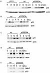
Induction of p53 and p21 in AY and p53S23A MEFs after DNA damage. Cell extracts were prepared from AY and p53S23A MEFs at the times indicated and analyzed for p53 expression by Western immunoblot analysis after exposure to 60 J of UV light/m2 (A) or 10 Gy of IR (B). The times after treatment (in hours) and genotypes are labeled at the top of the lanes. p53 and actin are indicated on the right. Shown is the induction of p21 (C) and Mdm2 (D) proteins in AY and p53S23A MEFs after 60- and 30-J/m2 UV treatment, respectively. The genotype and time points are indicated on top. p21, Mdm2, and actin are indicated on the right. (E) Immunoprecipitation and Western blot analysis of the p53-Mdm2 interaction in AY and p53S23A MEFs with or without UV radiation (30 J/m2). The genotypes and time points after radiation are labeled on top. p53 and Mdm2 are indicated on the right.
FIG. 5.
Induction of apoptosis in p53−/−, AY, and p53S23A ES cells after UV treatment. (A) p53 protein levels in AY and p53S23A ES cells at different times after UV treatment. Time after treatment is indicated at the top, genotypes are indicated on the left, and p53 and actin are indicated on the right. (B) Flow cytometric analysis of AY, p53−/−, and p53S23A ES cells harvested 16 h after exposure to 30 J of UV light/m2. Cell number is plotted as a function of the intensity of staining for Annexin V. Cells staining positive for Annexin V are apoptotic. The percentages of nonapoptotic cells are shown. (C) The percentage ratio of nonapoptotic cells in irradiated AY, p53−/−, and p53S23A ES cells relative to nonapoptotic cells in unirradiated controls 16 h after exposure to 10, 20, or 30 J of UV light/m2. Mean values from four independent experiments are presented with error bars. Percentages (y axis) were determined as follows: (no. of nonapoptotic cells in irradiated ES cells/no. of nonapoptotic cells in untreated control) × 100%.
FIG. 6.
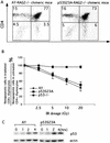
Induction of p53 and apoptosis in p53−/−, AY, and p53S23A mouse thymocytes after IR. (A) Mouse thymocytes were recovered from AY-RAG2−/− and p53S23A-RAG2−/− chimeric mice, stained for CD4 and CD8, and analyzed by flow cytometry. Cells residing in the lymphocyte gate were analyzed, and the percentages of total cells in a particular gate are indicated. (B) The percentage ratio of nonapoptotic CD4+ thymocytes in AY, p53−/−, and p53S23A thymocytes treated with 2.5, 5, 10, 15, and 20 Gy of IR to the nonapoptotic CD4+ thymocytes from untreated controls. Mean values from four independent experiments are presented with error bars. (C) p53 protein levels in AY and p53S23A thymocytes at different times after IR treatment were determined by Western blot analysis. Time after treatment and genotypes are indicated at the top; p53 and actin are indicated on the right.
Similar articles
- Critical role for Ser20 of human p53 in the negative regulation of p53 by Mdm2.
Unger T, Juven-Gershon T, Moallem E, Berger M, Vogt Sionov R, Lozano G, Oren M, Haupt Y. Unger T, et al. EMBO J. 1999 Apr 1;18(7):1805-14. doi: 10.1093/emboj/18.7.1805. EMBO J. 1999. PMID: 10202144 Free PMC article. - DNA damage-induced phosphorylation of p53 at serine 20 correlates with p21 and Mdm-2 induction in vivo.
Jabbur JR, Huang P, Zhang W. Jabbur JR, et al. Oncogene. 2000 Dec 14;19(54):6203-8. doi: 10.1038/sj.onc.1204017. Oncogene. 2000. PMID: 11175334 - Enhancement of the antiproliferative function of p53 by phosphorylation at serine 20: an inference from site-directed mutagenesis studies.
Jabbur JR, Huang P, Zhang W. Jabbur JR, et al. Int J Mol Med. 2001 Feb;7(2):163-8. doi: 10.3892/ijmm.7.2.163. Int J Mol Med. 2001. PMID: 11172619 - Mdm2 in the response to radiation.
Perry ME. Perry ME. Mol Cancer Res. 2004 Jan;2(1):9-19. Mol Cancer Res. 2004. PMID: 14757841 Review. - MEF immortalization to investigate the ins and outs of mutagenesis.
vom Brocke J, Schmeiser HH, Reinbold M, Hollstein M. vom Brocke J, et al. Carcinogenesis. 2006 Nov;27(11):2141-7. doi: 10.1093/carcin/bgl101. Epub 2006 Jun 15. Carcinogenesis. 2006. PMID: 16777987 Review.
Cited by
- p53 Stabilization and accumulation induced by human vaccinia-related kinase 1.
Vega FM, Sevilla A, Lazo PA. Vega FM, et al. Mol Cell Biol. 2004 Dec;24(23):10366-80. doi: 10.1128/MCB.24.23.10366-10380.2004. Mol Cell Biol. 2004. PMID: 15542844 Free PMC article. - Tumour suppression by p53: a role for the DNA damage response?
Meek DW. Meek DW. Nat Rev Cancer. 2009 Oct;9(10):714-23. doi: 10.1038/nrc2716. Epub 2009 Sep 4. Nat Rev Cancer. 2009. PMID: 19730431 Review. - ATM phosphorylation of Mdm2 Ser394 regulates the amplitude and duration of the DNA damage response in mice.
Gannon HS, Woda BA, Jones SN. Gannon HS, et al. Cancer Cell. 2012 May 15;21(5):668-679. doi: 10.1016/j.ccr.2012.04.011. Cancer Cell. 2012. PMID: 22624716 Free PMC article. - p53 modifications: exquisite decorations of the powerful guardian.
Liu Y, Tavana O, Gu W. Liu Y, et al. J Mol Cell Biol. 2019 Jul 19;11(7):564-577. doi: 10.1093/jmcb/mjz060. J Mol Cell Biol. 2019. PMID: 31282934 Free PMC article. Review. - Mdm2 Phosphorylation Regulates Its Stability and Has Contrasting Effects on Oncogene and Radiation-Induced Tumorigenesis.
Carr MI, Roderick JE, Gannon HS, Kelliher MA, Jones SN. Carr MI, et al. Cell Rep. 2016 Sep 6;16(10):2618-2629. doi: 10.1016/j.celrep.2016.08.014. Epub 2016 Aug 25. Cell Rep. 2016. PMID: 27568562 Free PMC article.
References
- Ahn, J. Y., J. K. Schwarz, H. Piwnica-Worms, and C. E. Canman. 2000. Threonine 68 phosphorylation by ataxia telangiectasia mutated is required for efficient activation of Chk2 in response to ionizing radiation. Cancer Res. 60:5934-5936. - PubMed
- Appella, E., and C. W. Anderson. 2001. Post-translational modifications and activation of p53 by genotoxic stresses. Eur. J. Biochem. 268:2764-2772. - PubMed
- Banin, S., L. Moyal, S. Shieh, Y. Taya, C. W. Anderson, L. Chessa, N. I. Smorodinsky, C. Prives, Y. Reiss, Y. Shiloh, and Y. Ziv. 1998. Enhanced phosphorylation of p53 by ATM in response to DNA damage. Science 281:1674-1677. - PubMed
Publication types
MeSH terms
Substances
LinkOut - more resources
Full Text Sources
Molecular Biology Databases
Research Materials
Miscellaneous


