Infectious cDNA clone of the epidemic west nile virus from New York City - PubMed (original) (raw)
Infectious cDNA clone of the epidemic west nile virus from New York City
Pei-Yong Shi et al. J Virol. 2002 Jun.
Abstract
We report the first full-length infectious clone of the current epidemic, lineage I, strain of West Nile virus (WNV). The full-length cDNA was constructed from reverse transcription-PCR products of viral RNA from an isolate collected during the year 2000 outbreak in New York City. It was cloned into plasmid pBR322 under the control of a T7 promoter and stably amplified in Escherichia coli HB101. RNA transcribed from the full-length cDNA clone was highly infectious upon transfection into BHK-21 cells, resulting in progeny virus with titers of 1 x 10(9) to 5 x 10(9) PFU/ml. The cDNA clone was engineered to contain three silent nucleotide changes to create a StyI site (C to A and A to G at nucleotides [nt] 8859 and 8862, respectively) and to knock out an EcoRI site (A to G at nt 8880). These genetic markers were retained in the recovered progeny virus. Deletion of the 3'-terminal 199 nt of the cDNA transcript abolished the infectivity of the RNA. The plaque morphology, in vitro growth characteristics in mammalian and insect cells, and virulence in adult mice were indistinguishable for the parental and recombinant viruses. The stable infectious cDNA clone of the epidemic lineage I strain will provide a valuable experimental system to study the pathogenesis and replication of WNV.
Figures
FIG. 1.
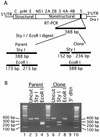
Construction of the full-length cDNA clone of WNV. Genome organization and unique restriction sites as well as their nucleotide numbers are shown at the top. Four cDNA fragments represented by thick lines were synthesized from genomic RNA through RT-PCR to cover the complete WNV genome. Individual fragments were assembled to form the full-length cDNA clone of WNV (pFLWNV) as described in Materials and Methods. The complete WNV cDNA is positioned under the control of T7 promoter elements for in vitro transcription. Three silent mutations (shown in lowercase) were engineered to create a _Sty_I site (∗) and to knock out an _Eco_RI site (Δ) in the NS5 gene. The numbers are the nucleotide positions based on the sequence from GenBank accession no. AF404756.
FIG. 2.
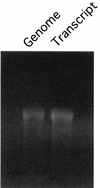
Transcription of WNV RNA. Formaldehyde-denaturing 1.0% agarose electrophoresis of RNA transcript together with genomic RNA purified from WNV.
FIG. 3.
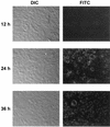
IFA of viral protein expression in cells transfected with full-length WNV RNA transcript. BHK-21 cells transfected with full-length WNV RNA transcript were analyzed by IFA at the indicated times posttransfection. Photomicrographs were taken at magnifications of ×400. The left and right panels represent the same field of view for each time point. The left panels were visualized with differential interference contrast (DIC), and the right panels were visualized with a fluorescein isothiocyanate (FITC) filter set. For the IFA, WNV immune mouse ascites fluid and goat anti-mouse immunoglobulin G antibody conjugated with FITC were used as primary and secondary antibodies, respectively.
FIG. 4.
Recombinant WNV retains the genetic markers engineered during cDNA construction. A _Sty_I site was created and an _Eco_RI site was knocked out in the NS5 gene of the recombinant virus to serve as genetic markers to distinguish recombinant virus from parental virus. A 388-bp fragment (from nt 8706 to 9093) spanning the _Sty_I or _Eco_RI site was amplified through RT-PCR from RNA extracted from either recombinant virus or parental virus. The RT-PCR fragments were subjected to _Sty_I and _Eco_RI digestion. The 388-bp fragment derived from recombinant virus should be cleaved by _Sty_I but not by _Eco_RI; the RT-PCR fragment amplified from parental viral RNA should be digested by _Eco_RI but not _Sty_I. (A) Schematic drawing of genetic marker analysis. The expected sizes of the digestion products are indicated. (B) Agarose gel analysis of genetic markers. Expected digestion pattern as depicted in panel A was observed. As a negative control, no RT-PCR products were detected from the extracted supernatant collected from cells 5 days after transfection with a mutant RNA containing a deletion of the 3′-terminal 199 nt of the genome (lane 9, 3′ dltn). A 100-bp ladder was loaded on lanes 1, 5, and 10 as a standard.
FIG. 5.
Plaque morphology of parental and clone-derived WNV on Vero cells. Vero cells in six-well plates were infected with 100 PFU of parental WNV 3356 (A) or 100 PFU of WNV derived from pFLWNV (B). Plaques were visualized 3 days postinoculation by staining for 24 h with neutral red.
FIG. 6.
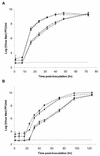
Comparison of the growth kinetics of recombinant and parental WNV. The growth of recombinant and parental viruses was compared at high and low MOIs on BHK-21 or Aedes albopictus C6/36 cells. (A) Growth in BHK-21 cells. (B) Growth in C6/36 cells. Viruses were inoculated at an MOI of 5.0 (filled symbols) or an MOI of 0.05 (open symbols) in triplicate in 12-well plates. Recombinant virus is designated by squares along a solid line. Parental virus is designated by triangles along a dashed line. Error bars represent ± standard deviation of triplicate wells. Dotted line indicates the limit of detection (500 PFU/ml).
Similar articles
- IS-98-ST1 West Nile virus derived from an infectious cDNA clone retains neuroinvasiveness and neurovirulence properties of the original virus.
Bahuon C, Desprès P, Pardigon N, Panthier JJ, Cordonnier N, Lowenski S, Richardson J, Zientara S, Lecollinet S. Bahuon C, et al. PLoS One. 2012;7(10):e47666. doi: 10.1371/journal.pone.0047666. Epub 2012 Oct 23. PLoS One. 2012. PMID: 23110088 Free PMC article. - Construction and characterization of subgenomic replicons of New York strain of West Nile virus.
Shi PY, Tilgner M, Lo MK. Shi PY, et al. Virology. 2002 May 10;296(2):219-33. doi: 10.1006/viro.2002.1453. Virology. 2002. PMID: 12069521 - Development and characterization of reverse genetics system for the Indian West Nile virus lineage 1 strain 68856.
Pavitrakar DV, Ayachit VM, Mundhra S, Bondre VP. Pavitrakar DV, et al. J Virol Methods. 2015 Dec 15;226:31-9. doi: 10.1016/j.jviromet.2015.09.008. Epub 2015 Sep 24. J Virol Methods. 2015. PMID: 26388421 - Discovery and molecular characterization of West Nile virus NY 1999.
Jordan I, Briese T, Lipkin WI. Jordan I, et al. Viral Immunol. 2000;13(4):435-46. doi: 10.1089/vim.2000.13.435. Viral Immunol. 2000. PMID: 11192290 Review. No abstract available. - Genetic systems of West Nile virus and their potential applications.
Shi PY. Shi PY. Curr Opin Investig Drugs. 2003 Aug;4(8):959-65. Curr Opin Investig Drugs. 2003. PMID: 14508880 Review.
Cited by
- IS-98-ST1 West Nile virus derived from an infectious cDNA clone retains neuroinvasiveness and neurovirulence properties of the original virus.
Bahuon C, Desprès P, Pardigon N, Panthier JJ, Cordonnier N, Lowenski S, Richardson J, Zientara S, Lecollinet S. Bahuon C, et al. PLoS One. 2012;7(10):e47666. doi: 10.1371/journal.pone.0047666. Epub 2012 Oct 23. PLoS One. 2012. PMID: 23110088 Free PMC article. - West Nile virus methyltransferase domain interacts with protein kinase G.
Keating JA, Bhattacharya D, Lim PY, Falk S, Weisblum B, Bernard KA, Sharma M, Kuhn RJ, Striker R. Keating JA, et al. Virol J. 2013 Jul 22;10:242. doi: 10.1186/1743-422X-10-242. Virol J. 2013. PMID: 23876037 Free PMC article. - In Vitro and in Vivo Evaluation of Mutations in the NS Region of Lineage 2 West Nile Virus Associated with Neuroinvasiveness in a Mammalian Model.
Szentpáli-Gavallér K, Lim SM, Dencső L, Bányai K, Koraka P, Osterhaus AD, Martina BE, Bakonyi T, Bálint Á. Szentpáli-Gavallér K, et al. Viruses. 2016 Feb 19;8(2):49. doi: 10.3390/v8020049. Viruses. 2016. PMID: 26907325 Free PMC article. - West Nile Virus NS1 Antagonizes Interferon Beta Production by Targeting RIG-I and MDA5.
Zhang HL, Ye HQ, Liu SQ, Deng CL, Li XD, Shi PY, Zhang B. Zhang HL, et al. J Virol. 2017 Aug 24;91(18):e02396-16. doi: 10.1128/JVI.02396-16. Print 2017 Sep 15. J Virol. 2017. PMID: 28659477 Free PMC article. - Human antibodies in Mexico and Brazil neutralizing tick-borne flaviviruses.
Cervantes Rincón T, Kapoor T, Keeffe JR, Simonelli L, Hoffmann HH, Agudelo M, Jurado A, Peace A, Lee YE, Gazumyan A, Guidetti F, Cantergiani J, Cena B, Bianchini F, Tamagnini E, Moro SG, Svoboda P, Costa F, Reis MG, Ko AI, Fallon BA, Avila-Rios S, Reyes-Téran G, Rice CM, Nussenzweig MC, Bjorkman PJ, Ruzek D, Varani L, MacDonald MR, Robbiani DF. Cervantes Rincón T, et al. Cell Rep. 2024 Jun 25;43(6):114298. doi: 10.1016/j.celrep.2024.114298. Epub 2024 May 29. Cell Rep. 2024. PMID: 38819991 Free PMC article.
References
- Ackermann, M., and R. Padmanabhan. 2001. De novo synthesis of RNA by the dengue virus RNA-dependent RNA polymerase exhibits temperature dependence at the initiation but not elongation phase. J. Biol. Chem. 276:39926-39937. - PubMed
- Anderson, J. F., T. G. Andreadis, C. R. Vossbrinck, S. Tirrell, E. M. Wakem, R. A. French, A. E. Garmendia, and H. J. Van Kruiningen. 1999. Isolation of West Nile virus from mosquitoes, crows, and a Cooper's hawk in Connecticut. Science 286:2331-2333. - PubMed
- Bernard, K. A., and L. D. Kramer. 2001. West Nile virus activity in the United States, 2001. Viral Immunol. 14:319-338. - PubMed
- Berthet, F. X., H. G. Zeller, M. T. Drouet, J. Rauzier, J. P. Digoutte, and V. Deubel. 1997. Extensive nucleotide changes and deletions within the envelope glycoprotein gene of Euro-African West Nile viruses. J. Gen. Virol. 78:2293-2297. - PubMed
Publication types
MeSH terms
Substances
LinkOut - more resources
Full Text Sources
Other Literature Sources
Medical
Research Materials


