The Ebola virus VP35 protein inhibits activation of interferon regulatory factor 3 - PubMed (original) (raw)
The Ebola virus VP35 protein inhibits activation of interferon regulatory factor 3
Christopher F Basler et al. J Virol. 2003 Jul.
Abstract
The Ebola virus VP35 protein was previously found to act as an interferon (IFN) antagonist which could complement growth of influenza delNS1 virus, a mutant influenza virus lacking the influenza virus IFN antagonist protein, NS1. The Ebola virus VP35 could also prevent the virus- or double-stranded RNA-mediated transcriptional activation of both the beta IFN (IFN-beta) promoter and the IFN-stimulated ISG54 promoter (C. Basler et al., Proc. Natl. Acad. Sci. USA 97:12289-12294, 2000). We now show that VP35 inhibits virus infection-induced transcriptional activation of IFN regulatory factor 3 (IRF-3)-responsive mammalian promoters and that VP35 does not block signaling from the IFN-alpha/beta receptor. The ability of VP35 to inhibit this virus-induced transcription correlates with its ability to block activation of IRF-3, a cellular transcription factor of central importance in initiating the host cell IFN response. We demonstrate that VP35 blocks the Sendai virus-induced activation of two promoters which can be directly activated by IRF-3, namely, the ISG54 promoter and the ISG56 promoter. Further, expression of VP35 prevents the IRF-3-dependent activation of the IFN-alpha4 promoter in response to viral infection. The inhibition of IRF-3 appears to occur through an inhibition of IRF-3 phosphorylation. VP35 blocks virus-induced IRF-3 phosphorylation and subsequent IRF-3 dimerization and nuclear translocation. Consistent with these observations, Ebola virus infection of Vero cells activated neither transcription from the ISG54 promoter nor nuclear accumulation of IRF-3. These data suggest that in Ebola virus-infected cells, VP35 inhibits the induction of antiviral genes, including the IFN-beta gene, by blocking IRF-3 activation.
Figures
FIG. 1.
Expression of the Ebola virus VP35 protein inhibits SeV-mediated activation but not the IFN-β-mediated activation of the ISG54 and ISG56 promoters. Shown are the levels of induction of an ISG54 promoter-driven CAT reporter gene (A and C) or of an ISG56 promoter-driven firefly luciferase reporter gene (B and D) in cells transfected with the indicated protein expression plasmids following SeV infection (A and B) or following treatment with IFN-β (C and D). Each transfection included an empty expression plasmid (empty vector) or an expression plasmid producing the viral proteins Ebola virus Zaire VP35 [VP35(Z)] or HA-tagged Ebola virus Reston VP35 [HA-VP35(R)], the influenza virus NS1 protein (NS1), or the Ebola virus Zaire NP. Vero cells were transfected with 4 μg of the indicated expression plasmid, namely, either 1 μg of an ISG-54 promoter-CAT reporter plasmid or 1 μg of an ISG56 promoter plasmid, plus 1 μg of the constitutively expressed Renilla luciferase reporter plasmid pRL-TK (an internal transfection control). At 24 h posttransfection, the cells were mock infected or infected with SeV at a multiplicity of 4 (A and B) or mock treated or treated with 1,000 units of human IFN-β/ml (C and D). Twenty-four hours postinfection, CAT and luciferase activities were determined. Activities of the inducible promoters were normalized to the luciferase activity from the Renilla luciferase plasmid. Fold induction refers to the level of induction relative to an empty plasmid-transfected, mock-infected transfection.
FIG. 2.
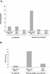
The VP35 protein of Ebola virus inhibits IRF-3-dependent induction of the mouse IFN-α4 promoter. The relative levels of IRF-3-dependent, virus-induced expression from a mouse IFN-α4 promoter reporter plasmid in the presence of empty vector, influenza virus NS1 protein, or Ebola virus VP35 are indicated following infection with SeV (A) or influenza virus delNS1 virus (B). 293T cells were calcium phosphate transfected with a mouse IFN-α4 promoter-driven CAT reporter plasmid and a constitutively expressed Renilla luciferase expression plasmid (pRL-tk), an IRF-3 expression plasmid, and an excess of either empty vector, an influenza virus NS1 expression plasmid, or VP35 expression plasmid. Eighteen hours posttransfection, the cells were infected with SeV (MOI = 1) (A) or influenza delNS1 virus (MOI = 1) (B). All transfections were normalized to the value for uninfected, empty vector-transfected cells that received the IRF-3 expression plasmid (left-most column). No reporter gene expression could be detected in cells in which IRF-3 was not overexpressed, either in mock-infected or in virus-infected cells.
FIG. 3.
The Ebola virus VP35 protein prevents the nuclear translocation of hIRF-3 after SeV infection. (A) Fluorescence images showing expression of HA-tagged VP35(R) protein (red) and the corresponding distribution of GFP-hIRF-3 (green). Cells expressing both HA-tagged VP35 and GFP-IRF-3 are indicated by the large white arrows. Examples of cells with nuclear GFP-IRF-3 are indicated by the small yellow arrows. Vero cells were transfected with 0.4 μg of VP35(R) expression plasmid plus 0.8 μg of pEGFP-C1-hIRF3 and infected 24 h later with SeV. Eight hours postinfection, cells were fixed and stained with anti-HA monoclonal antibody (red). (B) The percentage of GFP-IRF-3-expressing cells with nuclear GFP-IRF-3 in cells transfected with the indicated plasmids and either mock infected or infected with SeV is shown. Vero cells were transfected with 0.4 μg of empty vector or expression plasmids for Ebola virus Zaire VP35 [VP35(Z)], HA-tagged Ebola virus Reston VP35 [HAVP35(R)], influenza virus NS1 protein (NS1), or Ebola virus Zaire virus VP24 protein (VP24), plus 0.2 μg of pEGFP-C1-hIRF3. At 24 h posttransfection, the cells were mock infected or infected with SeV at an MOI of 10. Eight hours postinfection, the cells were examined for GFP localization. The results are the average of two independent experiments where 200 to 300 cells were counted per transfection.
FIG. 4.
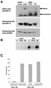
VP35 blocks dimerization and phosphorylation of IRF-3 in response to SeV infection. (A) Immunoblot analysis of IRF-3 in mock-infected (mock) and SeV-infected (SeV) 293 cells transfected with empty vector, VP35 expression plasmid, or NP expression plasmid. IRF-3 monomers and dimers were visualized following native gel electrophoresis, and different IRF-3 forms, including virus-induced, phosphorylated forms (indicated by asterisks), were visualized following SDS-PAGE of the same extracts used on the native gel. An anti-glyceraldehyde-3-phosphate dehydrogenase (GAPDH) blot is shown as a loading control. (B) Phosphorylated IRF-3 detected by [32P]orthophosphate labeling and immunoprecipitation of IRF-3 from mock- or SeV-infected 293 cells previously transfected with 1 μg of IRF-3 expression plasmid and 2 μg of empty vector, VP35 expression plasmid, or NP expression plasmid. (C) The relative ability of IRF-3 5D to activate an ISG56 promoter-driven reporter gene in Vero cells cotransfected with empty vector, VP35 expression plasmid, or NP expression plasmid. The values are reported as fold induction of the reporter gene relative to induction in cells receiving empty vector and no IRF-3 5D plasmid. All values were normalized to expression from a cotransfected, constitutively expressed Renilla luciferase reporter gene.
FIG. 5.
Ebola virus infection does not induce the activation of the ISG54 promoter or IRF-3 nuclear accumulation. (A) Ebola virus infection does not activate the ISG54 promoter. 293 cells were transfected with the IFN and IRF-3-responsive reporter plasmid pHIG54-CAT and, 24 h posttransfection, infected with the indicated viruses at an MOI of 3. Twenty-four hours posttransfection, the cells were either fixed and stained for the viral GP surface antigen (left panels) or lysed for CAT assays. The mock-infected immunofluorescence shows a background of red staining, but this is clearly distinguishable from the viral antigen staining in the infected cells. (B) Ebola virus infection did not induce GFP-IRF-3 nuclear accumulation. Cells were mock infected or infected with Ebola virus Zaire. Vero cells were transfected with the GFP-IRF-3 expression plasmid. Twenty-four hours posttransfection, the cells were mock infected or infected with Ebola virus Zaire at an MOI of 1. At 48 h postinfection, the cells were fixed and stained with an anti-GP antibody (red) and with DAPI stain (blue) to identify the nucleus. The top panels show GFP-IRF-3 and viral antigen staining; the bottom panels show the same fields as the top panels but with DAPI staining included to identify the nuclei. One field of uninfected cells is shown on the left. Two fields of virus-infected cells are shown, one in the center and one on the right.
Similar articles
- Ebola virus VP35 protein binds double-stranded RNA and inhibits alpha/beta interferon production induced by RIG-I signaling.
Cárdenas WB, Loo YM, Gale M Jr, Hartman AL, Kimberlin CR, Martínez-Sobrido L, Saphire EO, Basler CF. Cárdenas WB, et al. J Virol. 2006 Jun;80(11):5168-78. doi: 10.1128/JVI.02199-05. J Virol. 2006. PMID: 16698997 Free PMC article. - The VP35 protein of Ebola virus inhibits the antiviral effect mediated by double-stranded RNA-dependent protein kinase PKR.
Feng Z, Cerveny M, Yan Z, He B. Feng Z, et al. J Virol. 2007 Jan;81(1):182-92. doi: 10.1128/JVI.01006-06. Epub 2006 Oct 25. J Virol. 2007. PMID: 17065211 Free PMC article. - Evasion of interferon responses by Ebola and Marburg viruses.
Basler CF, Amarasinghe GK. Basler CF, et al. J Interferon Cytokine Res. 2009 Sep;29(9):511-20. doi: 10.1089/jir.2009.0076. J Interferon Cytokine Res. 2009. PMID: 19694547 Free PMC article. Review. - On the role of IRF in host defense.
Barnes B, Lubyova B, Pitha PM. Barnes B, et al. J Interferon Cytokine Res. 2002 Jan;22(1):59-71. doi: 10.1089/107999002753452665. J Interferon Cytokine Res. 2002. PMID: 11846976 Review.
Cited by
- Characterization of the RNA silencing suppression activity of the Ebola virus VP35 protein in plants and mammalian cells.
Zhu Y, Cherukuri NC, Jackel JN, Wu Z, Crary M, Buckley KJ, Bisaro DM, Parris DS. Zhu Y, et al. J Virol. 2012 Mar;86(6):3038-49. doi: 10.1128/JVI.05741-11. Epub 2012 Jan 11. J Virol. 2012. PMID: 22238300 Free PMC article. - Suppressor of Cytokine Signaling 3 Is an Inducible Host Factor That Regulates Virus Egress during Ebola Virus Infection.
Okumura A, Rasmussen AL, Halfmann P, Feldmann F, Yoshimura A, Feldmann H, Kawaoka Y, Harty RN, Katze MG. Okumura A, et al. J Virol. 2015 Oct;89(20):10399-406. doi: 10.1128/JVI.01736-15. Epub 2015 Aug 5. J Virol. 2015. PMID: 26246577 Free PMC article. - Human cytomegalovirus immediate-early 2 gene expression blocks virus-induced beta interferon production.
Taylor RT, Bresnahan WA. Taylor RT, et al. J Virol. 2005 Mar;79(6):3873-7. doi: 10.1128/JVI.79.6.3873-3877.2005. J Virol. 2005. PMID: 15731283 Free PMC article. - Innate cellular response to virus particle entry requires IRF3 but not virus replication.
Collins SE, Noyce RS, Mossman KL. Collins SE, et al. J Virol. 2004 Feb;78(4):1706-17. doi: 10.1128/jvi.78.4.1706-1717.2004. J Virol. 2004. PMID: 14747536 Free PMC article. - Molecular determinants of Ebola virus virulence in mice.
Ebihara H, Takada A, Kobasa D, Jones S, Neumann G, Theriault S, Bray M, Feldmann H, Kawaoka Y. Ebihara H, et al. PLoS Pathog. 2006 Jul;2(7):e73. doi: 10.1371/journal.ppat.0020073. PLoS Pathog. 2006. PMID: 16848640 Free PMC article.
References
- Andrejeva, J., D. F. Young, S. Goodbourn, and R. E. Randall. 2002. Degradation of STAT1 and STAT2 by the V proteins of simian virus 5 and human parainfluenza virus type 2, respectively: consequences for virus replication in the presence of alpha/beta and gamma interferons. J. Virol. 76:2159-2167. - PMC - PubMed
- Barnes, B., B. Lubyova, and P. M. Pitha. 2002. On the role of IRF in host defense. J. Interferon Cytokine Res. 22:59-71. - PubMed
- Barnes, B. J., P. A. Moore, and P. M. Pitha. 2001. Virus-specific activation of a novel interferon regulatory factor, IRF-5, results in the induction of distinct interferon alpha genes. J. Biol. Chem. 276:23382-23390. - PubMed
- Basler, C. F., and A. Garcia-Sastre. 2002. Viruses and the type I interferon antiviral system: induction and evasion. Int. Rev. Immunol., in press. - PubMed
Publication types
MeSH terms
Substances
LinkOut - more resources
Full Text Sources
Other Literature Sources
Medical
Molecular Biology Databases
Research Materials


